WWW.JNEUROSCI.ORG
-
The Journal of Neuroscience
 QUICK SEARCH:   [advanced]


-


HOME
  |  
SEARCH  |   ARCHIVE  |   SUBSCRIBE  |   CONTACT  |   HELP

This Article
Right arrow Abstract Freely available
Right arrow Full Text (PDF)
Right arrow Submit an eLetter
Right arrow Alert me when this article is cited
Right arrow Alert me when eLetters are posted
Right arrow Alert me if a correction is posted
Right arrow Citation Map
Services
Right arrow Email this article to a friend
Right arrow Similar articles in this journal
Right arrow Similar articles in Web of Science
Right arrow Similar articles in PubMed
Right arrow Alert me to new issues of the journal
Right arrow Download to citation manager
Right arrow reprints & permissions
Citing Articles
Right arrow Citing Articles via HighWire
Right arrow Citing Articles via Web of Science (602)
Right arrow Citing Articles via Google Scholar
Google Scholar
Right arrow Articles by Gould, E.
Right arrow Articles by Fuchs, E.
Right arrow Search for Related Content
PubMed
Right arrow PubMed Citation
Right arrow Articles by Gould, E.
Right arrow Articles by Fuchs, E.

 Previous Article  |  Next Article 

Volume 17, Number 7, Issue of April 1, 1997 pp. 2492-2498
Copyright ©1997 Society for Neuroscience

Neurogenesis in the Dentate Gyrus of the Adult Tree Shrew Is Regulated by Psychosocial Stress and NMDA Receptor Activation

Elizabeth Gould1, Bruce S. McEwen1, Patima Tanapat1, Liisa A. M. Galea1, and Eberhard Fuchs2

1 The Rockefeller University, New York, New York 10021, and 2 The German Primate Center, 37077 Göttingen, Germany

ABSTRACT
INTRODUCTION
MATERIALS AND METHODS
RESULTS
DISCUSSION
FOOTNOTES
REFERENCES


ABSTRACT

These studies were designed to determine whether adult neurogenesis occurs in the dentate gyrus of the tree shrew, an animal phylogenetically between insectivores and primates, and to explore the possibility that this process is regulated by stressful experiences and NMDA receptor activation. We performed immunohistochemistry for cell-specific markers and the thymidine analog bromodeoxyuridine (BrdU), a marker of DNA synthesis that labels proliferating cells and their progeny, on the brains of adult tree shrews subjected to psychosocial stress or NMDA receptor antagonist treatment. Cells that incorporated BrdU in the dentate gyrus of adult tree shrews were primarily located in the subgranular zone, had morphological characteristics of granule neuron precursors, and appeared to divide within 24 hr after BrdU injection. Three weeks after BrdU injection, BrdU-labeled cells had neuronal morphology, expressed the neuronal marker neuron specific enolase, and were incorporated into the granule cell layer. Vimentin-immunoreactive radial glia were observed in the dentate gyrus with cell bodies in the subgranular zone and processes extending into the granule cell layer.

Exposure to acute psychosocial stress resulted in a rapid decrease in the number of BrdU-labeled cells in the dentate gyrus. In contrast, blockade of NMDA receptors, with the NMDA receptor antagonist MK-801, resulted in an increase in the number of BrdU-labeled cells in the dentate gyrus. These results indicate that adult neurogenesis occurs in the tree shrew dentate gyrus and is regulated by a stressful experience and NMDA receptor activation. Furthermore, we suggest that these characteristics may be common to most mammalian species.

Key words: neurogenesis; tree shrew; psychosocial stress; granule neurons; dentate gyrus; NMDA receptors


INTRODUCTION

In most brain regions, the production of neurons is typically confined to a discrete developmental period. In contrast, granule neurons of the dentate gyrus are produced during an extended period that begins during gestation and spans well into the postnatal period in all mammalian species that have been examined, including the mouse, rat, guinea pig, rabbit, cat, and rhesus monkey (Angevine, 1965; Altman and Das, 1967; Schlessinger et al., 1975; Rakic and Nowakowski, 1981; Gueneau et al., 1982; Wyss and Sripanidkulchai, 1985). In the rat, granule neurons continue to be produced in adulthood from a pool of precursor cells that reside within the dentate gyrus (Kaplan and Hinds, 1977; Kaplan and Bell, 1984). The granule neurons produced in adulthood seem to migrate into the granule cell layer along existing radial glia, extend axons, make synaptic contacts, and express neuronal markers (Kaplan and Bell, 1984; Stanfield and Trice, 1988; Cameron et al., 1993; Okano et al., 1993; Kuhn et al., 1996).

The production of granule neurons in the adult rat dentate gyrus has been shown to be suppressed by adrenal steroids (Cameron and Gould, 1994) and excitatory input via the NMDA receptor subtype of glutamate receptors (Cameron et al., 1995). Because both circulating adrenal steroid levels and glutamate-mediated excitatory input to the hippocampus are enhanced by stress (Krugers et al., 1993; Moghaddam et al., 1994; Bartanusz et al., 1995), it is possible that stressful experiences naturally modulate the production of granule neurons in the dentate gyrus of adult animals. This possibility is supported by a recent study showing that cell proliferation in the dentate gyrus of adult rats can be suppressed by exposure to predator odor, a naturally aversive experience (Galea et al., 1996).

Although the existence of neurogenesis in the dentate gyrus of adult rhesus monkeys has been dismissed (Eckenhoff and Rakic, 1988), this phenomenon has not been investigated in other species of primates or tree shrews. The present studies were undertaken to determine whether granule cell production and its regulation by stressful experience and excitatory input occur in the dentate gyrus of the adult tree shrew Tupaia belangeri. Tree shrews, which are considered to be phylogenetically between insectivores and primates (Martin, 1990), have been studied extensively in a well characterized model of psychosocial stress. When paired with a same-sex conspecific, tree shrews rapidly establish a potent, enduring dominant/subordinate relationship that is particularly stressful to the subordinate animal (von Holst, 1972). To determine whether granule cell production occurs in the dentate gyrus of adult tree shrews, immunohistochemistry for cell-specific markers and the thymidine analog bromodeoxyuridine (BrdU), a marker of DNA synthesis, was performed on the brains of adult tree shrews exposed to psychosocial stress or NMDA receptor blockade.


MATERIALS AND METHODS

Animal care and treatment Adult (7 months to 2.5 years old) male tree shrews (Tupaia belangeri) from the breeding colony at the German Primate Center (Göttingen, Germany) were used in all experiments. Tree shrews are diurnal animals that reach sexual maturity between 4 and 5 months and have a lifespan of up to 12 years in captivity. All animal experimentation was conducted in accordance with the National Institutes of Health (NIH) Guide for the Care and Use of Laboratory Animals (NIH Publication 85-23, revised 1985) and were approved by the government of Lower Saxony, Germany. The animals were housed individually on a 12 hr/12 hr light/dark cycle with artificial illumination from 8:00 A.M. to 8:00 P.M. in air conditioned rooms (for details, see Fuchs and Schumacher, 1990). All treatments were performed during the day (lights on).

Experiment 1 To determine whether cells in the dentate gyrus of adult tree shrews incorporate BrdU and divide, tree shrews were given a single i.p. injection of BrdU [Sigma, St. Louis, MO; 75 mg/kg in saline and 0.007 N NaOH (this dose was used in all experiments)]. After a survival time of 2 or 24 hr, the tree shrews (n = 3 for each group) were anesthetized and perfused transcardially (see below). The 2 hr survival time allows for incorporation of BrdU by cells synthesizing DNA but not for completion of mitosis (Nowakowski et al., 1989). BrdU is available for uptake into cells synthesizing DNA for approximately 2 hr (Packard et al., 1973). The 24 hr survival time allows for the completion of at least one cell cycle by cells in S phase at the time of BrdU injection (Nowakowski et al., 1989).

Experiment 2 To determine whether cells that incorporate BrdU survive and express the neuronal marker neuron specific enolase (NSE), tree shrews (n = 3) were given a single i.p. injection of BrdU and were anesthetized and perfused transcardially 3 weeks later. In the dentate gyrus of the adult rat, mature granule neurons, but not glia, express NSE (Cameron et al., 1993). The majority of cells newly generated in the dentate gyrus of adult rats have been shown to express NSE by 3 weeks after incorporation of [3H]thymidine during DNA synthesis (Cameron et al., 1993).

Experiment 3 To determine whether cell proliferation can be modulated by a stressful experience, tree shrews exposed to acute psychosocial stress were examined (Fuchs et al., 1996). The experimental induction of psychosocial conflict was carried out as follows. An opaque partition between the neighboring cages of two males unknown to one another was removed. This resulted in active competition for control over the enlarged territory and the establishment of an obvious dominant/subordinate relationship. Under these conditions, the subordinate animal became virtually immobile, showed tail ruffling, and elicited alarm cries, whereas the dominant tree shrew maintained a normal level and sphere of activity. After 1 hr, the animals were separated again by the opaque partition and the subordinate tree shrews (n = 3) were given injections of BrdU. Subordinate tree shrews demonstrate elevated urinary cortisol levels at this time point compared with both control and dominant tree shrews (Magarinos et al., 1996). Two hours after BrdU injection, the animals were anesthetized and perfused transcardially. These animals were compared to tree shrews (n = 3) that were not exposed to a stressful experience but received a single BrdU injection, followed by a 2 hr survival time.

Experiment 4 To determine whether the production of cells in the dentate gyrus of adult tree shrews is affected by NMDA receptor activation, tree shrews were given injections of the specific noncompetitive NMDA receptor antagonist MK-801 (1.0 mg/kg in saline, i.p.; gift of Merck Research Laboratories, Rahway, NJ) or saline (n = 3 for each group). This dose has been shown to stimulate granule cell production in the dentate gyrus of adult rats (Cameron et al., 1995). Two hours after MK-801 treatment, the animals were given injections of BrdU and were anesthetized and perfused transcardially after a 2 hr survival time.

Histological procedures After treatments, the tree shrews received an overdose of Rompum/Ketanest and were perfused transcardially with 4.0% paraformaldehyde in 0.1 M phosphate buffer (PB). The heads were postfixed overnight, and the brains were removed from the skulls on the following day. Brain sections (40 µm) were cut on an oscillating tissue slicer in a bath of PB.

BrdU and vimentin immunohistochemistry For BrdU immunohistochemistry, the sections were permeabilized with 0.5% (v/v) Triton X-100 in PBS for 1 hr, incubated with Pronase E (3 µg/ml) in PBS at 37°C for 20 min, denatured in 2 N HCL for 20 min, rinsed twice in PBS, and incubated overnight at 4°C with monoclonal antibody (mAb) against BrdU (Novocastra, Newcastle upon Tyne, United Kingdom; 1:100 in PBS). The sections were rinsed in PBS, incubated for 1 hr in biotinylated mouse secondary antisera (Vector Laboratories, Burlingame, CA; 1:50 in PBS), rinsed in PBS, incubated for 1 hr in avidin-biotin-horseradish peroxidase (Vector, 1:50 in PBS), rinsed in PBS, and reacted with diaminobenzidine and hydrogen peroxide in PBS. After rinsing in PBS, the sections were mounted onto gelatinized glass slides and stained for Nissl using cresyl violet.

Some brain sections that were not stained for BrdU were incubated overnight in mAb against vimentin (Boehringer Mannheim, Indianapolis, IN; clone V9, 1:50 in PBS). Vimentin, a cytoskeletal protein of immature astroglia, is expressed by radial glia in the dentate gyrus of adult rats (Gould et al., 1992). After this, the sections were processed for avidin-biotin-horseradish peroxidase immunohistochemistry as described above for BrdU staining.

Combined BrdU and NSE immunohistochemistry Two different combined immunohistochemical methods were used to visualize BrdU and NSE on the same brain section. In the first method, the sections were reacted for BrdU immunohistochemistry as described above. After several rinses in PBS, the sections were incubated overnight in polyclonal antisera against NSE (Polysciences, Warrington, PA; 1:2000 in PBS). The sections were then rinsed in PBS and incubated for 2 hr in FITC-conjugated rabbit antisera (Vector, 1:50 in PBS) with goat normal serum, rinsed again, and mounted onto gelatinized slides and coverslipped under Crystal Mount (Biomeda Corp., Foster City CA). In the second method, the sections were reacted for BrdU immunohistochemistry as described above, with the exception that the solution of diaminobenzidine and hydrogen peroxide in PBS contained 2.5% nickel sulfate. The sections were then rinsed several times in PBS and incubated overnight in polyclonal antisera against NSE (Polysciences, 1:2000 in PBS). After several rinses in PBS, the sections were incubated for 1 hr in biotinylated rabbit antisera and reacted using the avidin-biotin-horseradish peroxidase protocol as described above.

Data analysis For each experiment, the slides were coded before quantitative analysis, and the code was not broken until the analysis was complete. For each brain, at least six sections were selected for analysis from middle to caudal dentate gyrus (between levels A 2.0 and A 3.0; see Tigges and Shantha, 1969). For each selected section, the number of BrdU-labeled cells was counted in the dentate gyrus [the granule cell layer (gcl) and hilus combined]. The cross-sectional area of the dentate gyrus was determined by use of a Zeiss Interactive Digitizing Analysis System (ZIDAS), and the data were expressed as densities (number of cells/mm2). Means were determined for these variables, and the data were subjected to two-tailed Student's t tests. For brain sections immunostained for both BrdU and NSE, the percentage of BrdU-labeled cells that were NSE-immunoreactive was determined.


RESULTS

BrdU incorporation in the dentate gyrus of adult tree shrews with 2 hr or 24 hr survival times

The dentate gyrus of tree shrews processed for BrdU immunohistochemistry at 2 and 24 hr survival times after BrdU injection revealed BrdU-labeled cells throughout the dentate gyrus at all levels examined (Figs. 1, 2, 3). These cells were typically observed in the subgranular zone (sgz), on the border of the gcl and the hilus (Figs. 1, 2, 3, 4), as well as within the gcl and hilus. There were no obvious differences in the distribution of BrdU-immunoreactive cells at different levels of the dentate gyrus for either time point (Figs. 1, 2, 3).
Fig. 1. 
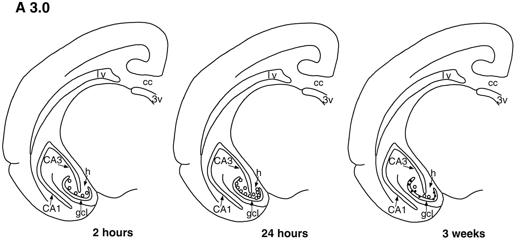
Template showing a coronal half-section of the tree shrew brain at level A 3.0 (see Tigges and Shantha, 1969), demonstrating that the number and distribution of BrdU-labeled cells (open circles) change with increased survival time after BrdU injection. Two hours after BrdU injection, most labeled cells are located in the sgz, on the border of the gcl and hilus. Twenty-four hours after BrdU injection, more than twice as many BrdU-labeled cells are observed in the dentate gyrus, predominantly in the sgz. By 3 weeks after BrdU injection, most BrdU-labeled cells are located in the gcl and express NSE (solid dots). cc, Corpus callosum; lv, lateral ventricle; 3V, third ventricle; h, hilus; CA1, CA1 region; CA3, CA3 region.
[View Larger Version of this Image (19K GIF file)]


Fig. 2. Template showing a coronal half-section of the tree shrew brain at level A 2.5 (see Tigges and Shantha, 1969), showing changes in the number and distribution of BrdU-labeled cells (open circles) with increased survival time after BrdU injection. Two hours after BrdU injection, most labeled cells are located in the sgz, between the gcl and hilus. Twenty-four hours after BrdU injection, more than two times as many BrdU-labeled cells are observed in the dentate gyrus, but their distribution remains similar to the 2 hr time point. Three weeks after BrdU injection, most BrdU-labeled cells are located in the gcl and are NSE-immunoreactive (solid dots). cc, Corpus callosum; lv, lateral ventricle; 3V, third ventricle; h, hilus; CA1, CA1 region; CA3, CA3 region.
[View Larger Version of this Image (20K GIF file)]


Fig. 3. Template showing a coronal half-section of the tree shrew brain at level A 2.0 (see Tigges and Shantha, 1969), indicating changes in the number and distribution of BrdU-labeled cells (open circles) with increased survival time after BrdU injection. Most BrdU-labeled cells are located in the sgz, between the gcl and hilus at the 2 and 24 hr time points. Three weeks after BrdU injection, most BrdU-labeled cells are located in the gcl and are NSE-immunoreactive (solid dots). cc, Corpus callosum; 3V, third ventricle; h, hilus; CA1, CA1 region; CA3, CA3 region.
[View Larger Version of this Image (23K GIF file)]


Fig. 4. Examples of cell types in the dentate gyrus of the adult tree shrew. A, BrdU-labeled cell (arrows) in the sgz of the dentate gyrus with morphological characteristics of granule cell precursor, i.e., round or oval medium-sized cell body. B, Cluster of BrdU-labeled cells (arrows) in the sgz of the dentate gyrus after MK-801 treatment. C, Vimentin-immunoreactive cell with radial glial morphology, i.e., irregular shaped cell body with radial process. The cell body is located on the border of the hilus and gcl, and the process extends through the gcl. g, Granule cell layer; h, hilus. Scale bar in B, 20 µm (applies to all frames).
[View Larger Version of this Image (99K GIF file)]

Most of the BrdU-labeled cells (~85%) had the morphological characteristics of granule cell precursors, i.e., round or oval, medium-sized cell bodies (Fig. 4). The remaining BrdU-immunoreactive cells had the morphological characteristics of glial cells, i.e., triangular or irregular, small cell bodies. Quantitative analysis revealed a significant increase in the number of BrdU-labeled cells in the dentate gyrus (>2 times) between 2 and 24 hr after BrdU injection [t(4) = -7.337; p < 0.005, see Table 1]. Despite this increase in cell number, the location of the BrdU-labeled cells did not change between 2 and 24 hr (Figs. 1, 2, 3). Moreover, the cross-sectional area of the dentate gyrus did not change between 2 and 24 hr after BrdU injection (2 hr = 1.5 ± 0.02 mm2, 24 hr = 1.5 ± 0.01 mm2; p < 0.4).

Table 1. The number of proliferating cells in the dentate gyrus of adult tree shrews at different survival times after BrdU injection, as well as after psychosocial stress or MK-801 treatment


Number of BrdU-labeled cells in the dentate gyrus/mm2
Time course experiment
Stress experiment
MK-801 experiment
2 hr 24 hr Control Subordinate Control MK-801

7.9  ± 1.5 22.7  ± 1.3* 6.8  ± 1.3* 1.6  ± 0.3* 11.8  ± 2.1 19.8  ± 1.7*

Values represent mean ± SEM each obtained from three tree shrews. Asterisks represent significant difference from 2 hr (time course experiment) or control (stress or MK-801 experiment); p < 0.05, unpaired Student's t tests.

Vimentin immunoreactivity in the dentate gyrus of adult tree shrews

Light microscopic examination of vimentin-immunoreactive tissue revealed numerous stained cells with the morphological characteristics of radial glia, i.e., triangular shaped cell bodies with radial processes (Fig. 4). These cells were usually oriented with the cell bodies in the sgz and the processes extending through the gcl (Fig. 4). There were no detectable differences in the distribution of vimentin-immunoreactive cells at different levels of the dentate gyrus.

Combined BrdU and NSE immunohistochemistry after a 3 week survival time

Three weeks after BrdU injection, numerous BrdU-labeled cells (~20 per section) remained detectable in the dentate gyrus (Figs. 1, 2, 3). These cells were located predominantly in the gcl, although a few remained in the sgz and hilus. Both immunohistochemical methods for double labeling BrdU and NSE yielded similar results; ~80% of the BrdU-labeled cells were NSE-immunoreactive. All of the cells that were labeled with BrdU and NSE were located in the gcl (Figs. 1, 2, 3) and had the morphological characteristics of granule neurons.

Effects of psychosocial stress on the number of BrdU-labeled cells in the dentate gyrus

Exposure to a single episode of acute psychosocial stress that lasted 1 hr resulted in a significant decrease in the density of BrdU-labeled cells in the dentate gyrus compared to unstressed controls [t(4) = -3.807; p < 0.05; Table 1]. No change in the cross-sectional area of the dentate gyrus was observed after acute psychosocial stress (control = 1.5 ± 0.5 mm2, stress = 1.2 ± 0.4 mm2; p < 0.4), indicating that this difference reflects a change in the number of proliferating cells. The morphological characteristics of BrdU-labeled cells did not differ between the tree shrews subjected to psychosocial stress and unstressed control animals.

Effects of NMDA receptor antagonist treatment on the number of BrdU-labeled cells in the dentate gyrus

Treatment with the NMDA receptor antagonist MK-801 resulted in a significant increase in the density of BrdU-labeled cells in the dentate gyrus [t(4) = -2.945, p < 0.05; Table 1). No change in the cross-sectional area of the dentate gyrus was observed after MK-801 treatment (control = 1.4 ± 0.3 mm2; MK-801-treated = 1.1 ± 0.2 mm2; p < 0.4). In MK-801-treated tree shrews, BrdU-labeled cells had morphological characteristics that were similar to those of controls, and these cells were often observed in clusters of 3-5 on the border of the gcl and hilus (Fig. 4).


DISCUSSION

The results of this report demonstrate that cells in the dentate gyrus of adult tree shrews incorporate BrdU, a marker of DNA synthesis that labels proliferating cells and their progeny. The observation that the number of BrdU-labeled cells more than doubles between 2 and 24 hr suggests that cells synthesizing DNA in the dentate gyrus divide at least once within a 24 hr period. Furthermore, the detection of BrdU-labeled cells that express a neuronal marker and have granule cell morphology 3 weeks after BrdU incorporation suggests that many proliferating cells in the dentate gyrus of adult tree shrews eventually become granule neurons. Moreover, the presence of radial glia with cell bodies in the sgz and processes extending through the gcl, as well as the change in location of BrdU-labeled cells from 24 hr to 3 weeks, suggests that newly generated cells migrate from the hilus or sgz to the gcl. Finally, these results also show that the production of cells in the dentate gyrus of adult tree shrews can be modulated rapidly by exposure to a stressful social encounter, as well as by changing the level of NMDA receptor activation.

Granule cell generation across mammalian species

Previous studies have demonstrated several characteristics of dentate gyrus granule cell production that are common to numerous mammalian species, from rats to rhesus monkeys. First, granule cell production occurs during an extended period that begins during gestation and continues into the postnatal period (Schlessinger et al., 1975; Rakic and Nowakowski, 1981; Altman and Bayer, 1990a,b). This feature seems to be unrelated to the degree of maturity at birth as both altricial species (e.g., the rat) and precocial species (e.g., the guinea pig) demonstrate this phenomenon (Altman and Das, 1967; Schlessinger et al., 1975). Second, suprapyramidal to infrapyramidal and outside-in gradients of granule cell generation exist (Schlessinger et al., 1975; Rakic and Nowakowski, 1981). Third, the location of granule cell precursors changes during development; precursor cells originate in the subependymal layer, next reside in the hilus, and finally in the sgz (Rakic and Nowakowski, 1981; Altman and Bayer, 1990a,b). However, there are characteristics of granule cell production that are not shared by all mammals examined. One prominent difference in granule cell generation between the rat and the monkey is in the developmental stage at which the majority of neurons are produced. In the rat, most (~80%) granule cells are produced postnatally, whereas in the rhesus monkey, most are produced prenatally (Schlessinger et al., 1975; Rakic and Nowakowski, 1981).

The extension of granule neuron production into adulthood has been well documented in the rat (Kaplan and Bell, 1984; Stanfield and Trice, 1988; Cameron et al., 1993; Okano et al., 1993) and recently demonstrated in the meadow vole, another species of rodent (Galea and McEwen, 1995). In contrast, few studies have investigated the possibility that adult neurogenesis occurs in the dentate gyrus of nonrodent mammalian species. One report dismissed the possibility that granule cells are produced in adulthood in the dentate gyrus of rhesus monkeys and suggested that adult neurogenesis is a phenomenon unique to rats, particularly strains that increase in size throughout life (Eckenhoff and Rakic, 1988). However, it is unlikely that considerable growth in adulthood is the basis for adult neurogenesis because the guinea pig, an animal that undergoes little brain growth during the postnatal period, appears to produce granule neurons in adulthood (Altman and Das, 1967). Rather, it appears that the mammalian dentate gyrus is a specialized brain region in which neurogenesis continues into adulthood regardless of the developmental pattern of the animal. In light of recent findings, the possibility that adult neurogenesis occurs in the dentate gyrus of most mammals, including other primate species, should be reconsidered. First, the results of this study have demonstrated granule neuron production and its regulation by neural activity and behavioral events in the dentate gyrus of the adult tree shrew. Second, we have observed BrdU-labeled cells with the morphology of immature granule neurons, as well as vimentin-immunoreactive radial glia, in the dentate gyrus of adult marmoset monkeys (Callithrix jacchus) and an adult cynomolgus (Macaca fascicularis) monkey (our unpublished observations). Moreover, the recent observations that precursor cells capable of generating neurons exist in the subependymal layer of adult humans (Kirschenbaum et al., 1994) suggest a reevaluation of the limits of neurogenesis across species in other regions as well.

Significance of radial glia in the adult brain

During development, radial glia are believed to participate in the migration of immature neurons from their site of origin to their final destination. The results of this study and our previous report (Gould et al., 1992) indicate that the dentate gyrus maintains a population of radial glia in adulthood in both the tree shrew and rat. In addition, cells produced in the dentate gyrus of both the tree shrew and the rat (Cameron et al., 1993) move from the hilus or sgz to the gcl after DNA synthesis, suggesting that radial glia guide migrating granule cells in adulthood. It should be noted, though, that the processes of radial glia are considerably longer than the distances immature granule cells appear to migrate in the adult. A recent report indicates that radial glia in the dentate gyrus of adult rats are associated with the dendrites of immature granule neurons (Seki and Arai, 1996), presenting the possibility that radial glia may serve other developmental functions, in addition to migration, such as supporting the growing dendritic tree.

NMDA receptor activation regulates cell proliferation

The results of this study also suggest that intrinsic factors regulating the production of granule neurons in the adult dentate gyrus may be common to mammalian species that undergo adult neurogenesis. Treatment with MK-801, a noncompetitive NMDA receptor antagonist, resulted in a rapid increase in the number of cells synthesizing DNA, suggesting that NMDA receptor activation normally inhibits cell proliferation in the dentate gyrus of the tree shrew. These results are consistent with our previous studies demonstrating that NMDA receptor blockade or activation increases or decreases, respectively, the production of granule cells in the dentate gyrus of adult rats (Cameron et al., 1995). Excitation may inhibit neurogenesis by preventing the synthesis or release of mitogenic factors from cells in the vicinity of precursor cells. Several studies have shown that stressful experiences rapidly stimulate glutamate release and alter the expression of NMDA receptors in the hippocampus of the rat (Krugers et al., 1993; Moghaddam et al., 1994; Bartanusz et al., 1995). Collectively, these results suggest that stressful experiences inhibit the production of granule neurons via actions on excitatory pathways to the dentate gyrus.

Psychosocial stress inhibits cell proliferation

The functional consequences of adult neurogenesis in the dentate gyrus are presently unknown. One approach to elucidating the function of granule neuron production in adulthood is to identify the factors that regulate its occurrence. The results of this study have shown that a brief exposure to psychosocial stress, during which a dominant/subordinate relationship is established, results in a rapid decrease in the number of cells that incorporate BrdU in the dentate gyrus of the subordinate animal. After a single encounter with a dominant tree shrew, the subordinate will remain in a constant state of arousal in the presence of the dominant animal. This stress reaction is characterized by a reduced sphere of activity, vigilance, alarm cries, and tail ruffling (von Holst, 1972). When removed from the presence of the dominant after the initial encounter, subordinate tree shrews will recover physically but will demonstrate immediate stress reactions after subsequent exposures to the dominant (von Holst, 1972), indicating that a potent memory of the experience has been formed. Because the hippocampus has been implicated in spatial learning and memory (Sutherland et al., 1983; Wishaw, 1987; McNaughton et al., 1989), as well as in contextual learning in conditioned fear paradigms (Phillips and LeDoux, 1992), it is possible that stress-induced stabilization of the granule cell population may be necessary for learning during a threatening social encounter. The production of new granule neurons for increased storage capacity may be permitted only during periods of low stress when learning is not critical.

In the tree shrew, the stress of subordination is mediated by visual and not olfactory cues; exposure to the olfactory markings or the odor of the dominant does not elicit a reaction (von Holst, 1972). We have shown that exposure of adult rats to fox odor, which increases both adrenal steroid levels and excitatory input (Heale et al., 1994; Vernet-Maury et al., 1984), rapidly inhibits cell proliferation in the dentate gyrus (Galea et al., 1996). Collectively, these findings suggest that rapid suppression of cell proliferation by a threatening experience, conveyed via cues from different sensory modalities, is a characteristic of the dentate gyrus that is common to mammalian species that undergo adult neurogenesis.


FOOTNOTES

Received Oct. 11, 1996; revised Dec. 3, 1996; accepted Jan. 10, 1997.


This work was supported by National Institute of Mental Health Grant MH52423 and a National Alliance for Research on Schizophrenia and Depression Young Investigator Award (E.G.). We thank Dr. Gabrielle Flugge for assistance with templates.

Correspondence should be addressed to Dr. Elizabeth Gould, Department of Psychology, Green Hall, Princeton University, Princeton, NJ 08544.


REFERENCES

  • Altman J, Bayer SA (1990a) Mosaic organization of the hippocampal neuroepithelium and the multiple germinal sources of dentate granule cells. J Comp Neurol 301:325-342 . [Web of Science][Medline]
  • Altman J, Bayer SA (1990b) Migration and distribution of two populations of hippocampal granule cell precursors during the perinatal and postnatal periods. J Comp Neurol 301:365-381 . [Web of Science][Medline]
  • Altman J, Das GD (1967) Postnatal neurogenesis in the guinea-pig. Nature 214:1098-1101 . [Medline]
  • Angevine JB (1965) Time of neuron origin in the hippocampal region: an autoradiographic study in the mouse. Exp Neurol 2:1-17.
  • Bartanusz V, Aubry J-M, Pagliusi S, Jezova D, Baffi J, Kiss JZ (1995) Stress-induced changes in messenger RNA levels of N-methyl-D-aspartate and AMPA receptor subunits in selected regions of the rat hippocampus and hypothalamus. Neuroscience 66:247-252 . [Web of Science][Medline]
  • Cameron HA, Gould E (1994) Adult neurogenesis is regulated by adrenal steroids in the dentate gyrus. Neuroscience 61:203-209 . [Web of Science][Medline]
  • Cameron HA, McEwen BS, Gould E (1995) Regulation of adult neurogenesis by excitatory input and NMDA receptor activation in the dentate gyrus. J Neurosci 15:4687-4692 . [Abstract]
  • Cameron HA, Woolley CS, McEwen BS, Gould E (1993) Differentiation of newly born neurons and glia in the dentate gyrus of the adult rat. Neuroscience 56:337-344 . [Web of Science][Medline]
  • Eckenhoff MF, Rakic P (1988) Nature and fate of proliferative cells in the hippocampal dentate gyrus during the life span of the rhesus monkey. J Neurosci 8:2729-2747 . [Abstract]
  • Fuchs E, Schumacher M (1990) Psychosocial stress affects pineal function in the tree shrew (Tupaia belangeri). Physiol Behav 47:713-717 . [Medline]
  • Fuchs E, Kramer M, Hermes B, Netter B, Hiemke C (1996) Psychosocial stress in tree shrews: clomipramine counteracts behavioral and endocrine changes. Pharmacol Biochem Behav 54:219-228 . [Web of Science][Medline]
  • Galea LAM, McEwen BS (1995) Sex differences in adult neurogenesis in the wild-trapped meadow vole. Soc Neurosci Abstr 21:1287.
  • Galea LAM, Tanapat P, Gould E (1996) Exposure to predator odor suppresses cell proliferation in the dentate gyrus of adult rats via a cholinergic mechanism. Soc Neurosci Abstr 22:1196.
  • Gould E, Cameron HA, Daniels DC, Woolley CS, McEwen BS (1992) Adrenal hormones suppress cell division in the adult rat dentate gyrus. J Neurosci 12:3642-3650 . [Abstract]
  • Gueneau G, Privat A, Drouet J, Court L (1982) Subgranular zone of the dentate gyrus of young rabbits as a secondary matrix. Dev Neurosci 5:345-358 . [Web of Science][Medline]
  • Heale VR, Vanderwolf CH, Kavaliers M (1994) Components of weasel and fox odors elicit fast wave bursts in the dentate gyrus of rats. Behav Brain Res 63:159-165 . [Web of Science][Medline]
  • Kaplan MS, Bell DH (1984) Mitotic neuroblasts in the 9-day-old and 11-month-old rodent hippocampus. J Neurosci 4:1429-1441 . [Abstract]
  • Kaplan MS, Hinds JW (1977) Neurogenesis in the adult rat: electron microscopic analysis of light radioautographs. Science 197:1092-1094 . [Abstract/Free Full Text]
  • Kirschenbaum B, Nedergaard M, Preuss A, Barami K, Fraser RA, Goldman SA (1994) In vitro neuronal production and differentiation by precursor cells derived from the adult human forebrain. Cereb Cortex 4:576-589 . [Abstract/Free Full Text]
  • Krugers HJ, Koolhaas JM, Bohua B, Korf J (1993) A single stress experience alters glutamate receptor-binding in rat hippocampal CA3 area. Neurosci Lett 154:73-77 . [Web of Science][Medline]
  • Kuhn HG, Dickinson-Anson H, Gage FH (1996) Neurogenesis in the dentate gyrus of the adult rat: age-related decrease in neuronal progenitor population. J Neurosci 16:2027-2033 . [Abstract/Free Full Text]
  • Magarinos AM, McEwen BS, Flugge G, Fuchs E (1996) Chronic psychosocial stress causes apical dendritic atrophy of hippocampal CA3 pyramidal neurons in subordinate tree shrews. J Neurosci 16:3534-3540 . [Abstract/Free Full Text]
  • Martin RD (1990) In: Primate origins and evolution. London: Chapman & Hall.
  • McNaughton BL, Barnes CA, Meltzer J, Sutherland RJ (1989) Hippocampal granule cells are necessary for normal spatial learning but not for spatially-selective pyramidal cell discharge. Exp Brain Res 76:485-496 . [Web of Science][Medline]
  • Moghaddam B, Boliano ML, Stein-Behrens B, Sapolsky R (1994) Glucocorticoids mediate the stress-induced extracellular accumulation of glutamate. Brain Res 655:251-154 . [Web of Science][Medline]
  • Nowakowski RS, Lewin SB, Miller MW (1989) Bromodeoxyuridine immunohistochemical determination of the lengths of the cell cycle and the DNA-synthetic phase for an anatomically defined population. J Neurocytol 18:311-318 . [Web of Science][Medline]
  • Okano HJ, Pfaff DW, Gibbs RB (1993) RB and Cdc2 expression in brain: correlations with [3H]thymidine incorporation and neurogenesis. J Neurosci 13:2930-2938 . [Abstract]
  • Packard DS, Menzies RA, Skalko RG (1973) Incorporation of thymidine and its analog, bromodeoxyuridine, into embryos and maternal tissues of the mouse. Differentiation 1:397-405 . [Medline]
  • Phillips RG, LeDoux JE (1992) Differential contribution of amygdala and hippocampus to cued and contextual fear conditioning. Behav Neurosci 106:274-285 . [Web of Science][Medline]
  • Rakic P, Nowakowski RS (1981) The time of origin of neurons in the hippocampal region of the rhesus monkey. J Comp Neurol 196:99-128 . [Web of Science][Medline]
  • Schlessinger AR, Cowan WM, Gottlieb DI (1975) An autoradiographic study of the time of origin and the pattern of granule cell migration in the dentate gyrus of the rat. J Comp Neurol 159:159-176.
  • Seki T, Arai Y (1996) Dendritic development of newly-generated granule cells in the adult hippocampus occurs upon contact with radial glia-like cells. Soc Neurosci Abstr 22:534.
  • Stanfield BB, Trice JE (1988) Evidence that granule cells generated in the dentate gyrus of adult rats extend axonal projections. Exp Brain Res 72:399-406 . [Web of Science][Medline]
  • Sutherland RJ, Whishaw IQ, Kolb B (1983) A behavioural analysis of spatial localization following electrolytic, kainate- or colchicine-induced damage to the hippocampal formation in the rat. Behav Brain Res 7:133-153 . [Web of Science][Medline]
  • Tigges J, Shantha TR (1969) In: A stereotaxic atlas of the tree shrew (Tupaia glis). Baltimore: Williams & Wilkins.
  • Vernet-Maury E, Polak EH, Demael A (1984) Structure-activity relationship of stress-inducing odorants in the rat. J Chem Ecol 10:1007-1019.
  • von Holst D (1972) Social stress in the tree-shrew: its causes and physiological and ethological consequences. In: Prosimian biology (Martin RD, Doyle GA, Walker AC, eds), pp 389-411. Philadelphia: University of Pittsburgh.
  • Whishaw IQ (1987) Hippocampal, granule cell and CA3-4 lesions impair formation of a place learning-set in the rat and induce reflex epilepsy. Behav Brain Res 24:59-72 . [Web of Science][Medline]
  • Wyss JM, Sripanidkulchai B (1985) The development of Ammon's horn and the fascia dentata in the cat: a [3H]thymidine analysis. Brain Res 350:185-198 . [Medline]

Copyright ©1997 Society for Neuroscience   0270-6474/1997/172492-07$05.00/0


This article has been cited by other articles:


Home page
J. Exp. Biol.Home page
K. Ghosal, M. Gupta, and K. A. Killian
Agonistic behavior enhances adult neurogenesis in male Acheta domesticus crickets
J. Exp. Biol., July 1, 2009; 212(13): 2045 - 2056.
[Abstract] [Full Text] [PDF]


Home page
J. Neurosci.Home page
L. Torner, S. Karg, A. Blume, M. Kandasamy, H.-G. Kuhn, J. Winkler, L. Aigner, and I. D. Neumann
Prolactin Prevents Chronic Stress-Induced Decrease of Adult Hippocampal Neurogenesis and Promotes Neuronal Fate
J. Neurosci., February 11, 2009; 29(6): 1826 - 1833.
[Abstract] [Full Text] [PDF]


Home page
J. Neurosci.Home page
A. J. Eisch, H. A. Cameron, J. M. Encinas, L. A. Meltzer, G.-L. Ming, and L. S. Overstreet-Wadiche
Adult Neurogenesis, Mental Health, and Mental Illness: Hope or Hype?
J. Neurosci., November 12, 2008; 28(46): 11785 - 11791.
[Abstract] [Full Text] [PDF]


Home page
FASEB J.Home page
L. G. Rabaneda, M. Carrasco, M. A. Lopez-Toledano, M. Murillo-Carretero, F. A. Ruiz, C. Estrada, and C. Castro
Homocysteine inhibits proliferation of neuronal precursors in the mouse adult brain by impairing the basic fibroblast growth factor signaling cascade and reducing extracellular regulated kinase 1/2-dependent cyclin E expression
FASEB J, November 1, 2008; 22(11): 3823 - 3835.
[Abstract] [Full Text] [PDF]


Home page
Physiol. Rev.Home page
B. S. McEwen
Physiology and Neurobiology of Stress and Adaptation: Central Role of the Brain
Physiol Rev, July 1, 2007; 87(3): 873 - 904.
[Abstract] [Full Text] [PDF]


Home page
J. Neurosci.Home page
T. D. Perera, J. D. Coplan, S. H. Lisanby, C. M. Lipira, M. Arif, C. Carpio, G. Spitzer, L. Santarelli, B. Scharf, R. Hen, et al.
Antidepressant-Induced Neurogenesis in the Hippocampus of Adult Nonhuman Primates
J. Neurosci., May 2, 2007; 27(18): 4894 - 4901.
[Abstract] [Full Text] [PDF]


Home page
J. Exp. Biol.Home page
C.-K. Song, L. M. Johnstone, M. Schmidt, C. D. Derby, and D. H. Edwards
Social domination increases neuronal survival in the brain of juvenile crayfish Procambarus clarkii
J. Exp. Biol., April 15, 2007; 210(8): 1311 - 1324.
[Abstract] [Full Text] [PDF]


Home page
J. Neurosci.Home page
R. M. Thomas, G. Hotsenpiller, and D. A. Peterson
Acute Psychosocial Stress Reduces Cell Survival in Adult Hippocampal Neurogenesis without Altering Proliferation
J. Neurosci., March 14, 2007; 27(11): 2734 - 2743.
[Abstract] [Full Text] [PDF]


Home page
GENES CELLSHome page
N. Kaneko, H. Okano, and K. Sawamoto
Role of the cholinergic system in regulating survival of newborn neurons in the adult mouse dentate gyrus and olfactory bulb.
Genes Cells, October 1, 2006; 11(10): 1145 - 1159.
[Abstract] [Full Text] [PDF]


Home page
Proc. Natl. Acad. Sci. USAHome page
A. Bick-Sander, B. Steiner, S. A. Wolf, H. Babu, and G. Kempermann
Running in pregnancy transiently increases postnatal hippocampal neurogenesis in the offspring
PNAS, March 7, 2006; 103(10): 3852 - 3857.
[Abstract] [Full Text] [PDF]


Home page
Proc. Natl. Acad. Sci. USAHome page
J. Wang, H. Rao, G. S. Wetmore, P. M. Furlan, M. Korczykowski, D. F. Dinges, and J. A. Detre
Perfusion functional MRI reveals cerebral blood flow pattern under psychological stress
PNAS, December 6, 2005; 102(49): 17804 - 17809.
[Abstract] [Full Text] [PDF]


Home page
Toxicol SciHome page
M. E. Gilbert, M. E. Kelly, T. E. Samsam, and J. H. Goodman
Chronic Developmental Lead Exposure Reduces Neurogenesis in Adult Rat Hippocampus but Does Not Impair Spatial Learning
Toxicol. Sci., August 1, 2005; 86(2): 365 - 374.
[Abstract] [Full Text] [PDF]


Home page
Arch Gen PsychiatryHome page
W. D. Taylor, D. C. Steffens, M. E. Payne, J. R. MacFall, D. A. Marchuk, I. K. Svenson, and K. R. R. Krishnan
Influence of Serotonin Transporter Promoter Region Polymorphisms on Hippocampal Volumes in Late-Life Depression
Arch Gen Psychiatry, May 1, 2005; 62(5): 537 - 544.
[Abstract] [Full Text] [PDF]


Home page
Physiol. Rev.Home page
D. N. Abrous, M. Koehl, and M. Le Moal
Adult Neurogenesis: From Precursors to Network and Physiology
Physiol Rev, April 1, 2005; 85(2): 523 - 569.
[Abstract] [Full Text] [PDF]


Home page
JCBHome page
A. G. Dayer, K. M. Cleaver, T. Abouantoun, and H. A. Cameron
New GABAergic interneurons in the adult neocortex and striatum are generated from different precursors
J. Cell Biol., January 31, 2005; 168(3): 415 - 427.
[Abstract] [Full Text] [PDF]


Home page
J. Neurosci.Home page
W. Sun, A. Winseck, S. Vinsant, O.-h. Park, H. Kim, and R. W. Oppenheim
Programmed Cell Death of Adult-Generated Hippocampal Neurons Is Mediated by the Proapoptotic Gene Bax
J. Neurosci., December 8, 2004; 24(49): 11205 - 11213.
[Abstract] [Full Text] [PDF]


Home page
J. Neurosci.Home page
Y. Kozorovitskiy and E. Gould
Dominance Hierarchy Influences Adult Neurogenesis in the Dentate Gyrus
J. Neurosci., July 28, 2004; 24(30): 6755 - 6759.
[Abstract] [Full Text] [PDF]


Home page
J. Physiol.Home page
S. Banjanin, A. Kapoor, and S. G. Matthews
Prenatal glucocorticoid exposure alters hypothalamic-pituitary-adrenal function and blood pressure in mature male guinea pigs
J. Physiol., July 1, 2004; 558(1): 305 - 318.
[Abstract] [Full Text] [PDF]


Home page
StrokeHome page
R. Tanaka, K. Yamashiro, H. Mochizuki, N. Cho, M. Onodera, Y. Mizuno, and T. Urabe
Neurogenesis After Transient Global Ischemia in the Adult Hippocampus Visualized by Improved Retroviral Vector
Stroke, June 1, 2004; 35(6): 1454 - 1459.
[Abstract] [Full Text] [PDF]


Home page
Am. J. PsychiatryHome page
J. D. Bremner, M. Vythilingam, E. Vermetten, V. Vaccarino, and D. S. Charney
Deficits in Hippocampal and Anterior Cingulate Functioning During Verbal Declarative Memory Encoding in Midlife Major Depression
Am J Psychiatry, April 1, 2004; 161(4): 637 - 645.
[Abstract] [Full Text] [PDF]


Home page
Proc. Natl. Acad. Sci. USAHome page
B. Alvarez-Borda, B. Haripal, and F. Nottebohm
Timing of brain-derived neurotrophic factor exposure affects life expectancy of new neurons
PNAS, March 16, 2004; 101(11): 3957 - 3961.
[Abstract] [Full Text] [PDF]


Home page
Cereb CortexHome page
C. Rocher, M. Spedding, C. Munoz, and T. M. Jay
Acute Stress-induced Changes in Hippocampal/Prefrontal Circuits in Rats: Effects of Antidepressants
Cereb Cortex, February 1, 2004; 14(2): 224 - 229.
[Abstract] [Full Text] [PDF]


Home page
J. Neurosci.Home page
T. Fujioka, A. Fujioka, and R. S. Duman
Activation of cAMP Signaling Facilitates the Morphological Maturation of Newborn Neurons in Adult Hippocampus
J. Neurosci., January 14, 2004; 24(2): 319 - 328.
[Abstract] [Full Text] [PDF]


Home page
ScienceHome page
M. L. Monje, H. Toda, and T. D. Palmer
Inflammatory Blockade Restores Adult Hippocampal Neurogenesis
Science, December 5, 2003; 302(5651): 1760 - 1765.
[Abstract] [Full Text] [PDF]


Home page
Mol. Interv.Home page
R. M. Thomas and D. A. Peterson
A Neurogenic Theory of Depression Gains Momentum
Mol. Interv., December 1, 2003; 3(8): 441 - 444.
[Abstract] [Full Text] [PDF]


Home page
J. Neurosci.Home page
S. S. Newton, E. F. Collier, J. Hunsberger, D. Adams, R. Terwilliger, E. Selvanayagam, and R. S. Duman
Gene Profile of Electroconvulsive Seizures: Induction of Neurotrophic and Angiogenic Factors
J. Neurosci., November 26, 2003; 23(34): 10841 - 10851.
[Abstract] [Full Text] [PDF]


Home page
J. Neurosci.Home page
S. Scotto-Lomassese, C. Strambi, A. Strambi, A. Aouane, R. Augier, G. Rougon, and M. Cayre
Suppression of Adult Neurogenesis Impairs Olfactory Learning and Memory in an Adult Insect
J. Neurosci., October 15, 2003; 23(28): 9289 - 9296.
[Abstract] [Full Text] [PDF]


Home page
Am. J. PsychiatryHome page
Y. I. Sheline, M. H. Gado, and H. C. Kraemer
Untreated Depression and Hippocampal Volume Loss
Am J Psychiatry, August 1, 2003; 160(8): 1516 - 1518.
[Abstract] [Full Text] [PDF]


Home page
Cancer Res.Home page
S. Mizumatsu, M. L. Monje, D. R. Morhardt, R. Rola, T. D. Palmer, and J. R. Fike
Extreme Sensitivity of Adult Neurogenesis to Low Doses of X-Irradiation
Cancer Res., July 15, 2003; 63(14): 4021 - 4027.
[Abstract] [Full Text] [PDF]


Home page
J. Physiol.Home page
R. guzman-marin, N. Suntsova, D. R Stewart, H. Gong, R. Szymusiak, and D. McGinty
Sleep deprivation reduces proliferation of cells in the dentate gyrus of the hippocampus in rats
J. Physiol., June 1, 2003; 549(2): 563 - 571.
[Abstract] [Full Text] [PDF]


Home page
Am. J. PsychiatryHome page
R. F. Deicken, M. P. Pegues, S. Anzalone, R. Feiwell, and B. Soher
Lower Concentration of Hippocampal N-Acetylaspartate in Familial Bipolar I Disorder
Am J Psychiatry, May 1, 2003; 160(5): 873 - 882.
[Abstract] [Full Text] [PDF]


Home page
Am. J. PsychiatryHome page
J. D. Bremner, M. Vythilingam, E. Vermetten, S. M. Southwick, T. McGlashan, A. Nazeer, S. Khan, L. V. Vaccarino, R. Soufer, P. K. Garg, et al.
MRI and PET Study of Deficits in Hippocampal Structure and Function in Women With Childhood Sexual Abuse and Posttraumatic Stress Disorder
Am J Psychiatry, May 1, 2003; 160(5): 924 - 932.
[Abstract] [Full Text] [PDF]


Home page
Am. J. PsychiatryHome page
M. Vythilingam, C. Heim, J. Newport, A. H. Miller, E. Anderson, R. Bronen, M. Brummer, L. Staib, E. Vermetten, D. S. Charney, et al.
Childhood Trauma Associated With Smaller Hippocampal Volume in Women With Major Depression
Am J Psychiatry, December 1, 2002; 159(12): 2072 - 2080.
[Abstract] [Full Text] [PDF]


Home page
Am. J. PsychiatryHome page
T. Nakano, M. Wenner, M. Inagaki, A. Kugaya, T. Akechi, Y. Matsuoka, Y. Sugahara, S. Imoto, K. Murakami, and Y. Uchitomi
Relationship Between Distressing Cancer-Related Recollections and Hippocampal Volume in Cancer Survivors
Am J Psychiatry, December 1, 2002; 159(12): 2087 - 2093.
[Abstract] [Full Text] [PDF]


Home page
NeuroscientistHome page
J. Allman, A. Hakeem, and K. Watson
Book Review: Two Phylogenetic Specializations in the Human Brain
Neuroscientist, August 1, 2002; 8(4): 335 - 346.
[Abstract] [PDF]


Home page
J. Neurosci.Home page
S. Nakagawa, J.-E. Kim, R. Lee, J. E. Malberg, J. Chen, C. Steffen, Y.-J. Zhang, E. J. Nestler, and R. S. Duman
Regulation of Neurogenesis in Adult Mouse Hippocampus by cAMP and the cAMP Response Element-Binding Protein
J. Neurosci., May 1, 2002; 22(9): 3673 - 3682.
[Abstract] [Full Text] [PDF]


Home page
J. Neurosci.Home page
S. C. Noctor, A. C. Flint, T. A. Weissman, W. S. Wong, B. K. Clinton, and A. R. Kriegstein
Dividing Precursor Cells of the Embryonic Cortical Ventricular Zone Have Morphological and Molecular Characteristics of Radial Glia
J. Neurosci., April 15, 2002; 22(8): 3161 - 3173.
[Abstract] [Full Text] [PDF]


Home page
J. Neurosci.Home page
E. Gould and C. G. Gross
Neurogenesis in Adult Mammals: Some Progress and Problems
J. Neurosci., February 1, 2002; 22(3): 619 - 623.
[Full Text] [PDF]


Home page
J. Neurosci.Home page
G. Kempermann
Why New Neurons? Possible Functions for Adult Hippocampal Neurogenesis
J. Neurosci., February 1, 2002; 22(3): 635 - 638.
[Full Text] [PDF]


Home page
J. Neurosci.Home page
L. Vallieres, I. L. Campbell, F. H. Gage, and P. E. Sawchenko
Reduced Hippocampal Neurogenesis in Adult Transgenic Mice with Chronic Astrocytic Production of Interleukin-6
J. Neurosci., January 15, 2002; 22(2): 486 - 492.
[Abstract] [Full Text] [PDF]


Home page
Proc. Natl. Acad. Sci. USAHome page
R. M. Sapolsky
Depression, antidepressants, and the shrinking hippocampus
PNAS, October 23, 2001; 98(22): 12320 - 12322.
[Full Text] [PDF]


Home page
Am. J. PsychiatryHome page
S. J. Mathew, J. D. Coplan, and J. M. Gorman
Neurobiological Mechanisms of Social Anxiety Disorder
Am J Psychiatry, October 1, 2001; 158(10): 1558 - 1567.
[Abstract] [Full Text] [PDF]


Home page
Proc. Natl. Acad. Sci. USAHome page
B. Czeh, T. Michaelis, T. Watanabe, J. Frahm, G. de Biurrun, M. van Kampen, A. Bartolomucci, and E. Fuchs
Stress-induced changes in cerebral metabolites, hippocampal volume, and cell proliferation are prevented by antidepressant treatment with tianeptine
PNAS, September 26, 2001; (2001) 211427898.
[Abstract] [Full Text] [PDF]


Home page
J. Neurosci.Home page
B. Seri, J. M. Garcia-Verdugo, B. S. McEwen, and A. Alvarez-Buylla
Astrocytes Give Rise to New Neurons in the Adult Mammalian Hippocampus
J. Neurosci., September 15, 2001; 21(18): 7153 - 7160.
[Abstract] [Full Text] [PDF]


Home page
Proc. Natl. Acad. Sci. USAHome page
A. C. DeVries, H.-D. Joh, O. Bernard, K. Hattori, P. D. Hurn, R. J. Traystman, and N. J. Alkayed
Social stress exacerbates stroke outcome by suppressing Bcl-2 expression
PNAS, September 5, 2001; (2001) 201215298.
[Abstract] [Full Text] [PDF]


Home page
Proc. Natl. Acad. Sci. USAHome page
E. Gould, N. Vail, M. Wagers, and C. G. Gross
Adult-generated hippocampal and neocortical neurons in macaques have a transient existence
PNAS, August 23, 2001; (2001) 181354698.
[Abstract] [Full Text] [PDF]


Home page
J. Neurosci.Home page
J. Katchanov, C. Harms, K. Gertz, L. Hauck, C. Waeber, L. Hirt, J. Priller, R. von Harsdorf, W. Bruck, H. Hortnagl, et al.
Mild Cerebral Ischemia Induces Loss of Cyclin-Dependent Kinase Inhibitors and Activation of Cell Cycle Machinery before Delayed Neuronal Cell Death
J. Neurosci., July 15, 2001; 21(14): 5045 - 5053.
[Abstract] [Full Text] [PDF]


Home page
J. Neurophysiol.Home page
J. S. Snyder, N. Kee, and J. M. Wojtowicz
Effects of Adult Neurogenesis on Synaptic Plasticity in the Rat Dentate Gyrus
J Neurophysiol, June 1, 2001; 85(6): 2423 - 2431.
[Abstract] [Full Text] [PDF]


Home page
J. Neurosci.Home page
B. K. McCabe, D. C. Silveira, M. R. Cilio, B. H. Cha, X. Liu, Y. Sogawa, and G. L. Holmes
Reduced Neurogenesis after Neonatal Seizures
J. Neurosci., March 15, 2001; 21(6): 2094 - 2103.
[Abstract] [Full Text] [PDF]


Home page
EndocrinologyHome page
P. Crisanti, B. Omri, E. J. Hughes, G. Meduri, C. Hery, E. Clauser, C. Jacquemin, and B. Saunier
The Expression of Thyrotropin Receptor in the Brain
Endocrinology, February 1, 2001; 142(2): 812 - 822.
[Abstract] [Full Text] [PDF]


Home page
J. Neurosci.Home page
J. E. Malberg, A. J. Eisch, E. J. Nestler, and R. S. Duman
Chronic Antidepressant Treatment Increases Neurogenesis in Adult Rat Hippocampus
J. Neurosci., December 15, 2000; 20(24): 9104 - 9110.
[Abstract] [Full Text] [PDF]


Home page
NeuroscientistHome page
N. B. Hastings, P. Tanapat, and E. Gould
Comparative Views of Adult Neurogenesis
Neuroscientist, October 1, 2000; 6(5): 315 - 325.
[Abstract] [PDF]


Home page
NeuroscientistHome page
J. Liu, R. Bernabeu, A. Lu, and F. R. Sharp
Neurogenesis and Gliogenesis in the Postischemic Brain
Neuroscientist, October 1, 2000; 6(5): 362 - 370.
[Abstract] [PDF]


Home page
J. Neurosci.Home page
S. J. Pleasure, A. E. Collins, and D. H. Lowenstein
Unique Expression Patterns of Cell Fate Molecules Delineate Sequential Stages of Dentate Gyrus Development
J. Neurosci., August 15, 2000; 20(16): 6095 - 6105.
[Abstract] [Full Text] [PDF]


Home page
J. Neurosci.Home page
H. E. Scharfman, J. H. Goodman, and A. L. Sollas
Granule-Like Neurons at the Hilar/CA3 Border after Status Epilepticus and Their Synchrony with Area CA3 Pyramidal Cells: Functional Implications of Seizure-Induced Neurogenesis
J. Neurosci., August 15, 2000; 20(16): 6144 - 6158.
[Abstract] [Full Text] [PDF]


Home page
Proc. Natl. Acad. Sci. USAHome page
X.-C. Li, E. D. Jarvis, B. Alvarez-Borda, D. A. Lim, and F. Nottebohm
A relationship between behavior, neurotrophin expression, and new neuron survival
PNAS, July 5, 2000; (2000) 140222497.
[Abstract] [Full Text]


Home page
J. Neurosci.Home page
M. A. I. Aberg, N. D. Aberg, H. Hedbacker, J. Oscarsson, and P. S. Eriksson
Peripheral Infusion of IGF-I Selectively Induces Neurogenesis in the Adult Rat Hippocampus
J. Neurosci., April 15, 2000; 20(8): 2896 - 2903.
[Abstract] [Full Text] [PDF]


Home page
ScienceHome page
E. Gould, A. J. Reeves, M. S. Graziano, and C. G. Gross
Neurogenesis in the Neocortex of Adult Primates
Science, October 15, 1999; 286(5439): 548 - 552.
[Abstract] [Full Text]


Home page
J. Neurosci.Home page
P. Tanapat, N. B. Hastings, A. J. Reeves, and E. Gould
Estrogen Stimulates a Transient Increase in the Number of New Neurons in the Dentate Gyrus of the Adult Female Rat
J. Neurosci., July 15, 1999; 19(14): 5792 - 5801.
[Abstract] [Full Text] [PDF]


Home page
J. Neurosci.Home page
J. P. Wagner, I. B. Black, and E. DiCicco-Bloom
Stimulation of Neonatal and Adult Brain Neurogenesis by Subcutaneous Injection of Basic Fibroblast Growth Factor
J. Neurosci., July 15, 1999; 19(14): 6006 - 6016.
[Abstract] [Full Text] [PDF]


Home page
Proc. Natl. Acad. Sci. USAHome page
B. S. McEwen
Permanence of brain sex differences and structural plasticity of the adult brain
PNAS, June 22, 1999; 96(13): 7128 - 7130.
[Full Text] [PDF]


Home page
Proc. Natl. Acad. Sci. USAHome page
D. R. Kornack and P. Rakic
Continuation of neurogenesis in the hippocampus of the adult macaque monkey
PNAS, May 11, 1999; 96(10): 5768 - 5773.
[Abstract] [Full Text] [PDF]


Home page
Proc. Natl. Acad. Sci. USAHome page
E. Gould, A. J. Reeves, M. Fallah, P. Tanapat, C. G. Gross, and E. Fuchs
Hippocampal neurogenesis in adult Old World primates
PNAS, April 27, 1999; 96(9): 5263 - 5267.
[Abstract] [Full Text] [PDF]


Home page
J. Neurosci.Home page
B. Kolb, B. Pedersen, M. Ballermann, R. Gibb, and I. Q. Whishaw
Embryonic and Postnatal Injections of Bromodeoxyuridine Produce Age-Dependent Morphological and Behavioral Abnormalities
J. Neurosci., March 15, 1999; 19(6): 2337 - 2346.
[Abstract] [Full Text] [PDF]


Home page
J. Neurosci.Home page
J. Liu, K. Solway, R. O. Messing, and F. R. Sharp
Increased Neurogenesis in the Dentate Gyrus After Transient Global Ischemia in Gerbils
J. Neurosci., October 1, 1998; 18(19): 7768 - 7778.
[Abstract] [Full Text] [PDF]


Home page
Endocr. Rev.Home page
E. R. de Kloet, E. Vreugdenhil, M. S. Oitzl, and M. Joëls
Brain Corticosteroid Receptor Balance in Health and Disease
Endocr. Rev., June 1, 1998; 19(3): 269 - 301.
[Abstract] [Full Text]


Home page
J. Neurosci.Home page
G. Kempermann, H. G. Kuhn, and F. H. Gage
Experience-Induced Neurogenesis in the Senescent Dentate Gyrus
J. Neurosci., May 1, 1998; 18(9): 3206 - 3212.
[Abstract] [Full Text] [PDF]


Home page
Arch Gen PsychiatryHome page
M. D. Nelson, A. J. Saykin, L. A. Flashman, and H. J. Riordan
Hippocampal Volume Reduction in Schizophrenia as Assessed by Magnetic Resonance Imaging: A Meta-analytic Study
Arch Gen Psychiatry, May 1, 1998; 55(5): 433 - 440.
[Abstract] [Full Text] [PDF]


Home page
Proc. Natl. Acad. Sci. USAHome page
E. Gould, P. Tanapat, B. S. McEwen, G. Flugge, and E. Fuchs
Proliferation of granule cell precursors in the dentate gyrus of adult monkeys is diminished by stress
PNAS, March 17, 1998; 95(6): 3168 - 3171.
[Abstract] [Full Text] [PDF]


Home page
Proc. Natl. Acad. Sci. USAHome page
G. Kempermann, H. G. Kuhn, and F. H. Gage
Genetic influence on neurogenesis in the dentate gyrus of adult mice
PNAS, September 16, 1997; 94(19): 10409 - 10414.
[Abstract] [Full Text] [PDF]


Home page
Proc. Natl. Acad. Sci. USAHome page
J. Bengzon, Z. Kokaia, E. Elmer, A. Nanobashvili, M. Kokaia, and O. Lindvall
Apoptosis and proliferation of dentate gyrus neurons after single and intermittent limbic seizures
PNAS, September 16, 1997; 94(19): 10432 - 10437.
[Abstract] [Full Text] [PDF]


Home page
J. Clin. Endocrinol. Metab.Home page
T. E. Seeman, B. S. McEwen, B. H. Singer, M. S. Albert, and J. W. Rowe
Increase in Urinary Cortisol Excretion and Memory Declines: MacArthur Studies of Successful Aging
J. Clin. Endocrinol. Metab., August 1, 1997; 82(8): 2458 - 2465.
[Abstract] [Full Text] [PDF]


Home page
Proc. Natl. Acad. Sci. USAHome page
X.-C. Li, E. D. Jarvis, B. Alvarez-Borda, D. A. Lim, and F. Nottebohm
A relationship between behavior, neurotrophin expression, and new neuron survival
PNAS, July 18, 2000; 97(15): 8584 - 8589.
[Abstract] [Full Text] [PDF]


Home page
Proc. Natl. Acad. Sci. USAHome page
E. Gould, N. Vail, M. Wagers, and C. G. Gross
Inaugural Article: Adult-generated hippocampal and neocortical neurons in macaques have a transient existence
PNAS, September 11, 2001; 98(19): 10910 - 10917.
[Abstract] [Full Text] [PDF]


Home page
Proc. Natl. Acad. Sci. USAHome page
A. C. DeVries, H.-D. Joh, O. Bernard, K. Hattori, P. D. Hurn, R. J. Traystman, and N. J. Alkayed
Social stress exacerbates stroke outcome by suppressing Bcl-2 expression
PNAS, September 25, 2001; 98(20): 11824 - 11828.
[Abstract] [Full Text] [PDF]


Home page
Proc. Natl. Acad. Sci. USAHome page
B. Czeh, T. Michaelis, T. Watanabe, J. Frahm, G. de Biurrun, M. van Kampen, A. Bartolomucci, and E. Fuchs
Stress-induced changes in cerebral metabolites, hippocampal volume, and cell proliferation are prevented by antidepressant treatment with tianeptine
PNAS, October 23, 2001; 98(22): 12796 - 12801.
[Abstract] [Full Text] [PDF]


This Article
Right arrow Abstract Freely available
Right arrow Full Text (PDF)
Right arrow Submit an eLetter
Right arrow Alert me when this article is cited
Right arrow Alert me when eLetters are posted
Right arrow Alert me if a correction is posted
Right arrow Citation Map
Services
Right arrow Email this article to a friend
Right arrow Similar articles in this journal
Right arrow Similar articles in Web of Science
Right arrow Similar articles in PubMed
Right arrow Alert me to new issues of the journal
Right arrow Download to citation manager
Right arrow reprints & permissions
Citing Articles
Right arrow Citing Articles via HighWire
Right arrow Citing Articles via Web of Science (602)
Right arrow Citing Articles via Google Scholar
Google Scholar
Right arrow Articles by Gould, E.
Right arrow Articles by Fuchs, E.
Right arrow Search for Related Content
PubMed
Right arrow PubMed Citation
Right arrow Articles by Gould, E.
Right arrow Articles by Fuchs, E.

-
-

Home  |   Search  |   Archive  |   Subscribe  |   Contact  |   Help

-
Copyright 2009 by Society for Neuroscience ONLINE ISSN: 1529-2401
-