

Article Figures & Data
Figures
- Fig. 1.
In the presence of carbachol, a nonhydrolyzable cholinergic agonist, depolarizing current injection resulted in either an sADP or a PP. A, Typical responses of a hippocampal CA1 pyramidal neuron to hyperpolarizing and depolarizing current injection. Neither an sADP nor a PP was observed in the absence of cholinergic agonists. The resting membrane potential of this neuron was −66 mV. After application of 20 μmcarbachol, the membrane potential depolarized to −60 mV, spike frequency adaptation was reduced, and the action potential afterhyperpolarization was abolished. In addition, an sADP(arrow) was now evident after cessation of the current stimulus. After a 20 min wash of carbachol, the membrane potential repolarized to −64 mV, spike frequency adaptation was again observed, and the sADP was absent. At least one component of action potential afterhyperpolarization, however, failed to return after wash of carbachol. B, In a different CA1 pyramidal neuron, 0.8 sec depolarizing current injection resulted in a typical pattern of repetitive action potentials. The membrane potential returned to the baseline level after cessation of the stimuli. In the presence of 20 μm carbachol, however, spike firing evoked by identical stimuli resulted in not only a slow afterdepolarization (sADP; arrow), but also a long-lasting plateau potential (PP; arrow). Notice the low-amplitude oscillations superimposed on the latter portion of the PP. Both the sADP and PP were reversed after carbachol had been washed from the slice for 20 min. Action potentials were truncated by the digitization rate.
- Fig. 2.
Both the sADP and PP were depressed by coapplication of the muscarinic receptor antagonist atropine. In control, the resting membrane potential of this neuron was −65 mV, and neither the sADP nor the PP was observed after action potential firing. The inset illustrates the typical firing pattern of this CA1 pyramidal neuron. The calibration bars in the insetrepresent 40 msec and 20 mV. In the presence of 20 μm carbachol, the membrane potential depolarized to −61 mV, and both the sADP and PP were observed after the spike firing. Coapplication of 1 μmatropine depressed both the sADP and PP, implicating the involvement of muscarinic receptors.
- Fig. 3.
The cholinergic-dependent PP consisted of at least four distinct phases, and the likelihood of generating a PP increased with the number of action potentials elicited. A, In the absence of cholinergic stimulation, the resting membrane potential of the neuron illustrated was −67 mV. Application of 20 μm carbachol depolarized this neuron to −62 mV. In addition, action potential firing evoked by depolarizing current injection (0.1 nA; 0.8 sec) elicited a long-lasting plateau potential in the CA1 pyramidal neuron illustrated. Notice that the spikes are superimposed on a depolarizing ramp that jumped suddenly to a depolarized membrane potential of −20 mV (phase 1;arrow). The PP remained at positive membrane potentials for a sustained period, long after cessation of the current injection (phase 2; arrow). After a prolonged period, the PP repolarized, surpassing the resting potential (phase 3; arrow). Finally, the membrane potential remained at a hyperpolarized potential for a period of ∼19 sec before a gradual return to the resting potential (phase 4; arrow). The PP was evoked five times and digitized on line at five different rates to illustrate not only the distinct phases of the PP, but also the reproducibility of the waveform. Action potentials were truncated by the digitization rate.B, In the presence of 20 μmcarbachol, the likelihood of generating a PP increased with the magnitude of depolarizing current injection. The responses to three amplitudes of current injection (0.1, 0.2, 0.3 nA; 0.8 sec) are illustrated. The resting membrane potential of the neuron before cholinergic stimulation was −68 mV, whereas in carbachol the neuron depolarized to −59 mV. C, The probability of generating a PP in carbachol also increased with the duration of depolarizing current injection. Depolarizing current injection for three durations (0.2, 0.4, 0.6 sec; 0.3 nA) is illustrated. The resting membrane potential of the neuron before cholinergic stimulation was −72 mV, whereas in carbachol the neuron depolarized to −65 mV.
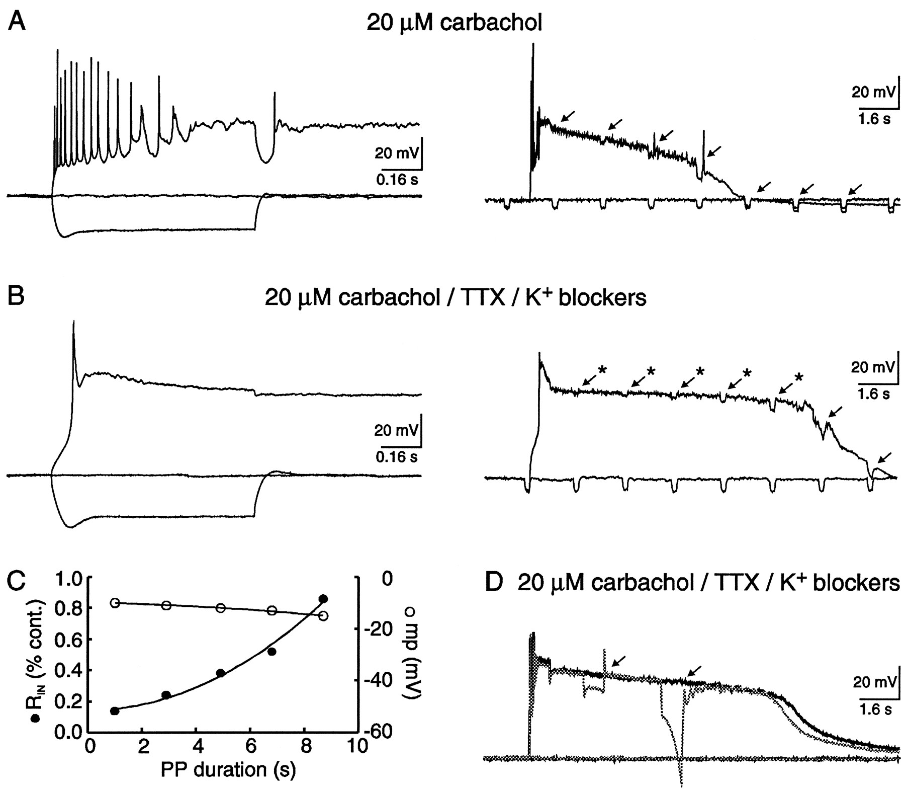
- Fig. 4.
The amplitude of the sADP and the duration of the PP decreased with negative membrane potentials. A, Typical response of a hippocampal pyramidal neuron to hyperpolarizing and depolarizing current injection. Neither an sADP nor a PP was observed in the absence of cholinergic stimulation. The resting membrane potential of this pyramidal neuron was −66 mV. B, In the presence of 20 μm carbachol, the neuron depolarized to −63 mV. After depolarizing current injection, both an sADP and a PP were evoked from a membrane potential of −60 mV.C–E, Decreasing the membrane potential with negative DC current injection decreased the amplitude of the sADP. In contrast, the PP was still generated, however, the duration decreased with lowered membrane potentials. F, A plot illustrating the PP duration and negative DC current injection versus membrane potential. The data were tabulated from three neurons.
- Fig. 5.
Both the sADP and PP were dependent on Na+ influx, independent of TTX-sensitive channels and Ca2+ influx via HVA channels. A, In 20 μm carbachol, depolarizing current injection evoked both an sADP and a PP. Lowering external Na+ from 152 to 26 mm reversibly depressed the sADP and abolished the PP, suggesting that Na+ influx was required for these afterpotentials. The resting potentials before and after cholinergic stimulation were −68 and −59 mV, respectively. The calibration bars in the inset represent 40 msec and 20 mV. B, In a different pyramidal neuron, both the sADP and PP were evoked by intracellular current injection. Lowering external Ca2+ from 2 to 0.1 mmreversibly abolished both the sADP and PP. The concentration of Mg2+ was simultaneously increased from 2 to 10 mm to maintain divalent cation charge screening. This finding suggests that Ca2+ influx was also necessary for these potentials. The resting potentials of this neuron before and after cholinergic stimulation were −67 and −60 mV, respectively. The calibration bars in the inset represent 40 msec and 20 mV. C, In 20 μmcarbachol, an sADP and a PP were observed after evoked action potentials. Coapplication of 1.2 μm TTX, a concentration sufficient to block action potentials, failed to depress either the sADP or the PP. Notice that the PP followed a slow regenerative potential, presumably Ca2+-dependent, whereas the sADP did not. Activation of voltage-dependent Na+ channels, therefore, was not required for either of these afterpotentials. The resting membrane potentials before and after carbachol were −65 and −62mV, respectively. D, In carbachol, an sADP and a PP were elicited by intracellular current injection. Coapplication of 100 μm Cd2+ abolished both the sADP and PP. Activation of HVA Ca2+ channels, therefore, was necessary to evoke these potentials. The resting membrane potential in control was −69 mV, whereas 20 μmcarbachol depolarized the membrane potential to −62 mV.
- Fig. 6.
The sADP and PP required Ca2+ influx through L- and N-type Ca2+ channels. A, In a CA1 pyramidal neuron, intracellular current injection revealed an sADP and a PP in the presence of 20 μm carbachol. The resting membrane potentials before and after cholinergic stimulation were −64 and −60 mV, respectively. The sADP was reduced and the PP could not be evoked after coapplication of the L-type channel blocker nimodipine.B, In another pyramidal neuron, both the cholinergic-dependent sADP and PP were also evoked by intracellular current injection. The resting membrane potentials before and after cholinergic stimulation were −66 and −61 mV, respectively. Both the sADP and PP were reversibly depressed by coapplication of the N-type channel blocker ω-conotoxin-GVIA.
- Fig. 7.
Both the sADP and PP were blocked by intracellular BAPTA. A, Typical responses of a hippocampal pyramidal neuron to hyperpolarizing and depolarizing current injection. Neither an sADP nor a PP was observed in the absence of cholinergic stimulation (A1). The calibration bars in the inset represent 40 msec and 20 mV. In the presence of carbachol, however, depolarizing current injection evoked a long-lasting PP (A2). This pyramidal neuron was loaded with pipette solution containing 1.1 mm EGTA and 0.1 mmCa2+ and, thus, intracellular Ca2+ was bufferred to ∼16 nm. B, A different pyramidal neuron, recorded from the same slice as the neuron illustrated in A, loaded with 10 mm BAPTA. In control aCSF, neither an sADP nor a PP was observed (B1). The calibration bars in the inset represent 40 msec and 20 mV. In the presence of carbachol, neither an sADP nor a PP could be evoked, suggesting that elevated [Ca2+]i was necessary for these potentials (B2).
- Fig. 8.
Blockers of K+ channels failed to unmask either the sADP or the PP in the absence of cholinergic stimulation. A, The sADP and PP were not observed after application of 1.2 μm TTX and K+ channel blockers (10 mmTEA, 5 mm 4-AP, 100 μmBa2+). Cholinergic stimulation, however, revealed a PP that was reversibly depressed by lowering Na+ from 152 to 26 mm in the external media (A1). Notice that robust Ca2+ spikes remained in low Na+, whereas the PP was reversibly depressed (A2). B, In addition, neither the sADP nor the PP was observed in pyramidal neurons loaded with 40 mm intracellular Cs+ and bathed in 1.2 μm TTX. As above, cholinergic stimulation revealed a PP that was reversibly depressed by lowering Na+ in the external media (B1). In this example, robust Ca2+ spikes also remained in low Na+, whereas the PP was reversibly depressed (B2).
- Fig. 9.
The sADP and PP were independent of a Na+/Ca2+ exchanger.A, Intracellular current injection revealed an sADP and a PP in the presence of 20 μm carbachol. The resting membrane potentials before and after cholinergic stimulation were −70 and −62 mV, respectively. Both the sADP and PP were still observed after substitution of 50 mmLi+ for equimolar Na+ in the external media. After Li+ substitution, the membrane depolarized an additional 9 mV; however, negative DC current was injected to maintain the membrane potential at the presubstitution value. B, In a different neuron, intracellular current injection also revealed an sADP and a PP in the presence of 20 μm carbachol. The resting membrane potentials before and after cholinergic stimulation were −66 and −61 mV, respectively. Both the sADP and PP were still observed after a reduction in bath temperature from 35 to 25°C. The neuron depolarized after temperature reduction, however, negative DC current was injected to maintain the membrane potential at the same value as at 35°C.
- Fig. 10.
Individual K+ channel blockers modulate the PP waveform. A, Action potential firing in the presence of 10 mm TEA resulted in a brief plateau potential that was presumably Ca2+-dependent (A1). Neither an sADP nor a PP was observed after cessation of the depolarizing stimulus. The calibration bars in the inset represent 40 msec and 20 mV. In a different neuron, both the sADP and PP were observed in the presence of 20 μm carbachol (A2). Coapplication of 10 mm TEA abolished the sADP and decreased the duration of the PP. B, In the presence of 5 mm 4-AP, individual action potentials activated brief afterdepolarizations that presumably were Ca2+-dependent (B1). After cessation of the depolarizing current stimuli, a brief afterpotential was commonly observed. Neither the sADP nor the PP was observed. The calibration bars in the inset represent 40 msec and 20 mV. In another neuron, a PP was evoked in the presence of 20 μm carbachol (B2). Coapplication of 5 mm 4-AP slightly prolonged the PP.C, Application of 100 μmBa2+ to the perfusate resulted in a brief afterpotential and subsequent spike broadening (C1). Brief afterpotentials were also observed after depolarizing current stimuli, however, neither the sADP nor the PP was evoked. In the presence of 20 μm carbachol, a different neuron exhibited an sADP and a PP after cessation of stimuli (C2). Coapplication of 100 μm Ba2+ to the perfusate increased the PP duration.
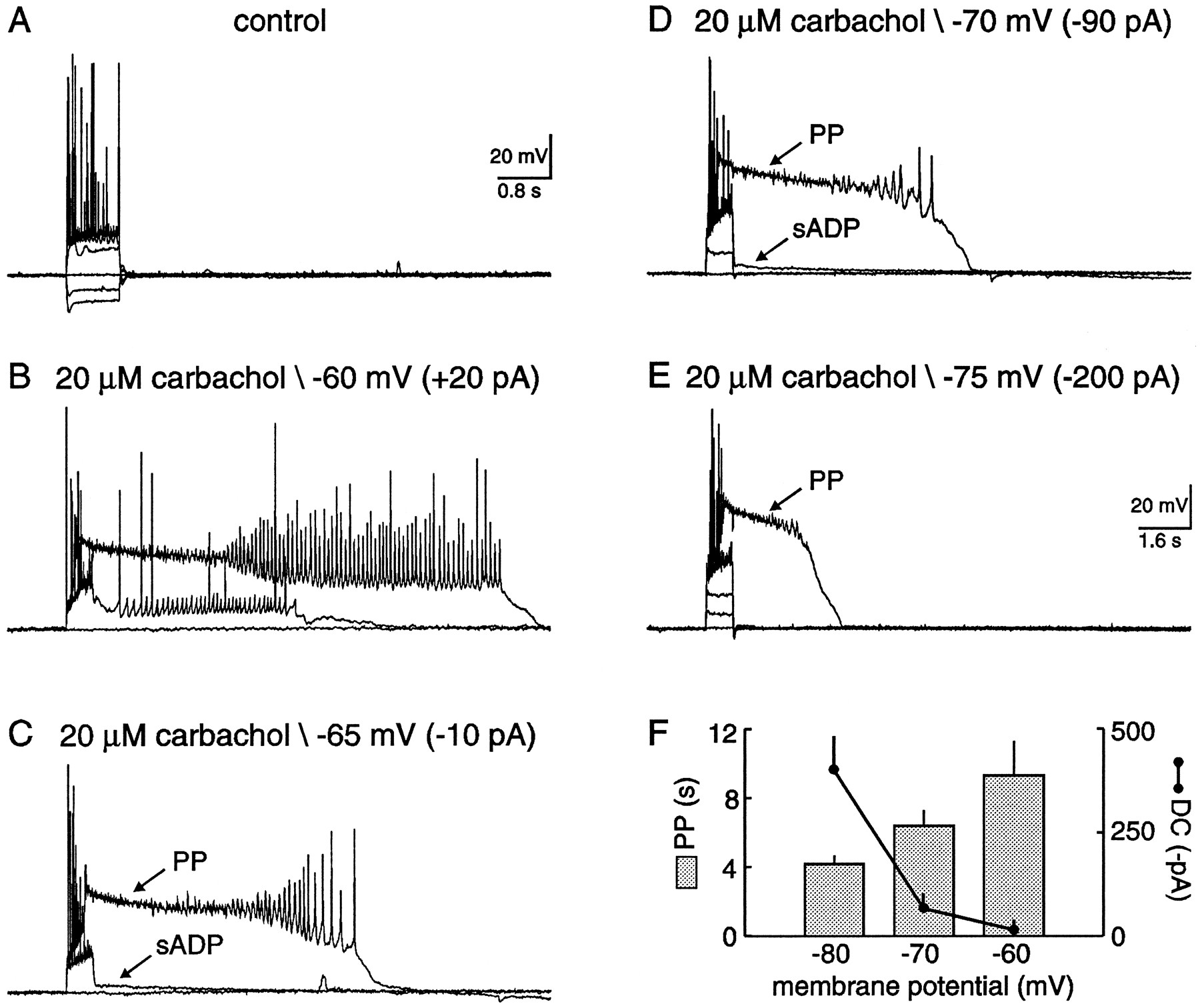
- Fig. 11.
The PP was associated with a largeRIN decrease and could not be terminated by hyperpolarizing pulses. A, Cholinergic-dependent PPs illustrated on two time scales. In the latter, brief current injection (−50 pA; 0.2 sec; 0.5 Hz) demonstrated a large decreasedRIN. B, PPs in the presence of carbachol, TTX, and K+ channel blockers (10 mm TEA, 5 mm 4-AP, 100 μm Ba2+). Under these conditions, brief current injection confirmed anRIN decrease in the absence of slow Na+ oscillations and suppression of K+ channels. C, A graph depicting the decreased RIN relative to control obtained at the resting potential, versus duration of the PP. In the same graph, membrane potential was also plotted versus PP duration. BothRIN and membrane potential values were obtained from the neuron illustrated in B (*). Notice that the change in membrane potential was minimal, whereas the increase inRIN was significant. D, Two superimposed PPs evoked from a CA1 pyramidal neuron. In the broken trace, hyperpolarizing current injection (−0.4 nA; 0.8 sec) failed to terminate the PP.


Tables
rp(mV) RIN(MΩ) APAMP(mV) APTH(mV) APDUR (msec) τ (msec) n K-gluconate1_a −65 ± 0.1 149 ± 3 92 ± 1 −47 ± 0.3 1.6 ± 0.01 20.7 ± 0.4 188 Cs-gluconate1_b −59 ± 0.7 183 ± 12 – – – 23.6 ± 1.8 11 KClc −65 ± 0.4 173 ± 24 92 ± 5 −48 ± 1.3 1.8 ± 0.06 22.8 ± 3.0 8 BAPTAd −67 ± 1.9 164 ± 47 88 ± 2 −45 ± 1.4 2.4 ± 0.15 21.6 ± 1.1 4 Abbreviations: rp, resting potential;RIN, input resistance;APAMP, action potential amplitude;APTH, action potential threshold;APDUR, action potential duration; τ, tau.
Intracellular solutions (in mm):
↵F1_a 140 K-gluconate; 1.1 EGTA; 0.1 CaCl2; 10 HEPES; 2 Mg-ATP; 0.3 Na-GTP.
↵F1_b 40 Cs-gluconate; 100 K-gluconate; 1.1 EGTA; 0.1 CaCl2; 10 HEPES; 2 Mg-ATP; 0.3 Na-GTP.
c70 KCl; 70 K-gluconate; 1.1 EGTA; 0.1 CaCl2; 10 HEPES; 2 Mg-ATP; 0.3 Na-GTP.
d100 K-gluconate; 10 K-BAPTA; 40 HEPES; 2 Mg-ATP; 0.3 Na-GTP.


