

Article Figures & Data
Figures
- Fig. 1.
Depolarizing concentrations of K+ promote survival of cerebellar granule neurons by preventing apoptosis. Granule neurons were cultured as described in serum-containing medium either in 5 or 25 mmK+. A, Viability kinetics over time in culture, measured by FDA staining. B, Kinetics of LDH activity released in the culture medium, normalized to the value obtained on DIV3 for the 25 mmK+ control culture. C, Quantitative DNA fragmentation (% fragmented vs total DNA) measured by Hoechst 33258 (0.5 μg/ml) fluorometry is not normalized to control. Bars represent mean ± SEM of three independent determinations at *p < 0.05 versus respective high K+controls by ANOVA and Scheffe’s F-test.
- Fig. 2.
TGF-β induces premature cell death in granule neurons grown in 5 mm K+. Cells were cultured as described in Materials and Methods. TGF-β2 (1 ng/ml) additions were made on DIV1, DIV4, and again on DIV7. A, Viability as assessed by FDA staining. B, Specific LDH release normalized to the value obtained for the 25 mm K+control cultures at DIV1. C, DNA fragmentation as described. Bars represent mean ± SEM of three independent determinations.
- Fig. 3.
Acceleration of apoptosis by TGF-β2 becomes apparent 24 hr before death of the untreated control cultures maintained in low K+. Phase-contrast microscopy shows that untreated low K+ controls (A) and TGF-β2 (1 ng/ml)-treated cultures (B) are indistinguishable in the first 6 d in vitro. By DIV7, the TGF-β2-treated neurons (D) show patches of dead cells throughout the culture, whereas untreated controls (C) display the macroscopic features of homogeneous and progressive apoptotic degeneration: appearance of apoptotic cell bodies, reduced cell density, and a thinning of the neurite network. On DIV8, TGF-β2-treated neurons (F) are dead compared with untreated low K+ controls (E). On DIV10, apoptotic cell death is completed in low K+ controls (G) compared with neurons maintained in high K+ (H). Magnification, 580×; scale bar (shown in H), 20 μm.
- Fig. 4.
TGF-β accelerates 5 mm K+-mediated apoptotic cell death of cerebellar granule neurons in vitro. Granule neurons were cultured and treated as described in Materials and Methods. All neuron preparations presented here were fixed and processed on DIV9. Nomarsky optics of cultures stained for in situ DNA end breaks shows that there is substantially more DNA fragmentation in low K+ than in high K+-grown neurons. The percentages of stained cells increase in low K+-grown neurons exposed to TGF-β2, whereas there is no difference between treated and untreated high K+ cultures.A, Low K+ control; B, low K+ exposed to TGF-β2 (1 ng/ml); E, high K+ control;F, high K+ exposed to TGF-β2 (1 ng/ml). For A andB, magnification is 480×; scale bar (shown inH), 13 μm. For E and F, magnification is 320×; scale bar (shown in H), 20 μm. Visualization under UV illumination of nuclei stained with the fluorescent dye Hoechst 33258 (5 μg/ml) reveals more condensed chromatin in low K+ than in high K+ culture conditions. TGF-β2 accelerates the rate of chromatin condensation in low K+-grown neurons, but it does not affect neurons maintained in high K+ culture conditions. C, Low K+ control;D, low K+ exposed to TGF-β2 (1 ng/ml); G, high K+ control; H, high K+ exposed to TGF-β2 (1 ng/ml). Magnification, 1100×; scale bar (shown in H), 6 μm.
- Fig. 5.
Granule neurons grown in 25 mm K+ are resistant to the proapoptotic effects of all three TGF-β isoforms. The neurons grown in either 5 or 25 mm K+conditions were exposed on DIV1, DIV4, and again on DIV7 to 10 ng/ml of either TGF-β1, TGF-β2, or TGF-β3. Neuronal survival as assessed by FDA staining (A) and LDH release (B) was measured upon death of the TGF-β3-treated low K+ cultures 36 hr before death of the untreated low K+ controls, whereas degeneration in either the TGF-β1- or TGF-β2-treated neurons was not yet completed. As determined by linear regression from the concentration–dependence curves, TGF-β3 scores the highest biological activity of the three isoforms with an EC50 = 1.87 ng/ml. Bars represent mean ± SEM of quadruplicate at *p < 0.05 versus respective 25 mmK+ controls by ANOVA and Scheffe’s F-test.
- Fig. 6.
CNTF and LIF, but not IGF-I, delay cell death in low K+ medium. Cultures were treated with the factors on DIV1, DIV4, and DIV7. Neuronal viability was assessed by LDH release on DIV3, DIV6, DIV8, and every day thereafter until death of the CNTF- and LIF-treated cultures occurred. IGF-I-treated cultures died at the same time as the untreated low K+controls on DIV10. CNTF and LIF were found to prolong survival over 2 din vitro. Neuronal survival is expressed as 100 − LDH release [%]. LDH values were calculated as described in Materials and Methods and normalized to the value obtained for the 25 mm K+ control on DIV3, set as 0% release. Bars represent mean ± SEM of two independent determinations.
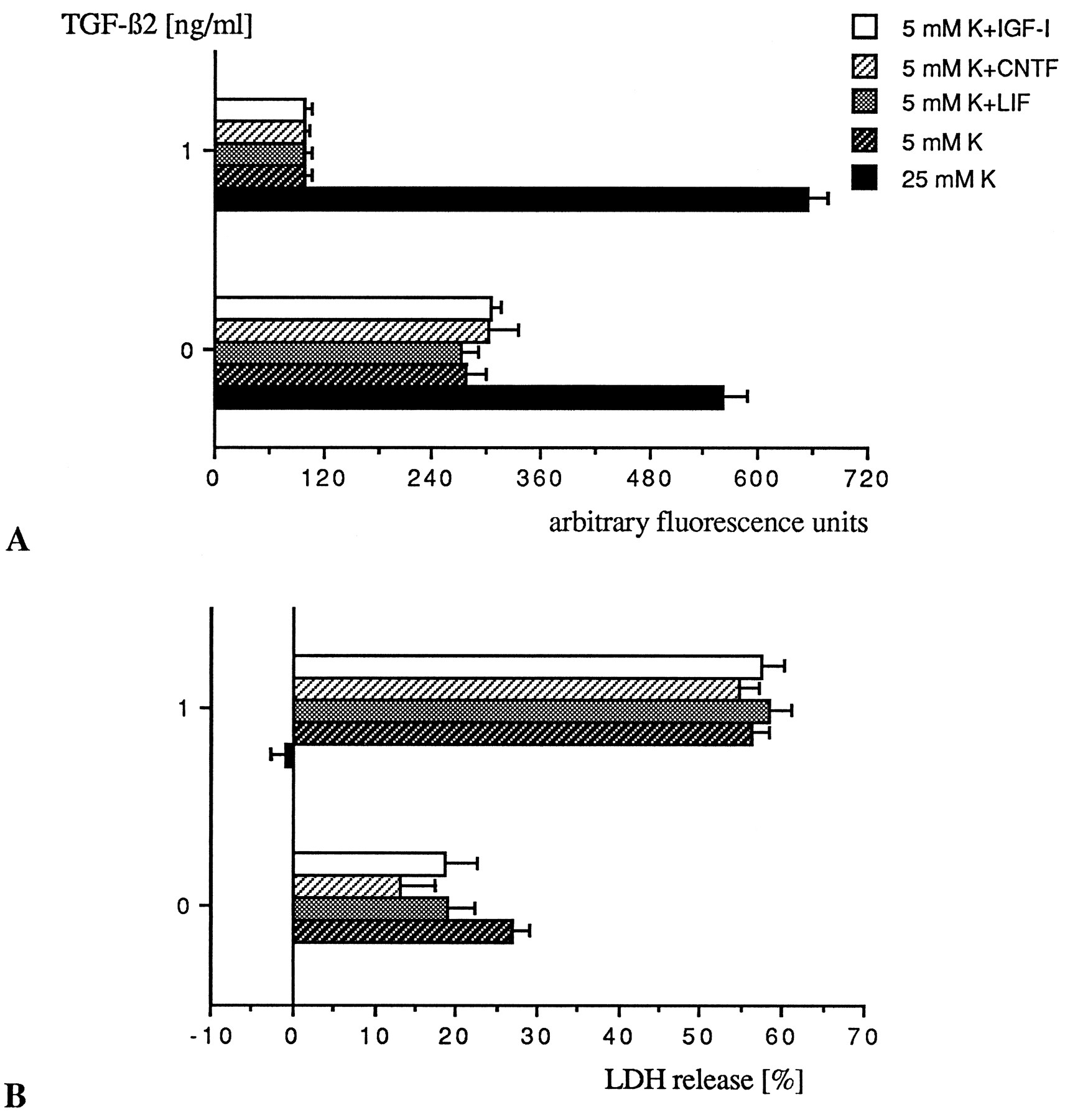
- Fig. 7.
CNTF, LIF, and IGF-I do not reverse the proapoptotic effect of TGF-β. Low K+-grown neurons were treated on DIV4 and DIV7 simultaneously with TGF-β2 (1 ng/ml) and one of the following cytokines: CNTF (10 ng/ml), LIF (10 ng/ml), or IGF-I (25 ng/ml). Assessment of neuronal survival by FDA staining (A) and measurement of LDH release (B) were performed upon death of the cultures exposed to TGF-β2, which occurred, regardless of the factor added, on DIV9, i.e., 24 hr before death of the untreated low K+ controls. At that time point (DIV9), the survival-promoting effect exerted by CNTF and LIF in the absence of TGF-β2 and measured in terms of LDH release represents an increase of viability of 13.7 and 7.9% for CNTF and LIF, respectively, over untreated control. LDH values were normalized to the value obtained for the untreated 25 mm K+ control, set as 0% release. Bars represent mean of triplicate ± SEM of a representative experiment.
- Fig. 8.
Cerebellar granule neurons maintained in either high or low K+ medium secrete latent TGF-β1 and TGF-β2. Cell-free conditioned media were transiently acidified as described in Materials and Methods, and both activated and nonactivated media were tested in triplicate for TGF-β1 and TGF-β2 in a quantitative sandwich enzyme immunoassay at the respective dilutions of 1:7 and 1:4. The detection limit of both immunoassays does not allow quantification of concentrations <60 pg/ml. A, Neurons grown in either low or high K+ concentrations produce equal amounts of latent TGF-β1, mostly in the first 24 hr of culture, with a slight increase over time in vitro. Naturally active TGF-β1 was not detected by the present technique. B, Neurons grown in either low or high K+ culture conditions release latent TGF-β2 with a continuous production over time in culture and a major contribution by high K+neurons. Naturally active TGF-β2 also was not detected.
- Fig. 9.
Antibodies to TGF-β do not prevent apoptotic cell death of cerebellar granule neurons. The neurons were set in culture in either 5 or 25 mmK+, as described. Upon seeding on DIV0 and every third day thereafter, on DIV3, DIV6, and DIV9, the 5 mm K+-grown neurons were exposed to either an anti-TGF-β2 (10 μg/ml) antibody (a-β2), a pan-specific anti-TGF-β (10 μg/ml) antibody (a-πβ), or an isotype IgG control (10 μg/ml). Viability checkings by FDA staining (A) and measurement of LDH release (B) were performed every third day. Bars represent mean of six different wells ± SEM of a representative experiment.


