

Article Figures & Data
Figures
- Fig. 1.
APPL immunoreactivity in wild-type larval CNS.A, An optical section of a Canton S(CS) larval CNS immunolabeled with Ab952M showing APPL immunoreactivity in the neuronal cell bodies in the eye disks (ed), ventral ganglion (VG), and brain lobes (BL). Neuroblasts in the optic proliferation centers are not APPL-immunoreactive (open arrow). Axons of photoreceptors traveling through the eye stalk (es) show intense APPL signal. B, An optical section through the CNS of anAppld larvae stained with Ab952M. Note the absence of APPL immunoreactivity. C, High magnification of APPL immunoreactivity in neuronal cell bodies of the ventral ganglion. APPL is concentrated in punctate structures surrounding nuclei.D, Horizontal optical section through a CS larval CNS showing APPL immunoreactivity in neuropil regions. In the ventral ganglion, APPL is concentrated in the neuropil of the three thoracic neuromeres (t1, t2, t3). In the connection between the ventral ganglion and brain lobes, APPL is detected along axonal tracks (thin arrow). In the central neuropil of the brain lobes, APPL is concentrated in distinct areas (thick arrows). Theinset shows the posterior tip of the ventral ganglion where APPL immunoreactivity is detected only in the neuropil of the eighth abdominal neuromere (a8, small arrows). Anterior is to thelower left for A and D, and to theleft for B. Scale bars: A, B, D, 50 μm; C, 10 μm.
- Fig. 2.
Localization of APPL relative to neuronal processes labeled with MAb 22C10. A, Larval CNS stained with anti-APPL (green) and 22C10 (red) antibodies. Boxed area including eye disk and brain lobe is shown at a higher magnification in B. B, In the eye stalk, APPL immunoreactivity colocalizes with 22C10 epitope, as revealed by the yellow color (open arrow). In the central neuropil of the brain lobe, APPL seems to be surrounding neuronal processes (white arrow). C, Optical section of a larval CNS stained with anti-APPL (green) and 22C10 (red) antibodies. Two boxed areas, comprising the optic lobe (D) in the brain lobe and the second thoracic neuromere (E) in the ventral ganglion, are shown at higher magnification in D and E, respectively.D, A section through the larval optic lobe stained with anti-APPL antibody (a), MAb 22C10 (b), and both antibodies (c). The central semicircular region stained with APPL corresponds to the neuropil of the medulla. Photoreceptor axons are intensely stained with both antibodies (small arrow). The thin arrow points to thick neuronal processes weakly stained with both antibodies. Thin fibers that run over the surface of the optic lobe are 22C10-immunoreactive and show APPL immunoreactivity concentrated in varicosities (arrowhead). E, A section through the second thoracic neuromere labeled with anti-APPL (a), 22C10 (b), and both (c). Notice that APPL immunoreactivity in this neuropil is not associated with 22C10-immunoreactive processes (arrow). In A–E, anterior is to the left. Scale bars: A, C, 50 μm; B, D, E, 25 μm.
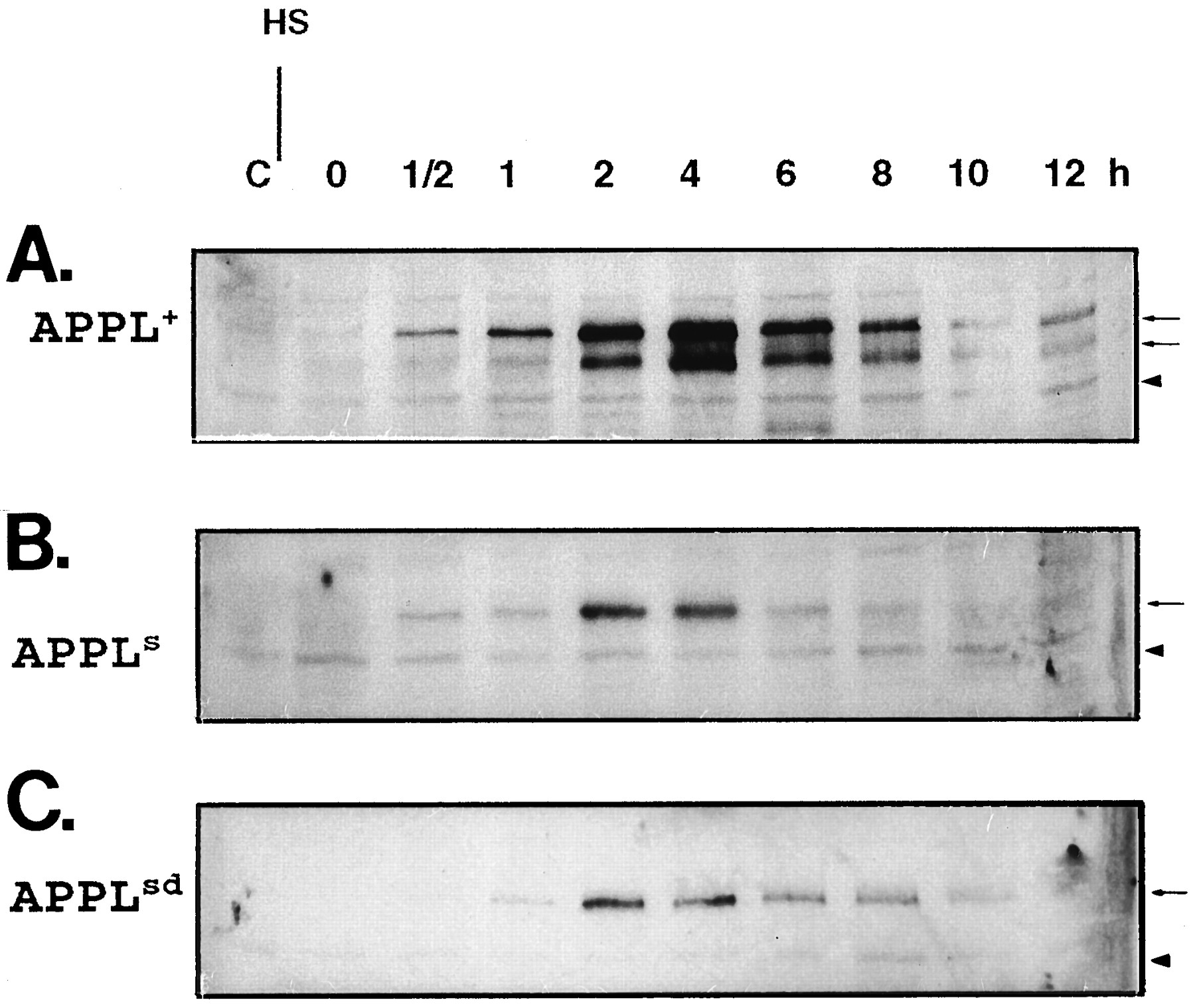
- Fig. 3.
Induction of hsp:Appl*transgenes. Immunoblot of head protein extracts probed with APPL-antibody. Adults were heat-shocked for 30 min at 37°C; protein extracts were made from fly heads at 0, 0.5, 1, 2, 4, 6, 8, 10, and 12 hr after the heat shock. Control extracts from flies before heat shock are loaded in the lane marked C. A,Appld; hsp:Appl+/+; B,Appld; hsp:Appls/+; C,Appld; hsp:Applsd/+. Same number of heads were homogenized and loaded in each lane. Thin arrows point to APPL bands. Arrowheads point to a cross-reactive band, present also before induction.
- Fig. 4.
Localization of the APPL+protein product of the induced hsp:Appl+transgene. Third-instar larvae of genotypeAppld; hsp:Appl+/+ were subjected to heat shock at 37°C for 1 hr, allowed to rest for 4 hr, and immunoprocessed as described (see Materials and Methods). A, Horizontal optical section through a CNS showing APPL distribution in the neuropil of the ventral ganglion: APPL is concentrated in the neuropil of the thoracic neuromeres (t1, t2); APPL is also found along axonal tracts connecting the ventral ganglion and the brain lobes (arrow). Compare with Figure 1D. B, Detail of APPL protein detected in the neuropil (arrowheads) of the eighth abdominal neuromere. Compare with Figure 1D,inset. C, High magnification of APPL vesicle-like signal observed in neuronal cell bodies. Compare with Figure1C. D, Z-series projection of horizontal optical sections through the ventral ganglion, exemplifying the uniform APPL staining observed in the cortex of the heat-shocked larval CNS. InA, B, and D, anterior is to theleft. Scale bars: A, B, D, 25 μm;C, 10 μm.
- Fig. 5.
Localization of APPL mutant proteins in larval CNS. Larval CNS of genotypes (A, D)Appld; hsp:Appl+/+, (B, E)Appld; hsp:Appls/+, and (C, F)Appld; hsp:Applsd/+ were heat-shocked at 37°C for 1 hr, allowed to rest for 4 hr, and immunoprocessed with anti-APPL antibody. A–C, A detail of APPL immunoreactivity in the thoracic neuromeres in the ventral ganglion showing how APPL+ (A) and APPLs (B) proteins are concentrated in this neuropil region (arrows), whereas APPLsd is not (C). D–F, A detail of APPL immunoreactivity in the optic lobe is shown. Although APPL+ (D) and APPLsd (F) proteins are found in the optic neuropil (big arrow; see Fig. 4D for endogenous APPL signal), APPLs protein (E) is not detected in this region. Anterior is to thelower left. Same magnification for A–F. Scale bar (shown in F): 25 μm.
- Fig. 6.
APPL immunoreactivity in the ventral ganglion during metamorphosis. Optical section through whole-mount preparations of ventral ganglions dissected from Canton S pupae at 0 hr (A), 6 hr (B), 12 hr (C), and 48 hr (D) after pupariation and stained with anti-APPL antibody. Signal in the thoracic neuromere (arrow in A andB) decreases after 6 hr, and by 12 hr APPL is distributed evenly in the neuropil of the ventral ganglion (C). By 48 hr, the level of APPL in the neuropil is low, and distinct processes (thin arrow) and varicosities (small arrow) are revealed. For A–D, anterior is to the left. Scale bars: A–D, 25 μm.
- Fig. 7.
APPL immunoreactivity in the optic lobe during metamorphosis. Horizontal paraffin section of heads of 0 hr (A), 25 hr (B), and 60 hr (C) pupae and adult (D) stained with anti-APPL antibody. A, 0 hr after pupariation, APPL-immunoreactive photoreceptor axons project from the eye disk (ed) through the eye stalk (es) into the optic lobe. The lamina (l), the outer and inner medulla neuropils (m), and the two neuropils in the lobula complex (loc) in the optic lobes are highly stained with APPL. B, By 25 hr after pupariation, APPL immunoreactivity in the medulla neuropil starts to split into three layers, the two outer ones being connected by processes. In the lobula complex, the lobula (lo) and lobula plate (lop) neuropils are intensely stained. C, 60 hr after pupariation, the optic neuropils have rotated. In the lamina (l), axons are distinguishable. In the medulla (m), APPL is arranged in three layers, showing a columnar organization. D, In adult, intense APPL signal remains only in the lamina neuropil (l). In the medulla, isolated processes are APPL-im-munoreactive (thin arrows). Some cell bodies show higher APPL immunoreactivity (arrowhead; lamina cortex). For A–D, anterior is at the top and lateral at the left. Same magnification was used for all. Scale bar (shown in D): 25 μm.
- Fig. 8.
APPL immunoreactivity in the adult mushroom bodies. A–D, Horizontal paraffin sections of Canton S adult heads stained with APPL antibody. A, Dorsal section of a brain showing APPL concentrated in the α lobes (α) and in the peduncle (p). In addition, APPL immunoreactivity is significantly higher in the central complex (cc) and in isolated processes (thin arrows).B, A section illustrating APPL immunoreactivity along the whole peduncle and in the β/γ lobes. APPL signal is higher in the cell bodies of the Kenyon cells (lower arrowhead) and in isolated neurons throughout the cortex (right arrowhead).C–D, Two adjacent paraffin sections showing APPL immunoreactivity absent from the dendritic fields in the calyces (open arrow) but present in axonal neuropils, such as the peduncle (p), and in Kenyon cell bodies (arrowhead). Small arrows in C point to Kenyon axons that converge to form the peduncle. E–F, An adult of the genotype Appld; hsp:Appl+/+ was heat-shocked for 30 min at 37°C. After a 4 hr resting period, the head was processed for paraffin sectioning and immunolabeled with anti-APPL antibody as described (see Materials and Methods). Two alternate sections are shown. E, Dendrites in the calyx do not show enrichment of APPL protein (open arrow), whereas the Ken- yon axons converging to form the peduncle do (small arrows).F, Intense APPL signal is seen in the lamina neuropil (l) and in the peduncle (p) of the mushroom body. For A–F, anterior is to the top. Same magnification was used for all. Scale bar (shown in F): 25 μm.
- Fig. 9.
APPL immunoreactivity in the larval mushroom bodies. A, A semi-horizontal optical section of a wild-type larval CNS, where APPL signal in the neuropil of the mushroom bodies [α (short arrow) and β/γ (long arrow) lobes] is evidenced. B–C, Larval CNSs of genotypes (B) Appld; hsp:Appls/+ and (C)Appld; hsp:Applsd/+ were heat-shocked at 37°C for 1 hr, allowed to rest for 4 hr, and immunoprocessed with anti-APPL antibody. Semi-horizontal optical sections are shown. B, APPLs protein is not enriched in the neuropil of the larval mushroom bodies. C, APPLsdprotein is highly concentrated in the neuropil of the larval mushroom bodies. For A–C, anterior is to the top. Same magnification was used for all. Scale bar (shown in B): 25 μm.


