

Article Figures & Data
Figures
- Fig. 1.
Ability of selective D1-like dopaminergic agonists to mediate reproductive behavior in ovariectomized rats with indwelling third ventricle cannulae. Microinjections of four selective D1-like agonists facilitated sex behavior in highly receptive rats primed with EB. Each experiment was repeated at least twice. The results are expressed as LQ (defined as percent of positive lordosis responses divided by number of mounts by a series of 4 male rats).Bars represent mean LQ ± SEM. Asterisksindicate a significant increase in LQ compared with the pretreatment response of the same rats 1 hr before D1-like agonist treatment (ANOVA,p ≤ 0.01) and/or the response in control females (one-way ANOVA followed by Mann–Whitney U test, p ≤ 0.01). Females were excluded if LQ exceeded 20% during tests for false-positive responses before receiving EB and for EB effect before agonist treatment. In another group of control rats, vehicle (0.1 ml, i.c.v.) was administered as a control for false results.
- Fig. 2.
Inhibition of SKF-induced lordosis behavior by selective D1-like antagonists. Chronically cannulated rats primed with EB were given microinjections of the D1-like antagonist SCH23390 1 hr before or the irreversible D1-like/D2 antagonist EEDQ 24 hr before challenge with SKF38393. The animals were screened for nonspecific effect of antagonist before agonist challenge and analyzed as described in Figure 1 and in Materials and Methods.
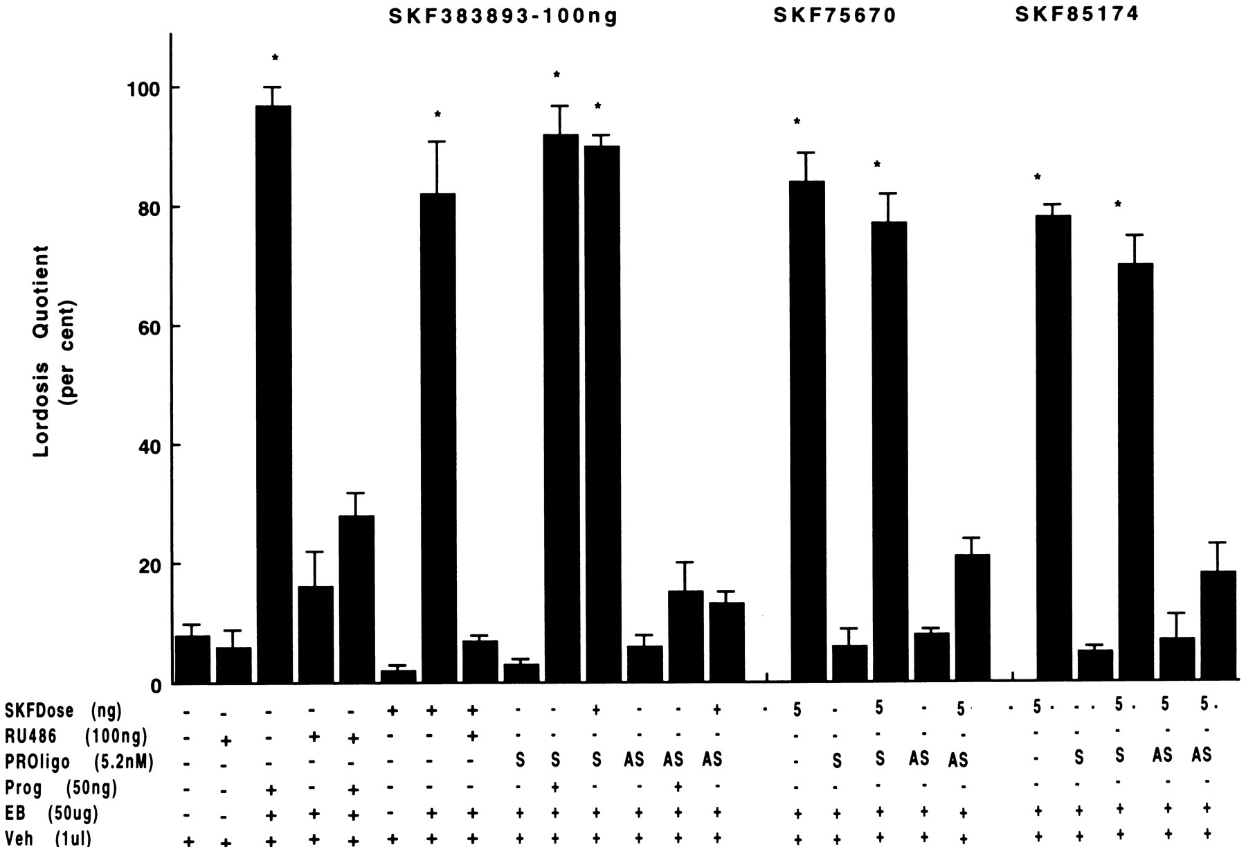
- Fig. 3.
Inhibition of SKF-facilitated sex behavior by phosphorothiolated antisense (AS) oligonucleotides to D5, but not D1, dopamine receptor mRNA. Oligonucleotides (2 and 4 nm for D5 and D1 receptors, respectively) were microinjected into indwelling third cerebroventricular cannulae 24 hr after EB priming. Twenty-four hours later, animals were tested for the behavioral effect of EB + oligonucleotide and then challenged with D1-like agonists. Random sense (RS) oligos were given to another group of rats as a control for nonspecific oligo effect.
- Fig. 4.
Specific effects of antisense (AS) and sense (S) oligonucleotides to D1 (A) and D5 (B) dopamine receptor mRNAs. A, Nuclear accumbens treatment with D1 antisense increases basal motor activity and blocks the induction of additional hyperactivity after SKF82958. A behavioral paradigm known for its D1 dopamine receptor specificity in targeted mutagenic mice (n = 40) was used as in Materials and Methods. Data are replicates of two experiments. B, Treatment with increasing levels of D5 antisense increases the inhibition of D1-like agonist-stimulated CAT gene expression. Mouse L(tk−) fibroblast cells stably expressing D5 dopamine receptors were transiently transfected with the rat ER expression vector and the ERE-elb-CAT reporter gene in the presence of increasing amounts (17–83 pmol/well) of antisense or random sense oligonucleotides to D5 dopamine receptors mRNA and subsequently treated with 10 μm SKF82958. Data are calculated as [1 − (%CAT activity in antisense oligo-treated cells/CAT activity in random sense cells)]. The presented results are the average of two experiments, each performed in duplicate. Values between experiments varied by <10%.
- Fig. 5.
Inhibition of SKF-induced lordosis response by antisense oligonucleotides to PRa mRNA and the antiprogestin RU38486 (RU486). Rats were administered RU38486 and challenged with D1-like agonist 1 hr later. In another group, oligonucleotides (2.6 nm, i.c.v.) were administered concurrently with EB and 24 hr later. At 48 hr after EB priming, animals received intracerebroventricular SKF challenge and were tested for behaviors as described in Materials and Methods.
- Fig. 6.
Facilitation of lordosis by microinjections of D1-like agonists into stereotaxically implanted cannulae in the ventromedial nucleus (VMN) (Panel A), but not arcuate nucleus (AN) (panel B) or preoptic area (POA) (panel B). Fourteen days after screening for false-positive and false-negative behaviors, animals were tested for behavioral responsiveness to SKF38393, SKF75640, or SKF85174. For blockade, D5 antisense oligonucleotide (2 nm total) was injected into VMN cannulae 24 hr before SKF38393 challenge. B presents the responses of those animals with either AN or POA cannulae. Animals served as their own controls for experimental results. Because all responded with LQ above 85%, the data are not shown from another group of animals with third ventricle cannulas that served to verify the effectiveness of D1-like agonists to induce responses on the day of intranuclear injections. Each experiment was repeated two to three times. Cannula placement was verified by microscopic examination at the end of experimentation. Data are shown for those animals with both cannula tips in target area.
- Fig. 7.
In situ hybridizations and dark-field autoradiograms of D1 (A) and D5 (B) mRNA. Modest distribution of D1, but not D5, dopamine receptor mRNA is visualized in the ventromedial nucleus of the hypothalamus (VMH) 48 hr after estrogen priming of ovariectomized female (E2F inD1A, B; D5A, B) and in the VMH of male (M in D1 C, D; D5C, D) rats. A D1 dopamine receptor cRNA probe transcribed from a rat D1 dopamine receptor cDNA and a D5 dopamine receptor riboprobe generated from a partial rat D5 dopamine receptor cDNA were used.A and C are at low magnification, andB and D are at higher magnification.HIP, Hippocampus; CPu, caudate putamen;3V, third ventricle; Re, reuniens thalamic nucleus; En, endopiriform nucleus.


