

Article Figures & Data
Figures
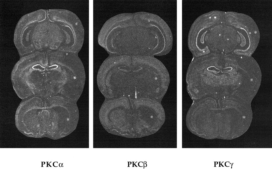
- Fig. 1.
In situ hybridization autoradiographs showing the distribution of PKCα, PKCβ, and PKCγ mRNAs in the rat brain at posterior hippocampal (top), dorsal hippocampal (middle), and caudate (bottom) levels 24 hr after 90 min of MCA occlusion. The loss of the signal is seen in the infarct core (left hemisphere). In this particular brain, the expression PKC-β mRNA has not changed in the cortex. No changes in the mRNA expression of PKCα, PKCβ, and PKCγ subspecies are seen in the perifocal region.Asterisks indicate infarcted areas. Magnification, 3.5×.
- Fig. 2.
In situ hybridization autoradiographs showing the distribution of PKCε and PKCζ mRNAs in the rat brain at posterior hippocampal (top), dorsal hippocampal (middle), and caudate (bottom) levels 24 hr after 90 min of MCA occlusion. The loss of the signal is seen in the infarct core (left hemisphere). No changes in the mRNA expression of PKCε and PKCζ subspecies are seen in the perifocal region. Asterisks indicate infarcted areas. Magnification, 5×.
- Fig. 3.
In situ hybridization autoradiographs showing the distribution and induction of PKCδ mRNA in the rat brain at posterior hippocampal (top), dorsal hippocampal (middle), and caudate (bottom) levels 4 hr (A), 24 hr (B), and 7 d (C) after 90 min of MCA occlusion. In the infarct core (right), the expression of PKCδ is decreased or lost at 4 hr (A), but is increased substantially 7 d (C) after 90 min of ischemia. Concomitantly, the perifocal and cortical expression is increased 4 hr (A) and 24 hr (B) (arrowheads) after the insult, but it is back to control levels 7 d (C) after the insult. The signal is decreased in the lateral section of the thalamus 7 d (C) after ischemia. Arrows(A–C) point to the upper margin of the infarcted areas, and arrowheads (A, B) point to the perifocal area with high expression of PKCδ mRNA.Stars (A–C) show the thalamic nuclei with high expression of PKCδ mRNA. Magnification, 8×.
- Fig. 4.
In situ hybridization autoradiographs showing inhibition of ischemia-induced perifocal PKCδ mRNA by administration of MK-801 (3 mg/kg) 30 min before 90 min of ischemia and followed by 12 hr of reperfusion. Sections at dorsal hippocampal (top) and caudate levels are shown. The ischemia-induced expression of PKCδ mRNA (arrowheads) is reduced significantly by MK-801 in perifocal regions, especially in the cortical area including the cingulate cortex. Ashows an ischemic animal pretreated with 0.9% NaCl; Bshows an ischemic animal pretreated with MK-801. In the brain shown inA, the lesion-induced PKCδ mRNA also encompasses the hippocampus. Arrows point to the infarct margins, andasterisks point to the thalamus that shows a high basal level of PKCδ mRNA expression. Magnification, 5×.
- Fig. 5.
Northern blotting analysis of total RNA from the rat brain 2 d after ischemia. A 32P-dATP-labeled PKCδ-oligonucleotide probe was used. The blot shows a band of ∼3.1 kb. C, Contralateral cortex; I, ischemic perifocal cortex; T, thalamus. The signal is weak in the cortical tissues but is increased in the ischemic cortex.
- Fig. 6.
Western blots of PKC subspecies in homogenates of tissues from the striatal core (A) and perifocal cortical (B) regions from the ischemic hemisphere (I) and of the corresponding region on the contralateral side (C). Arrowheadsindicate positions of PKCα, -β, -γ, -δ, and -ζ subspecies, and a small arrow points to PKCε, visible in the blots. The only clear change seen consistently in three separate experiments is the increase of PKCδ in the perifocal tissue from ischemic hemispheres (B, I, andC).
- Fig. 7.
PKCδ-immunoreactive cells in the rat brain 2 d after 90 min of ischemia. On the contralateral side of the frontoparietal (A) and cingulate (B,left) cortex, only a few immunoreactive neurons are seen, whereas in corresponding areas on the ipsilateral side (B, right; C), a large population of cortical neurons are immunoreactive. In cortical neurons, immunolabeling is extranuclear (D). In the contralateral striatum, numerous immunoreactive bundles of nerve fibers are seen (E). In the ipsilateral striatum (F), these nerve fibers have disappeared in the ischemic core (c), but have become strongly immunoreactive in the perifocal zone (p). In the infarct core, immunoreactive material is seen around small blood vessels, presumably in endothelial cells (G, curved arrows) and in glial-like cells (H). Scale bars: A–C, E, F, 250 μm; D, 25 μm; G, 125 μm; H, 50 μm.
- Fig. 8.
Immunofluorescence micrographs of focal (A, B) and perifocal (C–F) PKCδ immunoreactive cells double-stained with antiserum to complement C3 receptor, a marker for activated microglia and macrophages (A–D), and with an antiserum to GFAP (E, F), a marker for activated astrocytes, 3 d after 90 min of ischemia. Most of the PKCδ immunoreactive cells are macrophage-like cells with OX-42 immunoreactivity, round cell body, and few processes. GFAP-immunoreactive structures are not labeled with PKCδ antibody. Large arrows andarrowheads point to double-labeled macrophage-like cells. Small arrows show a small blood vessel surrounded by PKCδ-immmunoreactive cells and processes, some of which are also OX-42-immunoreactive. A field shown in E andF includes PKCδ-immunoreactive neuronal cell bodies (white stars) that are surrounded by GFAP-positive astrocyte processes (F). Scale bar (shown inA): 40 μm (A, B, E, F); 80 μm (C, D).


