

Article Figures & Data
Figures
- Fig. 10.
Light-induced Ca2+ signals and simultaneously recorded whole-cell currents (lower traces) measured in the absence of both extracellular Ca2+ and Na+. In contrast to Figure 9, the Ca2+ rise was virtually abolished when Na+ was substituted withN-methyl-d-glucamine (NMDG) with 0 Ca2+ and 2 mm EGTA applied by rapid perfusion from a puffer pipette. a, Response to a 1 sec saturating illumination in a cell perfused with NMDG solution, originally bathed in Ca2+-free, Na+-containing bath. b, Response from a photoreceptor perfused with NMDG, but this time after being initially bathed in normal Ringer’s (1.5 mmCa2+); again there was little or no increase in Ca2+. Both cells, clamped at −70 mV, produced small outward currents, as NMDG does not permeate the light-sensitive channels; c, Light-induced responses from the same cell as in Figure 11b to a weak 20 msec LED flash before (left), after (right), and during (middle) rapid perfusion with the Ca2+-free NMDG solution. The cell was clamped at −70 mV. After perfusion with NMDG, the response reversed and became slower (because of the absence of Ca2+-dependent feedback effects), but recovered completely after returning to normal Ringer’s. The Ca2+ measurement was made ∼60 sec after these responses were recorded, after the cell had been perfused again with NMDG for ∼30 sec.
- Fig. 1.
Indicator dyes do not significantly influence the light response. a, Quantum bumps recorded in response to a 1.5 sec stimulus of identical intensity in a cell loaded with 100 μmINDO-1 are similar to those recorded from another cell with control intracellular solutions containing no Ca2+ buffers.b, Similarly, Fluo-3 (100 μm) has little or no effect on responses to 20 msec flash or 500 msec step of light (the difference in absolute amplitudes is well within the experimental variability). Thedotted trace shows the response to a second 500 msec stimulus recorded 2 min after making a Ca2+measurement using a submaximal 100 msec flash containing ∼107 effective photons. All responses recorded in standard Ringer’s solution (1.5 mmCa2+) at a holding potential of −70 mV. Stimulus intensity in b was 3.8 × 10⁴effective photons · sec−1 (calibrated from bump counts as in a).
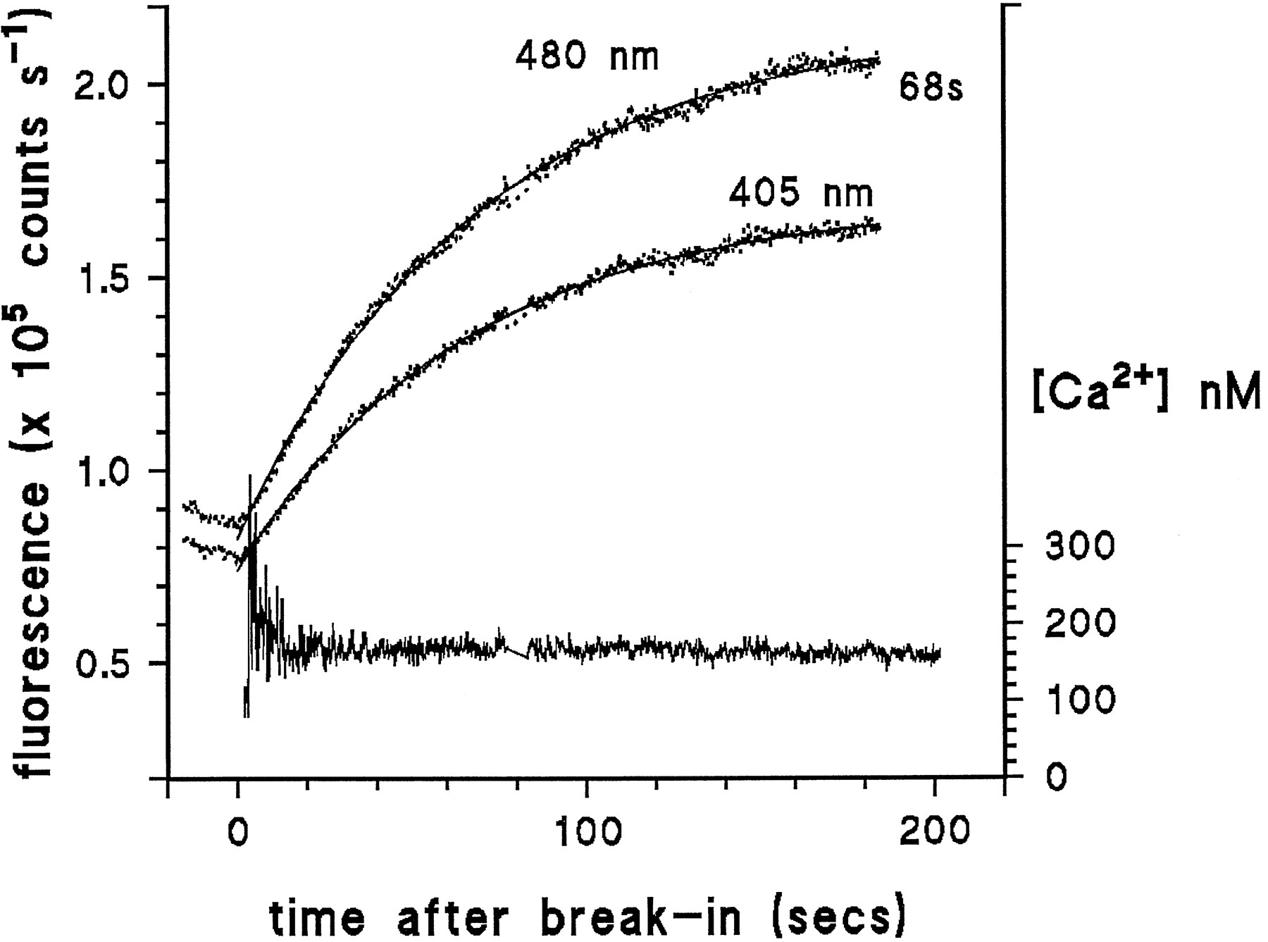
- Fig. 2.
Time course of dye loading in an oraphotoreceptor. Raw traces (dotted) show the INDO-1 fluorescence measured at 405 and 480 nm starting shortly after making a giga-seal. After establishing the whole-cell configuration (at time 0), fluorescence rises with a single exponential time course (smooth curves, single exponential fits to the traces with time constant of 68 sec). After subtraction of the background fluorescence, the ratioR between fluorescence at 405 and 480 nm was used to calculate cytosolic [Ca2+] according to Equation 1. Cytosolic [Ca2+] remained stable throughout the 200 sec recording, except for an apparent reduction during the first few seconds of recording. Recording made in normal (1.5 mm Ca2+) Ringer’s at −70 mV, briefly interrupted at ∼80 sec, for visual inspection of cell.
- Fig. 3.
Light-induced Ca2+ rises measured with INDO-1 in normal (1.5 mmCa2+) Ringer’s. a, Raw traces (raw) of fluorescence (counts per second) measured at 405 and 480 nm, sampled at 500 Hz;[Ca2+], after background subtraction, the time course of the rise in Ca2+is calculated from the ratio of the raw traces according to Equation 1(see Materials and Methods). The simultaneously recorded light-induced current (LIC, lower trace, holding potential −70 mV) saturated the amplifier (>10 nA). b shows the [Ca2+] signal and LIC on an expanded scale. Notice that the dark level can be readily measured during the latent period of the response (arrow). The 150 msec UV stimulus (360 nm) contained 3 × 10⁷ effective photons.
- Fig. 4.
Latency of the Ca2+ response determined using Fluo-3 in normal Ringer’s solution. The time course of the Ca2+ signal measured using the single-wavelength dye Fluo-3 was similar to that measured with INDO-1 (compare Fig. 3); however, a better signal-to-noise ratio can be obtained. The traces show the average of LIC (upper trace) and Ca2+ signal from eight photoreceptors in response to a 200 msec measuring flash (480 nm) containing ∼108 effective photons. The current trace has been inverted for easier comparison of time courses. The first detectable rise in Ca2+ always lagged the LIC by ∼3 msec (inset on expanded scale). Holding potential was −70 mV, data sampled at 1 kHz.
- Fig. 5.
Determination of Ca2+ levels in response to weak illumination in normal Ringer’s solution.a, A WT photoreceptor was first illuminated for 500 msec with a dim LED stimulus (∼2000 photons/sec) generating an inward current of ∼500 pA amplitude (dotted trace). The Ca2+ level reached during this period (530 nm) was then determined during the latent period of the response to a saturating UV measuring stimulus (50 msec, 3 × 10⁷ effective photons). The Ca2+ signal (solid trace) is replotted on an expanded time base below. b, Ca2+ levels obtained from 19 cells (filled squares), as in Figure 5a, plotted against the total charge flowing during the 500 msec adapting step. Open square, “Dark” Ca2+ concentration determined identically, but without preillumination with the LED (mean ± SD of 12 cells). The data have been fitted by a regression line of slope 2.7 nm/pC with an intercept (dark resting Ca2+ level) of 161 nm.Triangles represent data determined using measurements of light-induced Ca2+ rises in ora orninaE flies with small amounts of residual rhodopsin (Fig. 6).
- Fig. 6.
Ca2+ influx measured in real time in ora photoreceptors containing residual levels of rhodopsin. UV measuring flashes of 1 sec duration delivered toora (or ninaE) photoreceptors sometimes elicited small responses (lower traces) attributable to residual levels of rhodopsin, thus allowing direct measurement of Ca2+ influx (upper traces) during weak effective illumination. Traces from two different cells are shown using different intensities, generating responses of ∼20–40 pA (left) and 200 pA (right). Quantum bump noise can be clearly resolved in these small responses: as reported previously (Johnson and Pak, 1986), quantum bumps in ninaE mutants with greatly reduced rhodopsin levels were in fact typically larger than in WT. Substantial Ca2+ rises were detected in each case. The data from these and three other cells are plotted on Figure5b and show reasonable agreement with measurements made in WT photoreceptors using the two-flash paradigm.
- Fig. 7.
Light-induced Ca2+ rise in a WT photoreceptor loaded with the low-affinity indicator dyeMag-INDO-1. In response to a saturating UV stimulus, Ca2+ rose rapidly beyond 50 μm. Data were recorded at a holding potential of −70 mV in standard (1.5 mmCa2+) Ringer’s solution containing no Mg2+. Mg2+ was also omitted from the recording electrode solution. Similar results were obtained in four other cells.
- Fig. 8.
Ca2+ influx is reduced in the trp mutant. Measurements of Ca2+levels in the trp mutant were made using the two-flash paradigm (Fig. 5) and plotted against the total charge carried in response to the adapting flash. In the dark (open triangle), resting Ca2+ levels in trp were indistinguishable from WT; however, in response to the 500 msec LED-adapting flash, the Ca2+ rise was significantly less than in WT (dotted line replotted from Fig. 5). The regression line through the trp data had a slope of 1.09 nm/pC (i.e., ∼2.5× less than in WT).
- Fig. 9.
Light-induced Ca2+ signals measured in the absence of extracellular Ca2+. Substantial Ca2+ increases were detected in every cell in response to saturating UV-measuring stimuli: Ca2+ signals (upper traces), simultaneously recorded whole-cell currents at 0 and −70 mV (lower traces) (two different cells). The rise was at least as large in cells clamped at 0 mV as in those clamped at −70 mV, arguing against influx of residual Ca2+ as an explanation. Note also that the initial (dark) resting level of Ca2+ was higher in the cell clamped at 0 mV (see also Fig. 11). Bath contained 0 Ca2+, 2 mm EGTA, and 120 mmNaCl.


