

Article Figures & Data
Figures
- Fig. 1.
Timing of experiments in relation to the development of sensory neurons. A, For the analysis of trk expression and spinal cord projections, limb buds were removed before gangliogenesis and axon outgrowth. NT3 treatment began as soon as axons would normally invade the limb (stage 25) and continued until E10, which is near the end of the cell death period. B, For the analysis of the effects of NT3 on cell death and proliferation, limb buds were removed as described above. Embryos received two treatments with NT3 on E4 and E5, which is during the period of neuronal proliferation and during the early phase of cell death in the DRG. On E5, after a second dose of NT3, embryos were either fixed for counts of pyknotic nuclei or pulsed with BrdU for 4 hr and fixed for BrdU labeling.
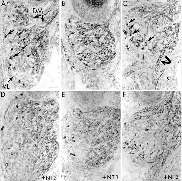
- Fig. 5.
TrkB+ neurons are insensitive to both limb bud deletion and NT3 treatment. Transverse sections of E10 DRG from control (A–C) and NT3-treated (D–F) embryos stained for trkB. A, In the normal lumbar DRG of control embryos, two distinct populations of trkB+ neurons can be identified: large diameter ventrolateral (VL) neurons (arrows) and smaller dorsomedial (DM) neurons (arrowheads).B, In lumbar DRG on the limb-deleted side, both classes of trkB+ neurons survived in control embryos. C, Thoracic DRG in control embryos contained more trkB+ VL neurons (arrows) than did lumbar ganglia. In thoracic ganglia, trkB+ fibers (curved arrow) can be seen extending into the sympathetic chain (s). D, In normal lumbar DRG, NT3 treatment (10 μg/d) had no obvious effect on the number of trkB neurons in either the VL or DM regions, but NT3 did cause an expansion of the trkB-negative portion of the VL region (compare A). E, In lumbar DRG on the deleted side, NT3 treatment also expanded the trkB-negative VL region without altering the survival of either population of trkB+ neurons.F, NT3 had a similar effect in thoracic DRG. In all panels, dorsal is up and lateral is to theleft. Scale bar, 50 μm.
- Fig. 2.
Specificity of antibodies to neurotrophin receptors. HEK 239 cells mock-transfected or transfected with chick cDNA encoding trkA, trkB, or trkC (columns). Transfected cells were immunolabeled with antibodies produced against the extracellular domains of chick trkA, trkB, and trkC (rows). Note that each antibody labels only cells transfected with the corresponding cDNA, and none of the antibodies cross-reacts detectably with cells expressing either of the other trk receptors.
- Fig. 3.
Effects of limb bud deletion and NT3 treatment on trkC+ sensory neurons. Transverse sections of E10 DRG from control (A–C) and NT3-treated (D–F) embryos are stained for trkC. A, In normal lumbar DRG of control embryos, trkC+ neurons (arrows) were mainly confined to the ventrolateral (VL) region of each ganglion. B, In lumbar DRG on the limb-deleted side, few trkC+ neurons (arrows) survived, and the VL aspect of the DRG was reduced (compare with A). C, In thoracic DRG of control embryos, few trkC+ neurons (arrows) were normally detected. As in lumbar segments, these neurons were localized primarily to the VL region.D, In normal lumbar DRG, NT3 treatment (10 μg/d) increased the number of trkC+ neurons (arrows) within the VL region (compare with A). E, In lumbar DRG on the limb-deleted side, NT3 treatment rescued the trkC+ population and maintained the normal size of the VL region (compare with A and B). F, In thoracic DRG, NT3 treatment increased the trkC+ population (arrows) and expanded the VL region of the DRG. In each panel, the orientation is the same: dorsal is up and lateral is to the left. VL indicates the ventrolateral region of the ganglion, and DM indicates the dorsomedial region. In this figure and in Figures 4 and 5, the lumbar ganglia shown are from LS3 and the thoracic ganglia shown are from T5 or T6. The ganglia shown are the same ganglia that were used for cell counts (Fig. 6). Scale bar, 50 μm.
- Fig. 6.
Quantitative analysis of sensory neuron survival on E10 after limb bud deletion and treatment with NT3 at 10 μg/d.A, Most trkC+ neurons were eliminated in lumbar DRG on the deleted side; the number of trkC+ neurons remaining was approximately the same as in normal thoracic DRG. NT3 treatment resulted in an approximately normal number of trkC+ neurons in lumbar DRG on the deleted side and doubled this population on the normal side. NT3 treatment also increased the trkC+ population in thoracic DRG to approximately the level in normal lumbar DRG. B, Limb bud deletion significantly reduced the trkA+ population by ∼40% in lumbar DRG on the deleted side. This population was not increased significantly by NT3 treatment. C, Limb bud deletion had only marginal effects on trkB+ neurons, which were not statistically significant. The numbers of trkB+ neurons in lumbar and thoracic DRG were not significantly altered by NT3 treatment. Values are mean ± SEM (5 embryos for each group); lumbar counts are all from LS3;asterisks indicate significant differences. In control embryos, means of cell counts in thoracic and limb-deleted lumbar DRG were compared statistically with those of normal lumbar DRG. In NT3-treated embryos, means were compared with the respective means for control DRG. All comparisons were made using a two-tailedt test.
- Fig. 4.
TrkA+ neurons are less sensitive to limb bud deletion, and their survival is not altered by NT3. Transverse sections of E10 DRG from control (A–C) and NT3-treated (D–F) embryos stained for trkA.A, In normal lumbar DRG of control embryos, trkA+ neurons (arrowheads) were confined mainly to the dorsomedial (DM) region of each ganglion.B, In lumbar DRG on the limb-deleted side, many trkA+ neurons (arrowheads) survived in control embryos despite the obvious reduction in the size of the ventrolateral (VL) region (compare with A).C, In thoracic DRG, trkA+ neurons (arrowheads) were also localized to the DM region.D, In normal lumbar DRG, NT3 treatment (10 μg/d) had no obvious effect on trkA+ neurons (arrowheads), but did cause an expansion of the trkA-negative VL region (compareA and Fig. 3D). E, In lumbar DRG on the limb-deleted side, NT3 treatment also expanded the trkA-negative VL region without altering the survival of trkA+ neurons (arrowheads; also see Fig. 6). F, NT3 had a similar effect in thoracic ganglia. In each panel, dorsal isup and lateral is to the left. Scale bar, 50 μm.
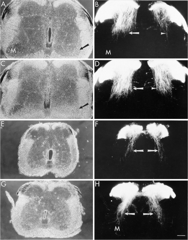
- Fig. 7.
NT3 increases the number of muscle spindle afferent collaterals in lumbar and thoracic segments. Transverse sections of E10 lumbar (A–D) and thoracic (E–H) spinal cord from control and NT3-treated (10 μg/d) embryos after DiI labeling of dorsal roots.A, Phase-contrast image of lumbar cord from a control embryo. The lateral motor column (M) is reduced ipsilateral (right, arrow) to the limb bud deletion.B, Fluorescence image of sensory fibers as revealed by DiI labeling. Contralateral to the deletion, the collateral fibers of muscle spindle afferents (Ia fibers, arrow) extend toward the motor column (M). This population of fibers was markedly reduced ipsilateral to the deletion (arrowhead). C, Lumbar spinal cord from an NT3-treated embryo. The lateral motor column remains reduced ipsilateral to the deletion (arrow). D, NT3 treatment restored a nearly normal population of Ia fibers ipsilateral to the deletion (arrowhead) and increased this population of fibers on the contralateral side (arrow). E, F, Thoracic spinal cord from a control embryo. In control cords, fewer Ia fibers (arrows) are present in thoracic segments than in normal lumbar segments (compare B, left). G, H, Thoracic spinal cord from an NT3-treated embryo. NT3 increased the number of Ia fibers (arrows) that project toward motoneurons (M) in thoracic segments. Dorsal isup in all panels. Scale bar, 100 μm.
- Fig. 8.
TrkC localization to the collateral fibers of muscle spindle afferents. Transverse sections of E10 lumbar (A, C) and thoracic (B, D) spinal cord from control and NT3-treated embryos after immunolabeling with antibodies to trkC.A, In control embryos, few trkC+ collateral fibers develop in the lumbar spinal cord on the deleted side (d). Contralateral to the deletion, trkC+ collaterals (arrows) of muscle spindle afferents project toward motoneurons (M). B, In control embryos, few trkC+ collaterals (arrowheads) are present in thoracic segments of the spinal cord. C, NT3 treatment restores the trkC+ collaterals (arrowheads) on the deleted side (d) of the lumbar spinal cord. NT3 treatment also increased the density of trkC+ fibers (arrows) that project toward motoneurons (M) on the contralateral side. D, NT3 treatment also resulted in an increase in the number of trkC+ collaterals (arrowheads) in thoracic segments. All panels are dark-field images of HRP labeling. Dorsal isup in all panels. Scale bar, 100 μm.


Tables
Control +NT3 Normal lumbar 256 ± 49 242 ± 46 Deleted lumbar 224 ± 43 274 ± 52 Neither limb bud deletion nor NT3 treatment had a significant effect on the proliferation of DRG precursers as assessed using a 4 hr exposure to BrdU on E5. BrdU-labeled cells were counted in every other section through the L3 DRG. Values are the mean (± SEM) number of labeled cells times 2 per 1000 healthy DRG cells. Means were compared using the Student’s t test. Data are from four embryos for each group.
Control +NT3 Normal lumbar 44 ± 9.0 7.5 ± 2.1* Deleted lumbar 354 ± 44.5 78.3 ± 12.61-160 Normal thoracic 229 ± 21.1 60.8 ± 12.61-160 NT3 treatment rescues DRG neurons from cell death due to target deprivation. Limb bud deletion results in excessive cell death on the deleted side, which is largely blocked by exogenous NT3. Treatment with NT3 also significantly reduced cell death in DRG with normal targets (i.e., contralateral lumbar and thoracic). Values are the mean number of pyknotic nuclei per DRG ± SEM. p values are from comparisons of control and respective NT3-treated means using a one-tailed Student’s t test. Data are from five to six embryos for each group.
↵* p < 0.0025;
↵F1-160 p < 0.0005.


