

Article Figures & Data
Figures
- Fig. 1.
Left, AdultMonodelphis together with a litter of neonates at P7, which was the age at which most animals were operated on. Adult females weigh ∼110 gm. Right, P7 animal at higher magnification; crown–rump length, 15 mm.
- Fig. 2.
Left, Control and operated (crush) animals walking in the runway used for “footprint” analysis. The feet are partly obscured by the edge of the runway. Walking patterns were quantitatively assessed by the use of footprint analysis (right), as described by de Medinacelli et al. (1982) and Kunkel-Bagden and Bregman (1990) and modified as outlined in Materials and Methods. A typical pattern of hindlimb footprints and the measurements made are illustrated. PL, Print length;TS, toe spread; IT, distance between intermediary toes; TOF, distance to opposite foot;DBF, base of support; LSL, left stride length (i.e., distance between consecutive left hindfoot prints);RSL, right stride length; r, rotation. Results from animals with crush lesions are summarized in Table 3 and Figure 5, and results from cut lesions are summarized in Table 4 and Figure 5.
- Fig. 3.
Typical examples of spinal cord lesions made in neonatal Monodelphis. These sections were prepared from animals that were terminally anesthetized within 1 hr (a–c, e) or 24 hr (d) of operation, and their spinal cords were removed for morphological examination as described in Materials and Methods. a, Low-power magnification of sagittal section of a P6 Monodelphis with a crush lesion of the spinal cord. The lesion is shown at higher magnification in c. Note that not only was the spinal cord lesion complete, but the crush was sufficiently deep to also disrupt the vertebral body at the level of the lesion [arrow in a; and note disrupted vertebral body (v) inc]. This was apparent in many of the crushes examined histologically. e, Low-power sagittal section of a P7Monodelphis with the cord transected by a cut. The lesion is illustrated at higher magnification in b.d, Crush lesion 24 hr after operation. Scale bars:a, e, 1 mm; b, 100 μm; b–d are at same magnification.
- Fig. 4.
a, Consecutive frames taken from video recordings of climbing in an adult Monodelphiswith spinal cord that had been crushed completely at P7 (operated) compared with an unoperated (control) adult. As described in Materials and Methods the animals climbed a narrow beam inclined at ∼45° to the horizontal. The time taken to climb and the number of errors made were recorded. b, Consecutive frames taken from video recordings of crossing a 1.5 cm grid in an adultMonodelphis with spinal cord that had been crushed completely at P7 (operated) compared with an unoperated control adult. As described in Materials and Methods the animals crossed a grid with bars 1.5 cm apart. The time taken to climb and the number of errors made were recorded (Fig. 5, Tables 3, 4) for the quantitative results of both tests). c, Consecutive frames taken from video recordings of swimming in an adult Monodelphis with spinal cord that had been completely crushed at P7 (operated) compared with an unoperated (control) adult. As described in Materials and Methods, animals made seven timed swims in a 1.2 m tank (Fig. 5, Tables 3, 4).
- Fig. 5.
Quantitative results from three of the behavioral tests made in adult animals with spinal cords that been crushed at P7 (left panels) or cut at P4 (right panels) and examined at 6 months of age. Top graphs, Comparison of individual control (open bars) and operated (filled bars) animals in the beam-climbing test (time taken and number of errors); middle panels, crossing the 1.5 cm grid (time taken and number of errors);bottom panels, Results of the swimming test (time taken to complete distance). Means from 10 trials (7 for swimming test) are shown; error bars indicate 1 SEM; where no bar is shown it was too small to be visible. The mean time for all animals with crush lesions to climb the narrow beam was 9.4 ± 2.4 sec, and it was 8.6 ± 0.6 sec in the controls (NS). The mean swimming time for animals with crush lesions was 4,5 ± 0.3 sec, and it was 4.3 ± 0.4 sec in the controls (NS). For the animals with cut lesions in the swimming test, the mean time for the controls was 2.7 ± 0.2 sec, and for the operated animals it was 5.3 ± 1.2 sec (NS). *Section of spinal cord from this animal are shown in Figure 8f; also compare Figure 6, SFI. ‡Sections of spinal cord from this animal are shown in Figure 8a. Note that for the tests on the animals with crush lesions, four controls were used except in the swimming test, in whichn = 3, because one of the controls was unable to swim.
- Fig. 6.
Results from calculation of SFI. The SFI is an estimate of locomotor ability calculated from measurements made in footprint analysis (Fig. 2; see Materials and Methods). The index is considered to be within normal limits if it is within 11% (note that this is not an SEM) on either side of the control mean. Open circles are mean values for individual animals from 10 trials in adults with spinal cords that had been crushed at P7–P8;filled circles are means of 10 trials for animals with spinal cords cut at P4. Error bars indicate 1 SEM. Mean values for each operated group (n = 10 for crush,n = 3 for cut) are shown by arrowsat right; the filled arrow is for cut data, and the open arrow is for crush data. Note that only one animal (with a crush lesion) falls outside of the normal range. The histological appearance of its spinal cord is shown in Figure 8f.
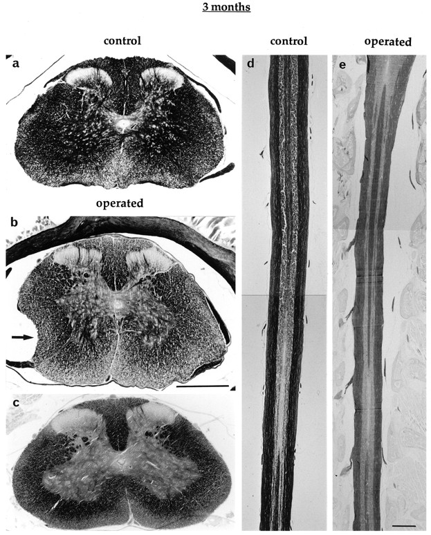
- Fig. 7.
Transverse (a–c) and longitudinal (horizontal) (d, e) silver-stained sections of spinal cords of 3-month-old Monodelphis at the level of a complete crush lesion made at P7–P8. a, From an unoperated control. In one animal (b) some deficit in the cross-sectional area was apparent (arrow). Otherwise the structure looked remarkably normal. Scale bar for a–c, 0.5 mm. d, From a control; e, from an operated (crush) animal. For these longitudinal sections, the rostral end of the spinal cord is attop. Scale bar for d, e, 1 mm. Lesions were made at T1–T2.
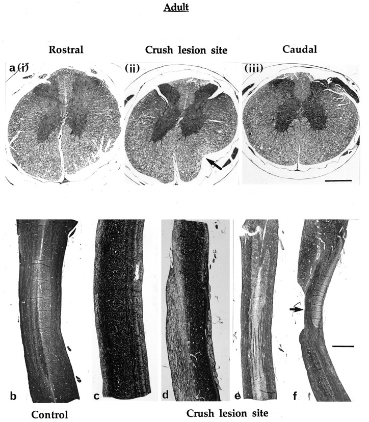
- Fig. 8.
Transverse [a, (i)–(iii)] and longitudinal (b–f) silver-stained sections from the spinal cord of adult Monodelphis that had been completely transected by a crush at P7. a, (i)–(iii)are from the same animal. (i), Rostral to the site of the lesion. (ii), At the level of the lesion (note the structural deficit indicated by the arrow).(iii), Caudal to the site of the lesion. Scale bar in(iii), 0.5 mm; (i) and(ii) are at same scale. Behavioral data for this animal are shown in Figure 5 (‡). b–f, Longitudinal sections of one control (b) and four operated (c–f) spinal cords of adultMonodelphis that were subjected to complete crushing of the cord (T1–T2) at P7–P8. In f there is an obvious deficit (arrow) in the gross structure, which correlated with a greater degree of impaired function on behavioral testing (this is the spinal cord from the only animal with a sciatic function index that was outside of the normal limits (Fig. 6). Scale bar inf, 1 mm; b–e are at same scale.
- Fig. 9.
Longitudinal sections of two 6-month-oldMonodelphis with spinal cords that had been transected completely by a cut (T1–T2) at P4. Note the considerable narrowing of the spinal cords at the site of the lesion; there is an artifactual break in section b. Numerous nerve fibers cross the site of the original lesion. Scale bar, 1 mm.
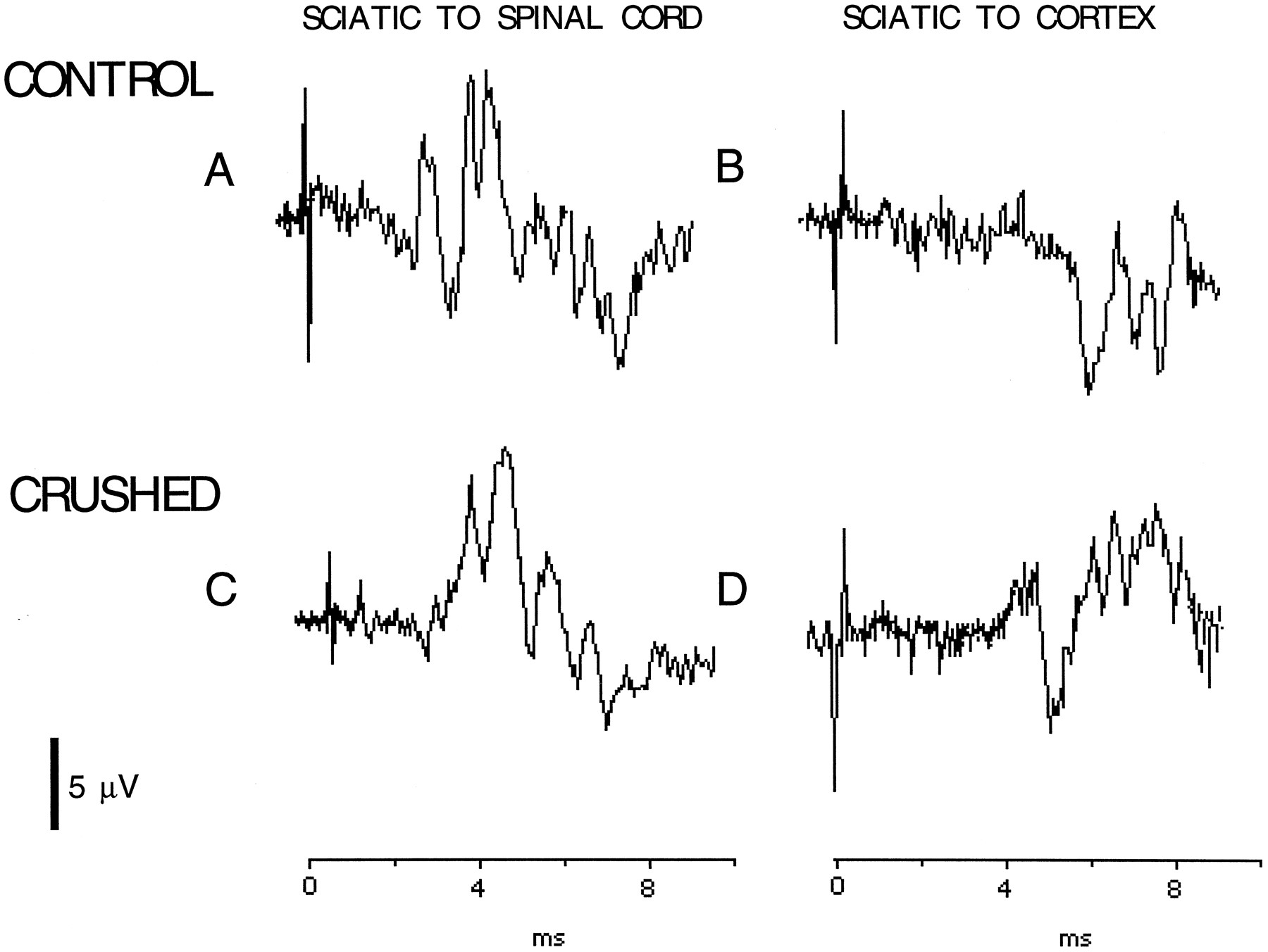
- Fig. 10.
Electrophysiological recordings in an anesthetized adult Monodelphis with spinal cord crush made at P7 (C, D) compared with an anesthetized control adult (A, B). Recording conditions were as described in Materials and Methods. In A andC the stimulating electrodes were placed on the sciatic nerve with recording electrodes on the spinal cord. In Band D the recording electrodes were moved to the sensory cortex. Similar records were obtained in all five operated and three control (unoperated) animals that were tested for impulse conduction across the site of the crush lesion.
- Fig. 11.
Spaced serial coronal sections showing the location of brainstem and midbrain neurons retrogradely labeled after injection of Fluororuby dextran amine into the lumbar spinal cord of adult Monodelphis. The series on the leftis from an animal in which the spinal cord was crushed on P7; on theright is an unoperated control. Inset, Micrographs show retrogradely labeled neuronal somata in the lateral vestibular nucleus (LV). Magnification, 5 × bar). CG, Midbrain central gray; DK, nucleus Darkschewitsch; DR, dorsal medullary reticular field; EW, Edinger Westphal nucleus; GR, gigantocellular reticular nucleus; IR, intermediate medullary reticular field; IN, interstitial nucleus of the medial longitudinal fasciculus; LC, locus coeruleus;LT, lateral tegmental area; RM, raphe magnus; RN, red nucleus; VR, ventral medullary reticular nucleus.


Tables
- Table 1.
Summary of spinal cord operations on Monodelphis domestica, indicating postnatal age at operation (day of birth = P0), nature of spinal lesion (crush or cut), and age at time of study
Postnatal age at operation (d) Operation Age at time of study n Methods B E M P 5–8 Crush P5–P9 (+0–24 hr) 25 11 18 6–7 Cut P6–P8 (+0–24 hr) 13 13 6 Cut 2 months 3 3 3 Controls 2 months 3 3 7–8 Crush 3 months 8 8 Controls 3 months 3 3 4 Cut 6 months 3 3 1 1 Controls 6 months 3 3 2 7–8 Crush 2–3 years 10 10 5 8 3 Controls 3 years 8 5 3 5 2 Methods of study: B, behavior; E, electrophysiology; M, morphology; P, pathway tracing (dextran amine injections);n, number of animals. Numbers in Methods column refer to the numbers of animals and preparations studied by the methods indicated; in some cases more than one method was applied to some preparations. A further 19 animals (P11–P27 at time of study) were used for electrophysiology studies of crush preparations (see Table 2).
- Table 2.
Conduction of action potentials across crush lesion of spinal cord at different times after making the lesion in postnatal animals aged P5–P8
Postnatal age at operation (d) Age at time of study No. conducting No. not conducting 5–6 P5 –P72-a 2 9 6–7 P11 –P16 2 6 7–8 P21 –P27 5 6 Spinal cords were dissected out from exsanguinated animals and tested as described in Materials and Methods. The potentials recorded in the two animals still conducting within 24 hr of crushing were just distinguishable from background noise. Histological examination was carried out in six of the cords tested at 0–24 hr after the lesion. All showed complete disruption of the cord at the site of crushing.
↵F2-a These animals were from the group of 25 P5–P8 animals listed in Table 1.
- Table 3.
Summary of quantitative data from behavioral testing ofMonodelphis with spinal cords that had been completely transected by crushing at P7–P8 (n = 10) compared with unoperated controls (n = 4–5)
Control (n = 4–5) Crushed (n = 10) Mean SEM Mean SEM Runway Stride length (cm) 11.8 0.6 10.2 0.2 Base of support (cm) 3.1 0.1 2.8 0.2 Print length (cm) 1.5 0.04 1.3* 0.01 Toe spread (cm) 1.6 0.04 1.5** 0.03 1.5 cm grid Time (secs) 3.3 0.7 3.0 0.4 Total errors HL 0.9 0.4 1.5*** 0.2 Total errors FL 0.7 0.3 0.4 0.1 3.5 cm grid Time (secs) 2.5 0.4 3.7 0.4 Total errors HL 0.7 0.4 1.6**** 0.5 Total errors FL 0.3 0.04 0.3 0.1 Swim + climb Time (sec) 4.3 0.7 4.3 0.3 After training each animal performed each test 10 times (7 times in the case of swimming tests), and the mean values were calculated (illustrated for some of the tests in Fig. 5). Here are presented the means and SEMs for all the control and for all the operated animals for the runway, grid-crossing, and swim + climb tests. For the runway tests the number of animals was n = 5 for the controls. For the other tests n = 4 animals for controls because one control animal died (cause not determined), except for the swimming tests in which the control n = 3, because one animal was unable to swim. Most differences between control and operated groups were not statistically significant. For this reason footprint analysis values for rotation, distance to opposite foot and distance between intermediary toes are not given. Note that the base of support did not change significantly. Mean values for the beam climb and swimming tests are given in the legend to Fig. 5. The only measures that showed significant differences between control and operated animals were print length (**p < 0.05) and toe spread (*p < 0.005). The significance level between the forelimb (FL) and hindlimb (HL) mean errors in the operated animals is indicated by ***p < 0.002; ****p < 0.04.
- Table 4.
Summary of quantitative data from behavioral testing of 6-month-old Monodelphis with spinal cords that had been transected completely by cutting at P4 (n = 3) compared with unoperated controls of the same age (n = 3)
Control (n = 3) Cut (n = 3) Mean SEM Mean SEM Runway Stride length (cm) 15.6 0.2 12.0 1.3 Base of support (cm) 2.8 0.2 2.6 0.2 Narrow beam Time (sec) 3.0 0.5 8.7* 1.7 Total errors 0.1 0.1 0.9 0.5 1.5 cm grid Time (sec) 1.7 0.3 3.1* 0.2 Total errors HL 0.7 0.3 1.5** 0.2 Total errors FL 0.7 0.2 0.4 0.3 3.5 cm grid Time (sec) 1.8 0.2 4.0*** 0.2 Total errors HL 0.6 0.3 1.6*,** 0.2 Total errors FL 0.6 0.2 0.2 0.1 Swim + climb Time (sec) 2.4 0.06 4.0 0.7 After training each animal performed each test 10 times (7 times for swimming), and the mean values were calculated (illustrated for some of the tests in Fig. 5). Here are presented the means and SEMs for all the control and for all the operated animals. None of the differences between control and operated in the runway footprint analysis was statistically significant; only stride length and base of support are shown. The operated opossums were significantly slower at climbing the beam and crossing both grids but did not make more errors than the controls. However, as with the animals with crush lesions (Table 3), there was a significant difference in error rate between forelimbs (FL) and hindlimbs (HL) of the operated animals when crossing the grids. *p < 0.05; ***p < 0.01 for differences between control and operated animals. **Significance level between the mean errors of the hind and forelimbs in the operated animals (p < 0.015).


