

Article Figures & Data
Figures
- Fig. 1.
A slice through an osmium-stained hair cell afferent synapse, reconstructed by electron tomography.A, An x–y plane through the reconstructed volume. B, Same plane asA, showing traced SB (thin dotted white line), SB-associated vesicles (dotted black lines), outlying vesicles (solid black lines), and the presynaptic (rightward arrowhead) and postsynaptic (leftward arrowhead) plasma membranes, which were rendered as a single object (thick dotted white line) joined at the edges of the plane. For clarity, tracing lines are shown thicker than they appeared in the tracing program. Also visible are coated vesicles (short arrow) and a tether attaching a vesicle to the SB (long arrow). Thewhite areas immediately adjacent to osmium-stained SBs were probably artifacts of the backprojection computation related to the steep contrast gradient at the border of the darkly stained SB. The effect was not seen near less electron-dense SBs stained with EPTA; synapse 1. Scale bar, 200 nm.
- Fig. 2.
Three-dimensional structure of synaptic organelles. A, Stereo pair rendering of the partially reconstructed synapse shown in Figure 1. The SB (blue) is open on the face that met the edge of the section. SB-associated vesicles (yellow) coat the surface of the SB and are visible through its semitransparent wall. Outlying vesicles (green) fill the adjacent presynaptic cytoplasm, which is delimited by the synaptic cleft (red), defined as the region in which pre- and postsynaptic membranes were in closest proximity. The slice through this synapse that is shown in Figure 1 was taken parallel to, and a short distance from, the open face of the SB. In black and white photocopies of these color figures the SB-associated vesicles appear lighter than the outlying vesicles. To view a stereo pair, hold the figure at forearm length and focus beyond the plane of the page; then merge the two panels until a central three-dimensional image appears. B–D, SBs were approximately spherical.B, Rendering of a semitransparent sphere (gold, arrow) fit to a partially reconstructed SB (blue). The region of the plasma membrane that formed the synaptic cleft is shown in red; vesicles were omitted for clarity. C, D, Shown is the fit of the sphere (smooth line,arrows) to the SB at each 1.3-nm-thick plane (dots) in the x–z(C) and y–z(D) planes passing through the center of the sphere. The dots are the edges of circles fit to the traced SB in each plane (see Materials and Methods); synapse 6.
- Fig. 3.
Distribution of vesicle diameters and volumes.A, Renderings of some of the vesicles described inB. B–D, Distributions of vesicle volumes (B) and diameters (C,D) fit by Gaussians (smooth lines). The smallest ticks on the abscissa denote the bin size. B, Vesicle volumes at a single synapse (synapse 6). C, All vesicle diameters at a single synapse (synapse 5). D, All vesicle diameters from all eight synapses. The vesicle total numbers six fewer here than in Table1 because these were shared by two SBs at synapse 4. Here they were counted only once.
- Fig. 4.
Spatial distribution of docked vesicles. SB-associated (yellow) and outlying (green) vesicles are shown at three synapses (A, B, synapse 1; C,D, synapse 3; E, synapse 2) in relation to the plasma membrane (gray), the SB (blue), and the presynaptic density (pink). Arrow pairs inA/B and C/D denote the same vesicle in both views of the same synapse. A, C,E, View through the mostly transparent plasma membrane of vesicles docked at it. Asterisks in Cand E show the locations of omega-shaped invaginations in the presynaptic membrane. Scale bar (applies to A,C, E), 100 nm. B, View of docked vesicles and the closed pole of the SB. D, Another tomogram of the synapse shown in C, revealing docked vesicles far from the SB, which is truncated by two edges of the volume. F, Nonsynaptic region of a hair cell showing vesicles in the hair cell cytoplasm (pale green) and those docked (dark blue) at the plasma membrane (red). Mitochondrial outer membranes (purple) are flat where truncated by the edge of the volume and reveal the wedge shape of this reconstruction.
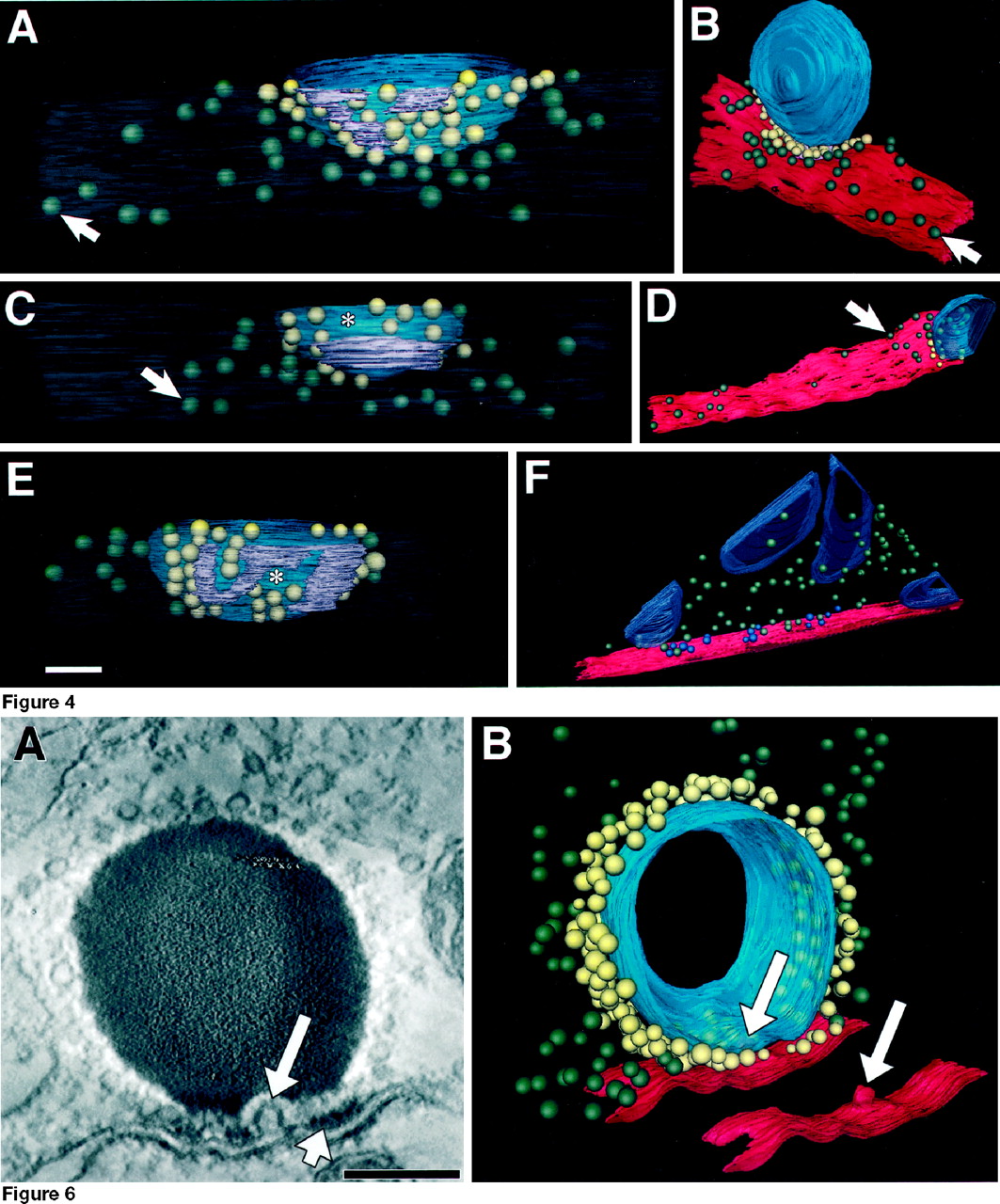
- Fig. 6.
Omega profile beneath the SB. A, Anx–y plane through an osmium-stained reconstructed synapse showing an omega-shaped invagination in the presynaptic plasma membrane that lies beneath the SB (long arrow) and adjacent to an area of presynaptic density (short arrow). White specks in thetop portion of the SB are saturated pixels and are artifact. Scale bar, 200 nm. B, Rendering of the same synapse showing the relationship of the omega profile (long arrows) to the partially reconstructed SB (blue), SB-associated vesicles (yellow), outlying vesicles (green), and the region of the plasma membrane that formed the synaptic cleft (red). The membrane is a subregion of that shown in Figure 4E. The SB is open on two sides in this reconstruction because it spanned the thickness of the section. The membrane and omega profile also are shown alone; synapse 2.
- Fig. 5.
Density distribution of docked vesicles. Vesicle concentration on the plasma membrane, shown as a percentage of the maximum packing density (see Materials and Methods), is plotted as a function of the radial distance from the projected center of the SB. The envelope of the bar graph shows the distribution of the 152 docked vesicles from the three synapses shown in Figure 4. White bars show docked SB-associated vesicles (n= 78); dark bars show docked outlying vesicles (n = 74). The rightmost bar shows the docked vesicle density computed from a tomogram that did not include a synapse (n = 15 vesicles).
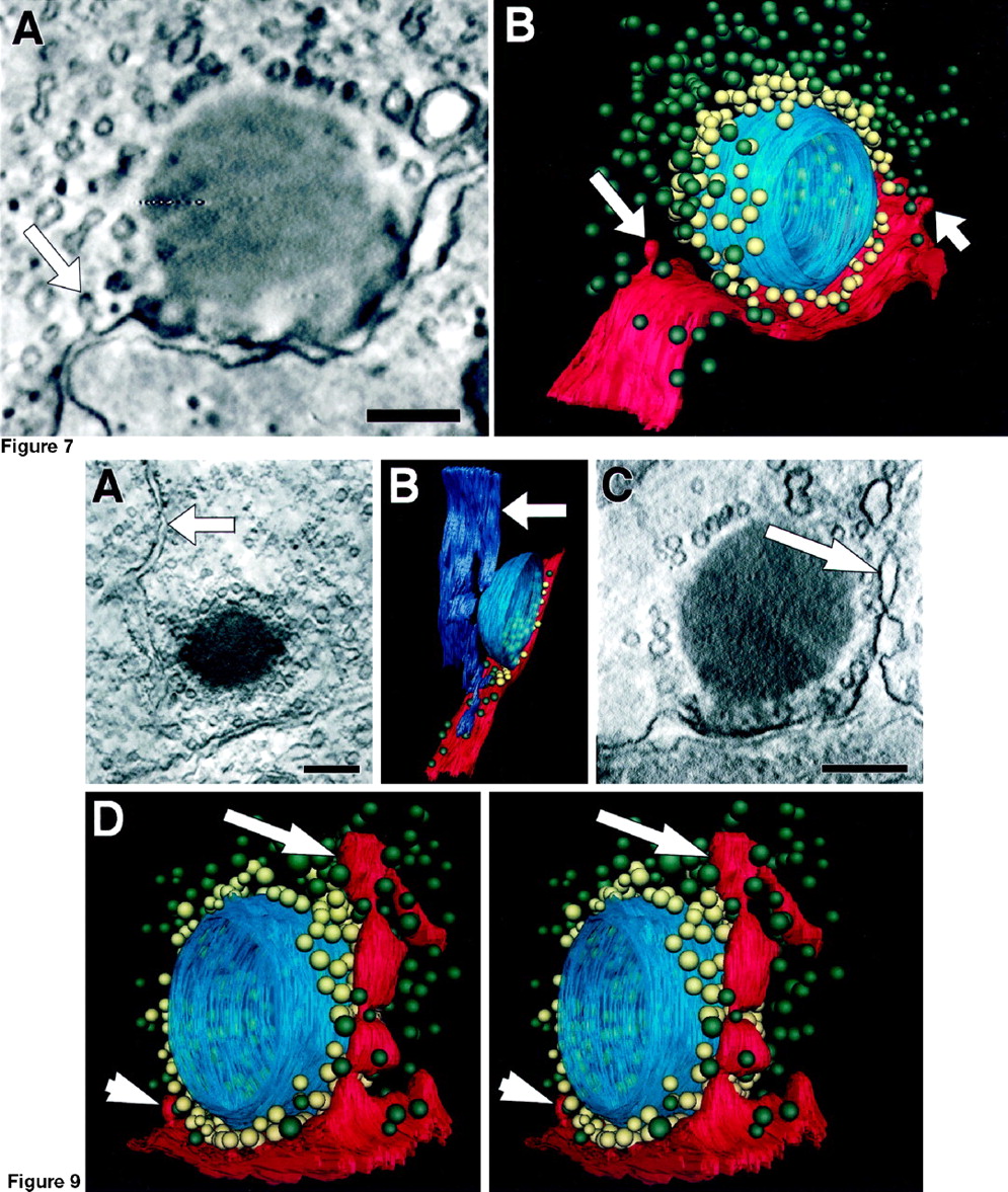
- Fig. 7.
Elongated omega profiles near the SB.A, An x–y plane through an EPTA-stained reconstructed synapse showing a vesicle formed from a stalk of the plasma membrane (long arrow) 165 nm from the SB. Scale bar, 200 nm. B, Rendering showing this invagination (long arrow) in relation to the SB (blue), SB-associated vesicles (yellow), outlying vesicles (green), and the plasma membrane (red). Another similar invagination occurred on the other side of the SB (short arrow); synapse 5.
- Fig. 9.
Large tubules near the SB. A,B, Membrane compartment (horizontal arrows) adjacent to the SB (synapse 1). A, Anx–y plane grazing an osmium-stained SB showing a membrane tubule extending toward the plasma membrane (arrow). B, Rendering of the same organelle (purple) showing its relationship to the SB (blue), plasma membrane (red), and docked vesicles (yellow andgreen). C, D, Deep plasma membrane invagination (long arrows) at another synapse (synapse 6). C, Plane through the EPTA-stained reconstructed synapse showing the invagination of the plasma membrane (arrow) adjacent to the SB. D, Stereo pair rendering of the same synapse revealing the lobular invagination (long arrow). A smaller invagination of the type shown in Figure 7 occurred on the other side of the SB (short arrow). Scale bars, 200 nm.
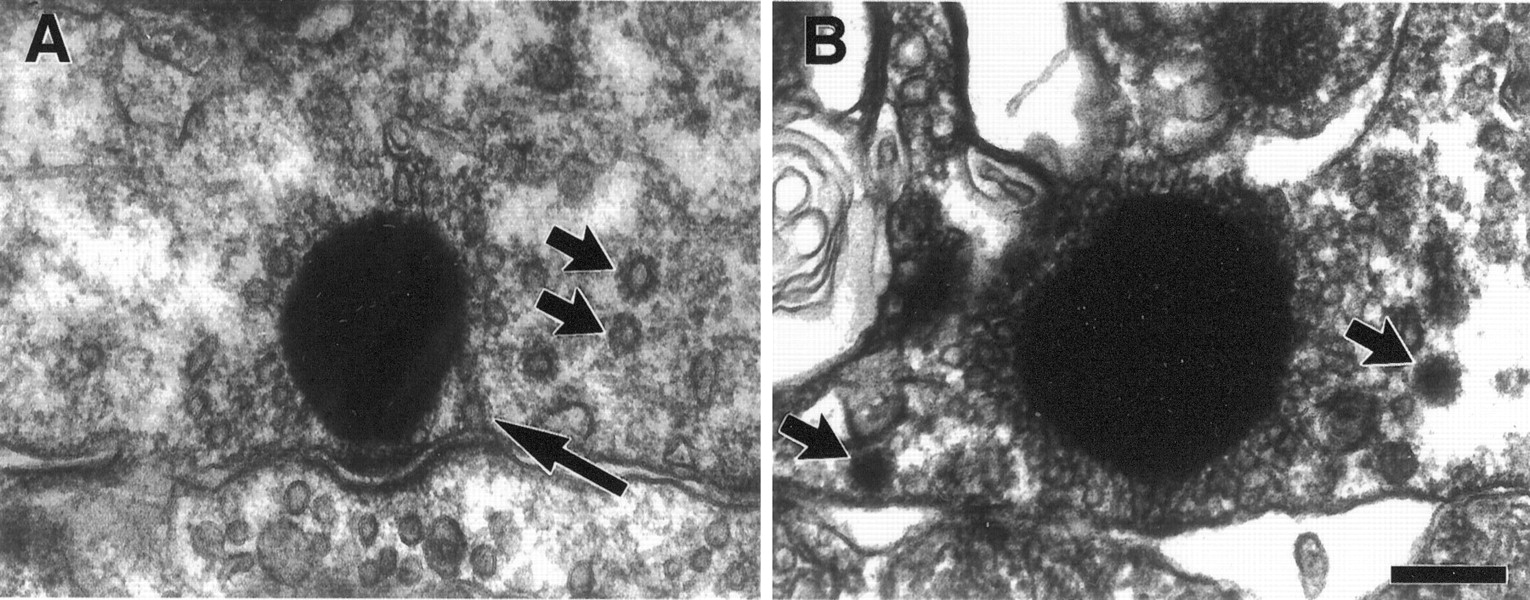
- Fig. 8.
Endocytosis machinery near the SB.A, Conventional transmission electron micrograph of a hair cell afferent synapse depolarized in 22 mmK+ saline. Numerous coated vesicles occur in the cytoplasm surrounding the SB (short arrows), and a coated pit in the plasma membrane is visible adjacent to the SB (long arrow). B, Conventional transmission electron micrograph of a synapse exposed to extracellular microperoxidase. Two coated vesicles contain the label (arrows). Scale bar, 200 nm.
- Fig. 10.
Tethers link the SB to vesicles and to the presynaptic density. A–D, Subregions ofx–y planes through osmium-stained reconstructed synapses. Each panel contains a portion of a SB. Scale bar, 100 nm. A, B, Tethers linking vesicles to the surface of the SB (arrows) and to other vesicles (arrowhead in A).C, Two tethers (arrows) linking one docked vesicle to the SB. The vesicle is in contact with the presynaptic plasma membrane, which forms the synaptic cleft (asterisks in C, D).D, Two filaments (arrows) join the SB to the presynaptic density on the plasma membrane. A,C, D, Synapse 2. B, Synapse 3. E, TEM micrograph of a synapse in a mechanically lysed hair cell. Although the presynaptic cytoplasm has lost most of its contents, the SB retains its monolayer of vesicles and remains at the active zone. Scale bar, 300 nm.


Tables
Synapse Stain Dimensions Synaptic body Outlying vesicles Voxel Edge (nm) Reconstructed volume (nm × nm × nm) Diameter (nm) Portion reconstructed (%) Vesicles counted (#) Total vesicles/SB (#) Packing density (%) Count (#) Packing density (%) 1 Os 3.3 845 × 845 × 200 524 29 162 562 68 164 6.5 2 Os 2.1 840 × 840 × 215 516 36 171 474 59 78 4.2 3 Os 3.0 900 × 900 × 205 362 34 89 265 60 67 4.0 4 EPTA 2.6 1470 × 1125 × 325 581 47 231 494 50 287 3.9 457 45 134 298 46 5 EPTA 2.9 1145 × 1145 × 380 477 75 139 186 26 208 3.4 6 EPTA 1.3 770 × 770 × 390 425 70 204 290 50 206 8.1 7 EPTA 1.6 770 × 630 × 155 440 20 103 506 83 — — 8 Os 1.4 780 × 745 × 255 431 56 171 307 52 78 6.4 468 ± 65 376 ± 133 55 ± 16 5.2 ± 0.67 (SD) (SD) (SD) (SEM) Voxels were cubes having the specified length along each edge. The listed dimensions are for the backprojections used to calculate SB size and vesicle concentrations. Data for the two SBs at synapse 4 are listed. At synapses 1 and 3, a second backprojection encompassing a larger volume was computed also (data not shown).


