

Article Figures & Data
Figures
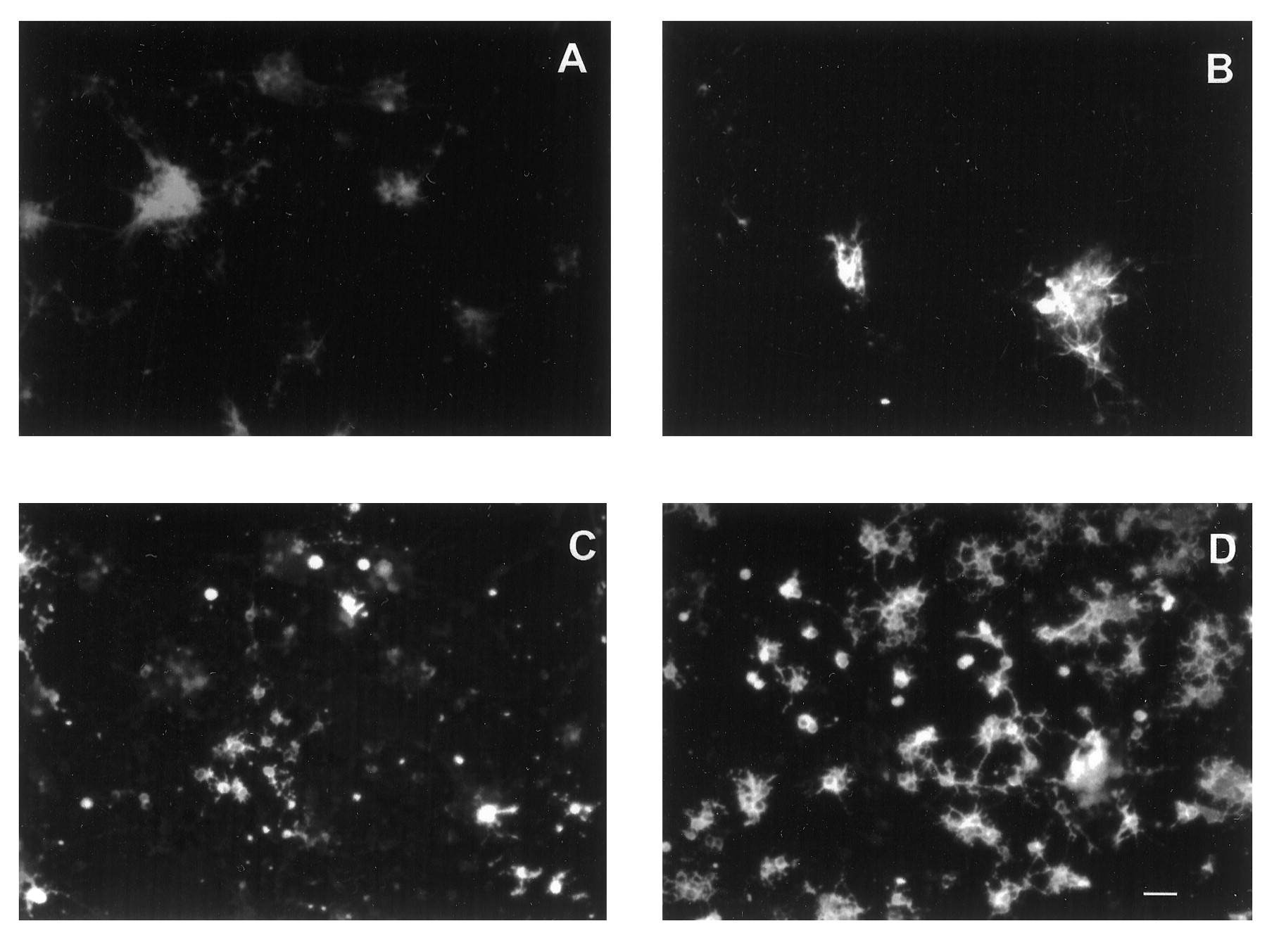
- Fig. 1.
Effects of BMP2 on the growth of cortical VZ cells plated at low density with FGF2 10 ng/ml. Photomicrographs of rat E13 cortical cells grown with FGF2 alone (A,D, G, J), FGF2 plus BMP2 10 ng/ml (B, E, H,K), or FGF2 plus BMP2 100 ng/ml (C, F, I,L). A–C, Phase-contrast images at 5 div.D–F, Bisbenzimide staining at 8 div.G–I, β tubulin III staining at 8 div.J–L, Nestin staining at 8 div. The same photographic field is shown for each condition in D,G, J; E, H,K; and F, I,L at 8 div. Arrows indicate examples of dying cells with apoptotic bodies (A–C), cells expressing β tubulin III-immunoreactivity (G–I), and nestin immunoreactivity (J–L). In each condition, an occasional cell coexpressed both β tubulin III and nestin (for example, thetop cell indicated with an arrow inI and L). Scale bar: A–C, 8 μm; D–L, 12 μm.
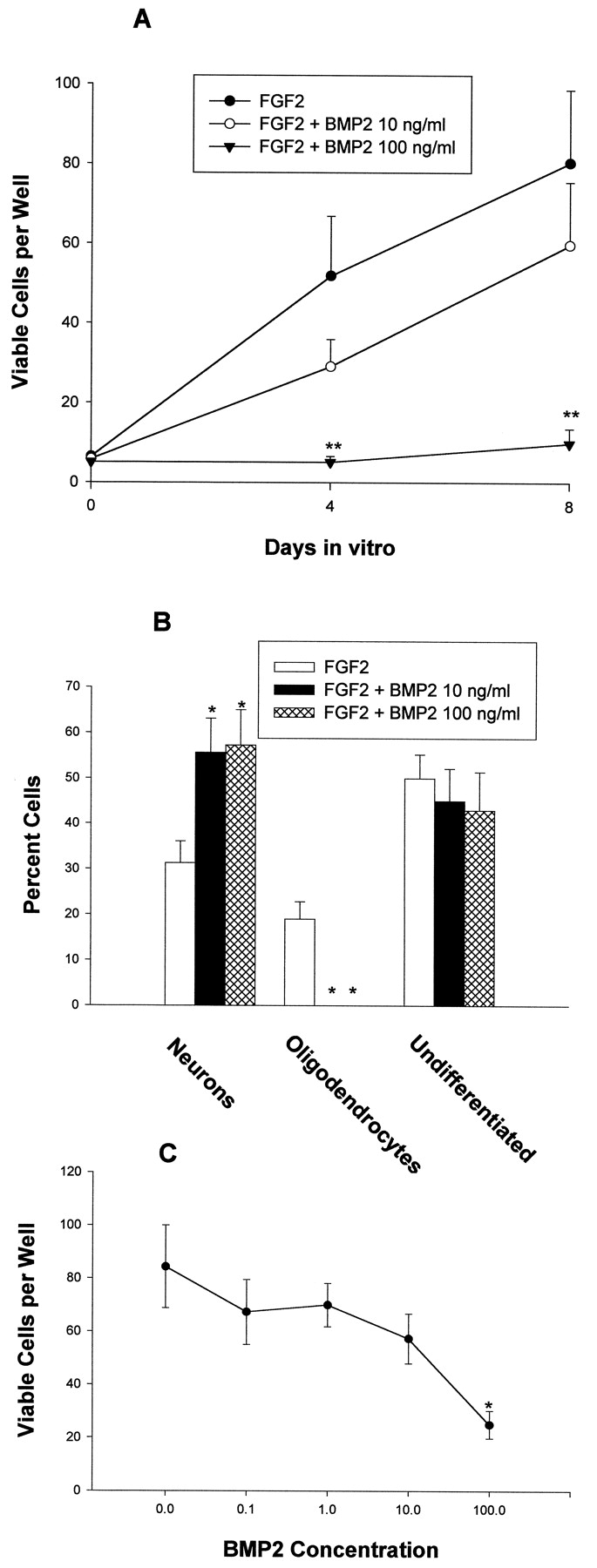
- Fig. 2.
Effects of BMP2 on the growth of cortical VZ cells. The number of total viable cells was quantitated at the indicated time points. A, Effects of BMP2 10–100 ng/ml on E13 cells grown at low density with FGF2 10 ng/ml for 8 div.B, Effects of BMP2 10 ng/ml on cells grown at moderate density with FGF2 0.1–10 ng/ml for 9 div. C, Effects of BMP2 0.1–100 ng/ml on E13 cortical cells grown at moderate density without FGF2 for 5 div. *p < 0.05; **p < 0.01.
- Fig. 3.
Effects of BMP2 on E16 cortical cells grown at low density with FGF2 10 ng/ml. Photomicrographs of cells grown with FGF2 alone (A, C, E) or FGF2 plus BMP2 10 ng/ml (B, D,F) for 8 div. A, B, Bisbenzimide staining. C, D, β tubulin III staining. E, F, O4 staining. The same photographic field is shown for each condition. Scale bar, 12 μm.
- Fig. 4.
Effects of BMP2 on E16 cortical cells. The number of total viable cells and cells expressing neuronal (β tubulin III), oligodendroglial (O4), and neuroepithelial (nestin) markers was quantitated at the indicated time points. A, Effects of BMP2 on E16 cells grown at low density with FGF2 10 ng/ml.B, Effects of BMP2 on the differentiation of E16 cortical grown at low density with FGF2 10 ng/ml for 8 div.C, Effects of BMP2 0.1–100 ng/ml on E16 cortical cells grown at moderate density without FGF2. *p < 0.05; **p < 0.01.
- Fig. 5.
Effects of delaying the addition of BMP2 on E16 cortical cell differentiation. Cells were grown at low density with FGF2 alone for the first 4 div, and then BMP2 was added at 10 or 100 ng/ml. The number of cells expressing neuronal (β tubulin III), oligodendroglial (O4), astrocytic (GFAP), and neuroepithelial (nestin) markers was quantitated at 8 div. *p < 0.05; **p < 0.01.
- Fig. 6.
Effects of noggin on E16 cortical cells grown at low density with FGF2 10 ng/ml. Photomicrographs of cells grown with FGF2 alone (A, C) or FGF2 plus noggin 100 ng/ml (B, D) for 8 div. A,B, β tubulin III staining. C,D, O4 staining. Scale bar, 15 μm.
- Fig. 7.
Effects of noggin on E16 cortical cells grown at low density with FGF2 10 ng/ml. The number of cells expressing neuronal (β tubulin III), oligodendroglial (O4), and neuroepithelial (nestin) markers was quantitated at 8 div. *p < 0.05; **p < 0.01.
- Fig. 8.
BMP2 and noggin expression in the developing cortex. Photomicrographs of E16 rat (A) and neonatal (B, C) murine cortex stained with a monoclonal antibody to BMP2 (A, B) and a monoclonal antibody to noggin (C). The arrows in A indicate the pial (top) and ventricular (bottom) surfaces. For neonatal coronal sections (B, C), thearrows in C indicate the position of (from top to bottom) the pial surface, the border between cortical gray matter and developing subcortical white matter, the border between subcortical white matter and subventricular zone, and the lateral ventricle.


Tables
FGF2 FGF2 + BMP2 Fate of individual cells: n = 663 n = 816 Nonviable 276 (41.6) 378 (46.3) Single cells 252 (38) 252 (30.9) Neurons 224 (88.9) 240 (95.2) Clones containing 2–4 cells 39 (5.9) 36 (4.4) with neurons 26 (66.7) 32 (88.9) Clones containing >4 cells 96 (14.5) 150 (18.4) with neurons 9 (10.7) 109 (72.7) with neurons and OLs 41 (42.7) 0 (0) with OLs 37 (38.5) 0 (0) without neurons or OLs 9 (10.7) 41 (27.3) Cellular composition within clones: 2–4 Cell clones with neurons, % neurons 100 96.9 >4 Cell clones with neurons, % neurons 3.6 ± 1.2 20.5 ± 3.4** >4 Cell clones with OLs, % OLs 16.4 ± 2.8 1-a Viability within clones Average # of viable cells per >4 cell clone 102.9 ± 11.9 70.3 ± 10.8 Average # of nonviable cells per >4 cell clone 14.4 ± 2.0 18.2 ± 2.0 Clonal analysis of E16 cortical cells grown in FGF2 alone or FGF2 plus 1 ng/ml BMP2. Fate of individual cells, Each absolute value is followed by a percentage in parentheses, which reflects the percentage of all cells in that particular category. Cellular composition within clones, Viability within clones, Each value represents the mean ± SEM. Neurons and oligodendroglia were identified by β tubulin III and O4 expression, respectively. Cells expressing the astrocytic marker GFAP were not included because they were never seen as single cells or in small clones and were present in <1% of clones of more than four cells.
↵F1-a No clones containing oligodendroglia were seen with BMP2 treatment.
*p < 0.05; **p < 0.01.


