

Article Figures & Data
Figures
- Fig. 1.
Neuron-specific induction of c-fosexpression by C-Ag4,8. a, Right, Cortical cultures were incubated for 10 min with C-Ag4,8 at a concentration determined previously to provide maximal induction of AChR clusters on cultured chick myotubes.Left, Control cultures were treated with an equivalent concentration of medium from sham-transfected COS-7 cells. Treated cultures were returned to cNBM, incubated for 2 hr at 37°C, and then fixed and labeled using the anti-Fos antibody Ab-2. Whereas basal levels of c-fos expression in control cultures are low, staining for Fos is markedly increased after treatment with C-Ag4,8. The majority of cells are darkly stained with prominent round nuclei and neuronal morphology (arrows). Cells that appear unaffected by C-Ag4,8 treatment have more irregularly shaped nuclei and non-neuronal morphology (arrowheads). b, Cultures were treated with either control (left) or C-Ag4,8–containing (middle,right) medium followed by double labeling for either Fos (fluorescein channel) and MAP-2 (Texas Red channel) or Fos and GFAP (Texas Red channel). In C-Ag4,8–treated cultures, levels of Fos expression evident in MAP-2–labeled neurons (arrows) are high, whereas Fos levels of MAP-2–negative non-neuronal cells (arrowheads) are comparable with basal levels seen in control cultures. Conversely, after treatment with C-Ag4,8, levels of c-fos expression are low in GFAP-labeled non-neuronal cells but high in the GFAP-negative neurons. Scale bars, 50 μm.
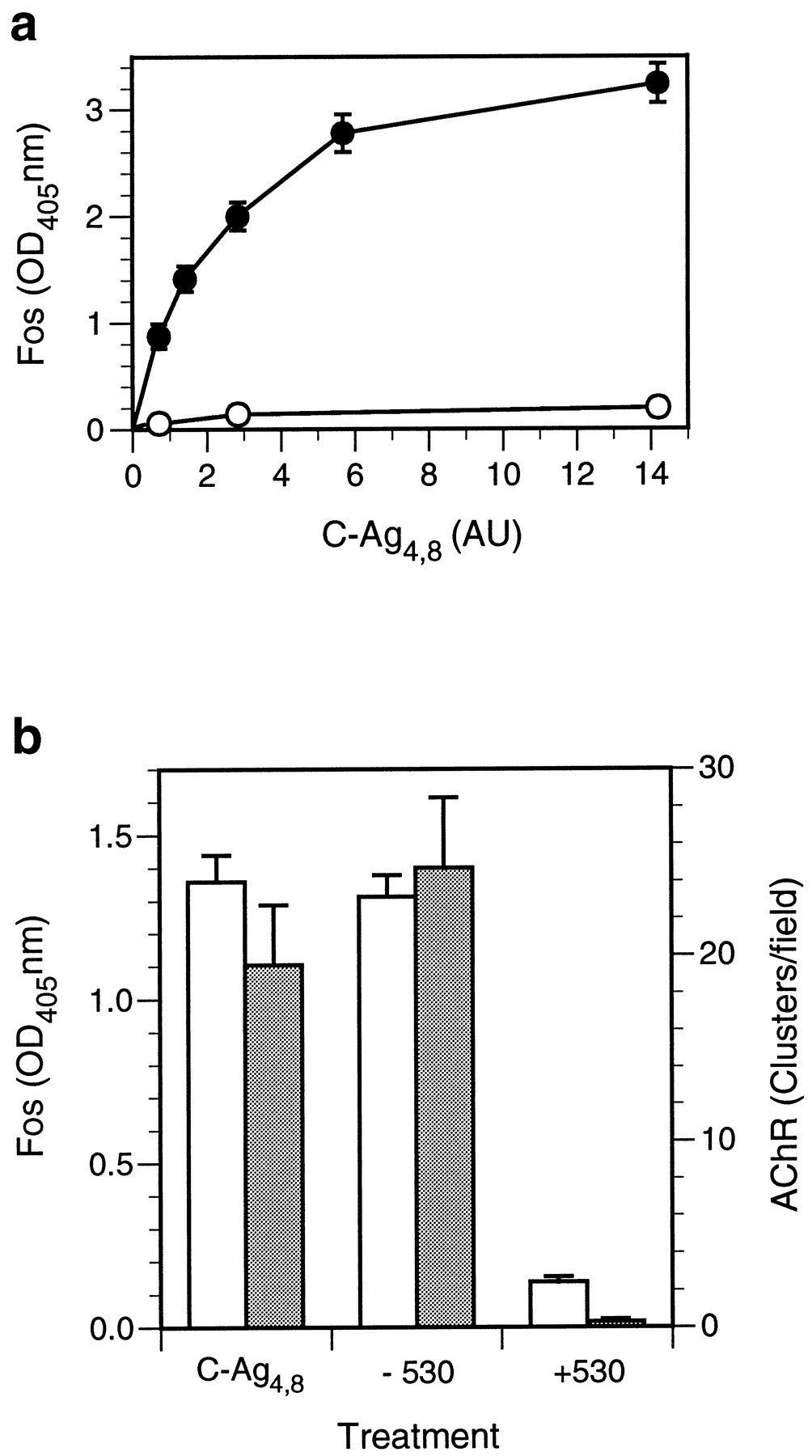
- Fig. 2.
c-fos induction in cultured cortical neurons by C-Ag4,8 is concentration dependent and saturable.a, Cortical cultures were treated with C-Ag4,8 or control medium in NBM for 10 min at room temperature, returned to cNBM, and incubated for 2 hr at 37°C after which Fos levels were determined using the quantitative assay described in Materials and Methods. Levels of Fos expression, shown in arbitrary OD405 units, increased in a C-Ag4,8concentration–dependent manner (filled circles). Basal levels of Fos expression (open circles) were low and did not change significantly with an increasing concentration of control medium added to the culture. The graph shows Ab-2–specific binding; nonspecific binding, determined by labeling C-Ag4,8– or control medium–treated cultures with Ab-2 preadsorbed to an excess of peptide antigen, was <5% of total binding and has been subtracted. The C-Ag4,8 concentration is given in AU where one unit is the amount of C-Ag4,8 required to induce a half-maximal increase in AChR clusters assayed on cultured chick myotubes. Each point represents the mean ± SEM for triplicate determinations. Similar results were seen in two other experiments. b, To test the specificity of C-Ag4,8 induction of Fos expression, we immunoprecipitated an aliquot of C-Ag4,8–containing medium with the anti-agrin monoclonal antibody Agr 530. The chart showsc-fos–inducing (open bars; left scale) or AChR-clustering (shaded bars;right scale) activity measured in cortical neuron cultures and cultured chick myotubes, respectively, after treatment with a saturating concentration of C-Ag4,8medium (C-Ag4,8) or an equivalent amount of C-Ag4,8 medium immunoprecipitated in the absence (−530) or presence (+530) of Agr 530. Greater than 90% of thec-fos–inducing and AChR-clustering activities are removed from the C-Ag4,8 medium by immunoprecipitation with Agr 530, confirming that C-Ag4,8 is responsible forc-fos induction in cortical neurons. Basal levels of Fos observed in cortical cultures treated with sham-conditioned medium and spontaneous AChR clusters have been subtracted. Error bars represent the mean ± SEM for triplicate determinations. Similar results were also seen in a second experiment.
- Fig. 3.
Time course of C-Ag4,8 induction ofc-fos. Cortical cultures were incubated in a saturating concentration of C-Ag4,8 for the indicated length of time and then washed and returned to the incubator in cNBM for 2 hr before levels of Fos expression were assayed. Induction ofc-fos is relatively rapid and half-maximal after only 5 min of exposure to C-Ag4,8. The graph shows C-Ag4,8–specific c-fos induction; levels of Fos in sister wells treated with sham-conditioned medium have been subtracted. Error bars represent the mean ± SEM for triplicate determinations. Similar results were also seen in a second experiment.
- Fig. 4.
C-Ag4,8 induction ofc-fos is Ca2+-dependent and blocked by heparin. Cortical cultures were incubated for 10 min in a saturating level of C-Ag4,8 in the presence of Ca2+(a) or heparin (b) at the indicated concentrations and then returned to the incubator in cNBM for 2 hr before determination of Fos levels by enzyme-linked assay as described. Graphs show C-Ag4,8–specificc-fos induction (circles); levels of Fos in sister wells treated with sham-conditioned medium have been subtracted. Induction of c-fos is Ca2+-dependent and blocked by heparin, similar to agrin-induced AChR clustering in muscle. Treatment with heparin alone (b; square) at 500 μg/ml had no significant effect. Each data pointrepresents the mean ± SEM for triplicate determinations.
- Fig. 5.
Intracellular Ca2+ and protein kinase activation are required for C-Ag4,8signaling. Cortical cultures were equilibrated for 1 hr in BAPTA- AM (a) or 10 min in staurosporine (b) followed by coincubation for 10 min with a saturating concentration of C-Ag4,8. a, BAPTA-AM–treated cultures were subsequently washed with NBM and then returned to cNBM media for 2 hr at 37°C before determination of Fos levels. b, Cultures treated with staurosporine were incubated with staurosporine alone for an additional 10 min after removal of C-Ag4,8 before being returned to cNBM.Graphs show C-Ag4,8–specific Fos expression (circles); Fos induction in sister cultures treated with sham-conditioned medium containing BAPTA-AM or sham-conditioned medium alone has been subtracted. Treatment with staurosporine alone at the highest concentration used (b; square) had no significant effect. Each data point represents the mean ± SEM for triplicate determinations. Similar results were seen in at least one other experiment for each treatment.
- Fig. 6.
Agrin isoforms that lack AChR-clustering activity induce c-fos expression in cortical neurons.a, Cortical cultures were exposed to C-Ag4,0(open circles)– or C-Ag0,0(filled circles)–containing media for 10 min, and c-fos expression was assayed. Nonspecific induction of c-fos determined in control cultures treated with sham-conditioned medium has been subtracted. Induction of c-fos by both C-Ag4,0 and C-Ag0,0 is concentration-dependent and saturable. Regardless of their AChR-clustering activities, the different agrin isoforms exhibit similar specific activities in terms ofc-fos induction in cortical neurons. b,To confirm the specificity of action of C-Ag4,0(open bars)– and C-Ag0,0(shaded bars)–containing media, the ability of Agr 530 to immunoprecipitate thec-fos–inducing activity was tested in a manner similar to that described in Figure 2. Greater than 80% of thec-fos–inducing activity was precipitated by the antibody. To facilitate comparison, we expressed results as the percent of the maximal level of Fos induction for a given isoform within each experiment. Data show the mean ± SEM for triplicate determinations. Similar results were obtained in at least one other experiment for each group.
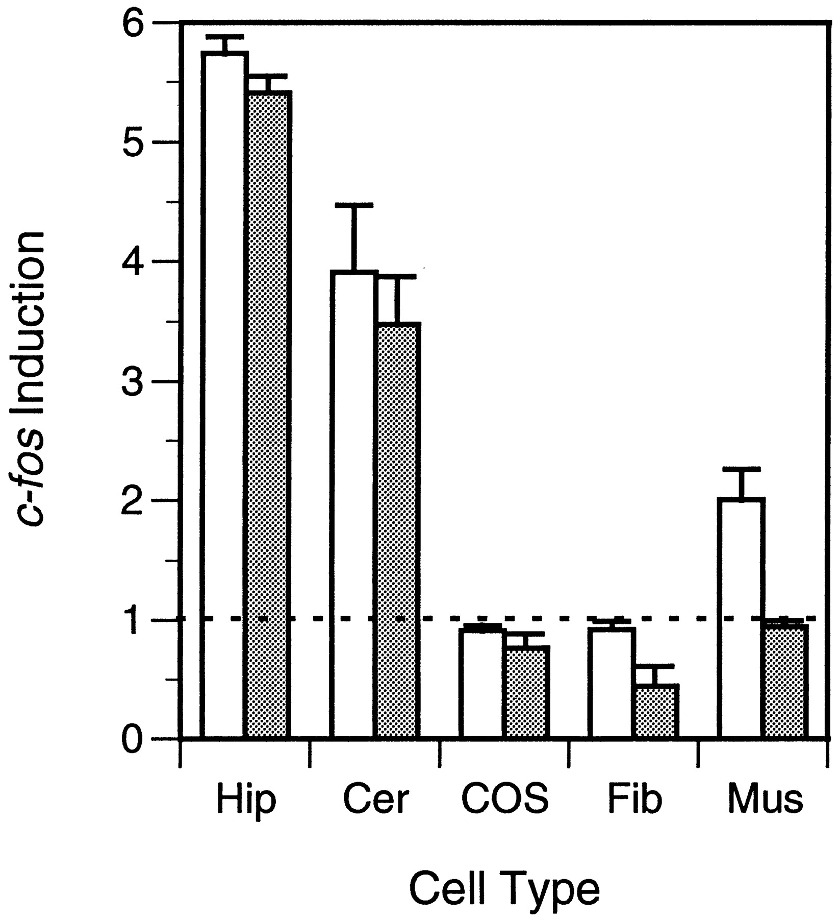
- Fig. 7.
Induction of c-fos by different agrin isoforms is cell-specific. Fos levels were measured in cultured hippocampal (Hip), cerebellar (Cer), COS-7 (COS), chick fibroblast (Fib), and muscle (Mus) cells at 2 hr after a 10 min treatment with a saturating concentration of C-Ag4,8 (open bars)– or C-Ag0,0 (shaded bars)–containing media. Induction ofc-fos is expressed as the ratio of Fos expressed in agrin-treated cultures over that observed in control cultures treated with sham-conditioned medium. A value of 1 represents no induction. Fos expression in hippocampal and cerebellar cultures was increased by a similar amount after treatment with either agrin isoform. In contrast, only C-Ag4,8 induced c-fos in muscle, and neither isoform was effective in fibroblasts or COS-7 cells. Error bars represent the mean ± SEM for triplicate determinations. Similar results were obtained in one other experiment for each group.
- Fig. 8.
Biochemical properties of c-fosinduction by C-Agz0 isoforms. Extracellular Ca2+ dependence (a), blockade by heparin (b), the requirement for intracellular Ca2+ (c), and inhibition by staurosporine (d) of C-Ag4,0(open circles)– and C-Ag0,0(filled circles)–dependent induction of c-fos were examined. With the exception of a minor heparin-resistant component of the C-Ag0,0–dependent increase in Fos expression, marked similarity is evident in the biochemical profiles of the two isoforms. For comparison, results are expressed as the percent of the maximal level of Fos induction for a given isoform within each experiment. Data represent the mean ± SEM for triplicate determinations. Similar results were obtained in at least one other experiment in each group.


