

Article Figures & Data
Figures
- Fig. 1.
Dissociated neurons on a coronal forebrain section. A, The density of fluorescence-labeled neurons is greatest on gray matter, such as the neocortex (ctx) and caudate-putamen (cp); intermediate levels of attachment are found near the corpus callosum (cc) midline (white arrow); and virtually no neurons are attached to more lateral portions of the corpus callosum (white asterisk). The dense plexus of neurites on gray matter is sharply inhibited at the border with the corpus callosum (white arrowheads). From neurons attached to the corpus callosum, neurite outgrowth is oriented in a direction parallel to the longitudinal axis of the tract (see C) in contrast to the complex pattern of neurite outgrowth on gray matter (see also Fig. 2A). B, The same field shown in A is shown with fluorescence in combination with phase-contrast optics. Note the low optical density (black arrow) near the midline of the corpus callosum corresponding to the relatively parallel orientation of the fibers here, in contrast to the darker regions (white asterisk) found laterally where fibers run more obliquely.C, Higher power photomicrograph of the center of fieldA showing neurite outgrowth that is primarily in parallel with the underlying fiber tract is shown. Scale bars:A, B, 300 μm; C, 100 μm.
- Fig. 2.
Dissociated neurons on a coronal forebrain section. A, Neurite outgrowth from fluorescence-labeled neurons attached to the caudate-putamen (cp) shows no preferred orientation. A single neuron (white asterisk) is attached near the border between the caudate-putamen and the corpus callosum (cc) with a neurite (white arrowhead) extending parallel to, but not crossing onto, the corpus callosum. Several neurites (white arrows) extend on the caudate-putamen but not on the corpus callosum.B, The same field with epifluorescence in combination with phase-contrast optics shows the border (black arrows) between the caudate-putamen and the corpus callosum and the location of the neuron featured in A (white asterisk). Scale bar, 100 μm.
- Fig. 3.
Dissociated neurons on GFAP-stained sections.A, Many fluorescence-labeled neurons are attached to the corpus callosum (cc) with neurite outgrowth oriented in parallel with the longitudinal axis of the tract. Few neurons, in contrast, are attached to the transversely sectioned cingulum (cg). B, Higher power photomicrograph of an area indicated by the white arrows inA and C is shown. C, GFAP immunoreactivity is densely represented in all regions of the forebrain. Within the corpus callosum, GFAP-immunoreactive processes are primarily oriented in parallel with the longitudinal axis of the tract. D, Area corresponding to B shows GFAP staining. ctx, Neocortex; hf,hippocampal formation. Scale bars: A, C, 300 μm;B, D, 100 μm.
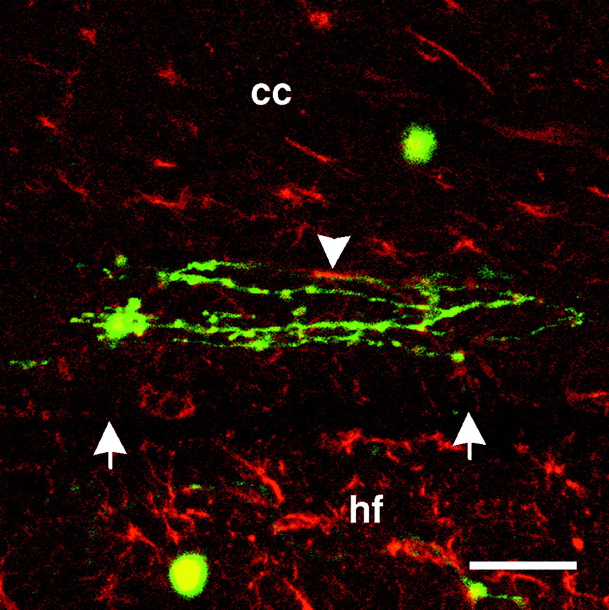
- Fig. 4.
Composite confocal photomicrograph of dissociated neurons on GFAP-labeled sections. Dissociated neurons and neurites (green) on corpus callosum (cc) near the border (white arrows) with the underlying hippocampal formation (hf) are shown. GFAP immunoreactivity is shown in red. Colocalization of neurites and GFAP immunoreactivity is limited (white arrowhead). Scale bar, 50 μm.
- Fig. 5.
Explants on coronal forebrain sections.A, A fluorescence-labeled explant is shown attached to the amygdala (amg) with its neurite halo sharply inhibited at the border (black arrows) with the optic tract (ot). A single neuron is shown (black arrowhead) with the neurite extending on, and in parallel with, the optic tract. B, Fluorescence-labeled explants are shown attached directly to the optic tract with extensive outgrowth on, and in parallel with, the tract (black arrows). Outgrowth from explants on the optic tract is comparable in length with that of explants on neocortex (ctx; black arrowhead). Neurite outgrowth on portions of the optic tract sectioned transversely is more mixed in orientation (white arrow). The outgrowth halo of a detached explant (white arrowhead) is shown having grown on the amygdala but inhibited by the adjacent optic tract. C, A fluorescence-labeled explant is shown attached to the corpus callosum (cc) with extensive outgrowth extending on, and in parallel with, the tract. As neurites approach the cingulum (cg), they become increasingly mixed in orientation. D, The same field shown in C is shown with phase-contrast optics.cp, Caudate-putamen. Scale bars: A, 100 μm; B, 250 μm; C, D, 100 μm.
- Fig. 6.
Explants on CNS tissue sections. A,A horizontal section of cervical spinal cord showing a fluorescence-labeled explant attached to the lateral funiculus with extensive neurite outgrowth on, and predominantly in parallel with, the fiber tract. A few neurites are oriented in nonparallel directions (black arrow) but heavily fasciculated.B, A coronal section of forebrain showing a fluorescence-labeled explant attached to the caudate-putamen (cp). Some neurites (black arrowhead) cross directly over white matter (black asterisks), but these are generally fasciculated, whereas nonfasciculated neurites (black arrows) generally avoid white matter.C, A coronal section of forebrain showing a fluorescence-labeled explant attached to the corpus callosum (cc) with some fascicles (black arrowhead) oriented nonparallel to the fiber tract. Defasciculation (black arrows) is coincident with reorientation in the parallel direction. D, The same field shown in C with phase-contrast optics showing the border (black arrows) between the neocortex (ctx) and the corpus callosum. Scale bars:A, 150 μm; B, 100 μm; C, D, 100 μm.
- Fig. 7.
Rate of neurite outgrowth on corpus callosum, assessed both in parallel and perpendicular to the longitudinal axis, and on representative gray matter regions. After 8 d in culture, parallel neurite growth on the corpus callosum (white triangles) is comparable with that on the hippocampus (white squares) and significantly greater than neurite growth on neocortex (white circles). Neurite outgrowth on neocortex, in turn, is significantly greater than growth on the corpus callosum, assessed perpendicular to the longitudinal axis (black triangles). Values are expressed as mean ± SEM. Asterisks indicate statistical significance after 8 d in culture (p < 0.001).


