

Article Figures & Data
Figures
- Fig. 1.
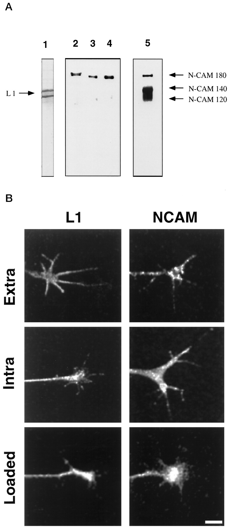
Immunocytochemistry, trituration loading, and specificity of L1 and NCAM antibodies in chick DRG neurons.A, Specificity of 8D9, 4d, and 5e is demonstrated by Western blot analysis. Lane 1, DRG lysate probed with 8D9. Two bands are observed, likely because of proteolysis. Lane 2, DRG lysate probed with 4d recognizes only NCAM-180;lane 3, purified NCAM-180 probed with 4d; lane 4, whole chick brain lysate probed with 4d; lane 5, DRG lysate probed with 5e, which recognizes all three NCAM isoforms. Note that different gels were used for lane 1 and for lanes 2–5 so that the migration distances do not correspond between these lanes. B,Trituration loading of intra-L1 followed by fixation and staining with secondary antibody (L1, Loaded) results in a similar pattern to that observed by immunocytochemistry with intra-L1 (L1, Intra) and with 8D9, which shows expression of L1 in growth cones (L1, Extra). Trituration loading of 4d followed by fixation and staining with secondary antibody (NCAM, Loaded) shows similar specific staining compared with indirect immunocytochemistry using 4d (NCAM, Intra) or with 5e (NCAM, Extra).
- Fig. 2.
CALI of purified L1 inhibits its neurite-promoting activity. CALI directed against L1 using intra-L1 or 8D9 resulted in marked inhibition of the percentage of cells with neurites.Lanes A–D show CALI of mouse L1 substrate using MG-labeled intra-L1: lane A, no treatment; lane B, laser irradiation without MG-labeled antibody; lane C, MG-labeled intra-L1 without laser irradiation; lane D, MG-labeled intra-L1 with laser irradiation. Lanes E–H show CALI of chick L1 substrate using MG-labeled 8D9:lane E, no treatment; lane F, laser irradiation without MG-labeled antibody; lane G, MG-labeled 8D9 without laser irradiation; lane H, MG-labeled 8D9 with laser irradiation. Lanes I–K show that laser irradiation using MG-8D9 did not affect neurite outgrowth-promoting activity of laminin. Lane I, No treatment; lane J, irradiation without MG-labeled 8D9;lane K, MG-labeled 8D9 with laser irradiation. The data are representative of three experiments with n > 100 neurons for each treatment.
- Fig. 3.
CALI of NCAM-180 intracellular domain disrupts brain spectrin binding in vitro. Purified chick NCAM (all isoforms) was incubated with purified bovine brain spectrin after CALI using 4d, 5e, or nonspecific IgG. The complex was immunoprecipitated with anti-NCAM, dissociated in Laemmli sample buffer, fractionated by SDS-PAGE, and immunoblotted with anti-spectrin antibodies to assay NCAM-180/spectrin binding. A shows spectrin immunoblot, whereas B shows the same filter after being stripped and probed with anti-NCAM (all isoforms), showing that there were equivalent amounts of NCAM present in each lane.Lane 1, Anti-NCAM immunoprecipitation; lane 2, no anti-NCAM control; lane 3, preincubation of NCAM with MG-labeled nonspecific antibody (IgG); lane 4, the same conditions as lane 3 except subjected to laser irradiation; lane 5, preincubation with MG-labeled 4d (intracellular domain of NCAM-180); lane 6, same conditions as lane 5 except subjected to laser irradiation (CALI using 4d); lane 7, preincubation with MG-labeled 5e (extracellular domain of all NCAM isoforms); lane 8, same conditions as lane 7, except subjected to laser irradiation (CALI using 5e).
- Fig. 4.
Micro-CALI of L1 causes neurite retraction. Micro-CALI of L1 causes neurite retraction but does not inhibit filopodial motility. A chick DRG growth cone was incubated with MG-labeled 8D9, and motility was observed by time-lapse video microscopy before laser irradiation (A). Laser irradiation of the growth cone (B, C) caused neurite retraction (D) after 10 min and subsequent recovery and growth along a different path (E, F) after 30 min. Filopodial motility is not affected. Scale bar, 10 μm.
- Fig. 5.
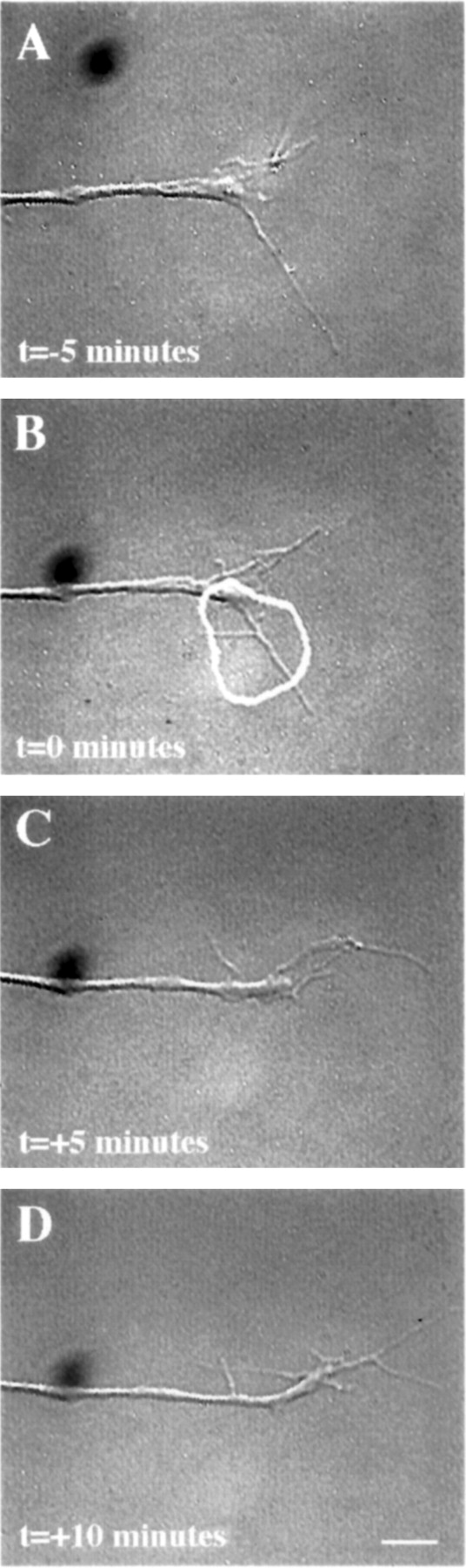
Micro-CALI of the intracellular domain of NCAM 180 using MG-labeled 4d caused regional growth cone retraction.A, A growth cone was first observed for 5 min. A region of the growth cone was chosen for micro-CALI (white outline), laser irradiation was initiated att = 0 (B) and continued untilt = +5 min (C), and observed for 5 more minutes (D). In C, the filopodia and lamellipodia of the irradiated region retracted from the laser spot, whereas the rest of the growth cone did not appear to be affected. In D, the growth cone began to grow in another direction, causing a visible bend in the neurite. The time-lapse imaging was done using DIC microscopy instead of phase contrast as was used for Figures 4 and 6. Scale bar, 10 μm.
- Fig. 6.
Micro-CALI of the extracellular domain of all isoforms of NCAM using MG-labeled 5e does not affect growth cone behavior. A growth cone was incubated with MG-labeled 5e and observed by video time-lapse microscopy (A). A region of the growth cone is chosen for micro-CALI (white outline) and laser irradiated for 5 min (B, C). D,The growth cone is observed for an additional 5 min. Micro-CALI did not cause filopodial or lamellipodial retraction. Scale bar, 10 μm.
- Fig. 7.
Micro-CALI of NCAM-180 and L1 simultaneously causes both neurite retraction and growth cone collapse. DRG neurons were loaded with MG-labeled 4d and incubated with MG-labeled 8D9. After observation for 10 min (a), the growth cone was irradiated for 5 min, starting at t = 0 (b, black outline in b) and ended at t = +5 min (c,white outline). Shortly thereafter, the filopodia and lamellipodia retract, and the growth cone continues to collapse, but the neurite does not decrease in length (d). Over the next 10 min the neurite retracts (e) and begins to recover after an additional 10 min (f). Scale bar, 10 μm.
- Fig. 8.
Quantitative analysis of micro-CALI of NCAM and L1. Average values for neurite extension, changes in filopodial length, and absolute rates of filopodial motility were determined for each experimental treatment, before (B) and after (A) laser irradiation.NCAM/L1, Simultaneous micro-CALI of NCAM and L1 using MG-labeled 4d and 8D9; NCAM, micro-CALI using MG-labeled 4d; L1, micro-CALI of L1 using MG-labeled 8D9;IgG, laser irradiation with MG-labeled nonimmune IgG;None, laser irradiation in the absence of MG-labeled reagents. Values that are significantly different (p < 0.01) than control values (None) are marked with double asterisks.


Tables
Treatment Neurite retraction Local growth cone collapse CALI of L1 (8D9) 10 /12 1 /12 CALI of L1 (intra L1) 8 /12 0 /12 CALI of NCAM-180 (4d) 0 /25 21 /25 CALI of NCAM (5e) 0 /20 3 /20 CALI of L1 & NCAM-180 (8D9 & 4d) 12 /13 10 /13 CALI using IgG 0 /20 2 /20 CALI using BSA 0 /12 1 /12 No treatment 0 /13 0 /13 DRG neurites were treated with micro-CALI of L1, NCAM-180, both together, or a variety of controls. Growth cone motility and neurite outgrowth were assessed, and neurons were deemed to undergo neurite retraction if the neurite decreased in length after laser irradiation and increased in length before irradiation. Neurons were deemed to show local growth cone collapse when a net decrease in filopodial length was observed within the laser spot, and there was a net increase in filopodial length in the rest of the growth cone and within the chosen laser spot before irradiation.
Behavior Frequency observed for each treatment Treatment Micro-CALI using 4d Micro-CALI using 5e Micro-CALI Control IgG Regional retraction 21/25* 3/20 2/20 Lateral growth cone movement 16/25 4/20 3/20 Movement away 14/16** 2/4 1/3 Movement toward 2/16 2/4 2/3 Odds ratio of moving away 7.0*** 1.0 0.5 Comparison of growth cone retraction and turning observed during micro-CALI of NCAM experiments. Regional retraction is defined as a decrease in length of the cord drawn from the neurite neck to the furthest extent of the leading edge within the area of irradiation during CALI. Lateral growth cone movement (defined operationally as components of leading edge extension that are perpendicular to the midline of the growth cone) was observed before and after micro-CALI, and changes in lateral movement were scored. “Moving away” means that the leading edge of the non-laser irradiated side is extending away from the laser spot and does not simply denote lamellipodial retraction. Of the growth cones that moved away from the laser spot, 93% also exhibited filopodial retraction for micro-CALI experiments targeting the intracellular domain of N-CAM 180 of cells growing on laminin. The odds ratio assumes a 50% probability for a change in lateral growth cone movement to be away from the laser spot (odds ratio = 1).
*p < 0.002 by Poisson's test.
**p < 0.02, significantly associated with filopodial retraction by McNemar's exact test.
***p < 0.02 by one-sample binomial test.


