

Article Figures & Data
Figures
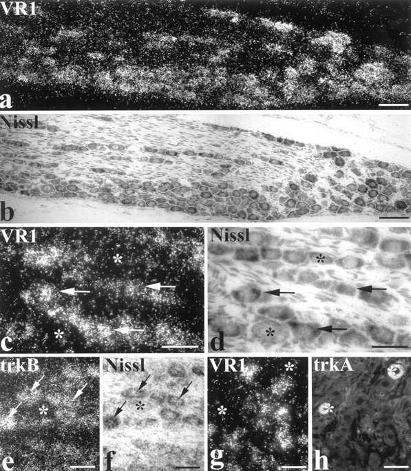
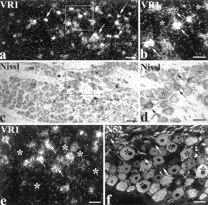
- Fig. 1.
Expression of VR1 mRNA in the dorsal root ganglion is confined primarily to small- to medium-sized cells that do not stain for heavy chain neurofilament. In situ hybridization for VR1 mRNA (a, b) with counterstaining of Nissl substance in cells with toluidine blue (c,d). VR1 mRNA is expressed by only a subset of DRG cells (a, c, arrows). Higher magnification photomicrographs (b, d) of the area indicated show that expression is restricted to small- to medium-sized cells, with the levels of VR1 mRNA differing considerably between cells (arrows). Large cells do not express this mRNA (asterisks). Combined in situhybridization for VR1 mRNA (e) and immunofluorescence for heavy chain neurofilament using antibody N52 (f). Expression of VR1 mRNA is observed in many cells that are negative for neurofilament immunoreactivity (e, f, arrows). Almost all neurofilament-positive cells are devoid of labeling for VR1 mRNA (e, f, asterisks). Scale bars: a, c, 100 μm; b,d–f, 50 μm.
- Fig. 2.
VR1 mRNA is expressed by both the NGF- and GDNF-responsive small cell populations. In situhybridization for VR1 mRNA (a, c,e) combined with fluorescence histochemistry (b, d, f).a, b, trkA. Most small- to medium-sized trkA-immunoreactive cells express VR1 mRNA (thin arrows). A few small trkA cells (thick white arrows) and large-diameter cells (asterisks) have little or no detectable VR1 mRNA. c,d, IB4. As with the trkA subpopulation of small- to medium-sized cells, most IB4-labeled cells express VR1 (thin arrows). Some IB4 cells, however, do not possess levels of VR1 mRNA above background (thick white arrows). Some very small-diameter IB4-negative profiles have high levels of VR1 mRNA (thick black arrows). e,f, RET. Whereas small RET-positive cells are often labeled for VR1 mRNA (thin arrows), large RET-positive cells and other large cells do not express VR1 (asterisks). A very small-diameter RET-positive cell is shown that has very high levels of VR1 mRNA (double arrows and insert in f). Scale bars: a–f, 50 μm; inset, 10 μm.
- Fig. 3.
Scatterplot diagrams of VR1 expression in selected populations of dorsal root ganglion cells. In each graph, individual profiles are plotted according to their diameter (in micrometers; along the x-axis) and the percentage of the profile area that is covered by silver grains as a measure of VR1 mRNA expression (along the y-axis). The dashed lines represent the criteria above which cells are considered labeled for VR1 mRNA. The symbols on the far left of each graph represent the mean ± SEM percentage area for each population. a, TrkA, Large cells (>40 μm in diameter) do not express VR1 mRNA. Grain density over smaller profiles varies considerably. b,IB4, Profiles generally tend to have less VR1 signal than the trkA identified profiles. Quite a few profiles have little or no VR1 mRNA. c, Trk/IB4, This double-labeled population has a characteristic size range (from ∼20 to <30 μm) and shows consistent VR1 labeling above threshold.d, TrkA-neg/IB4 neg, These profiles on the whole are very small (ranging from 15 to 25 μm) and possess very high levels of VR1 mRNA. The mean percentage area labeling for this population was significantly higher than all other populations atp = 0.01. e,Somatostatin, This subpopulation of IB4 cells exhibits characteristically low or beneath threshold levels of VR1 mRNA signal. The mean percentage area labeling was significantly lower than the trkA, trkA-negative/IB4-negative (p = 0.01), and trkA/IB4 (p = 0.05) populations.
- Fig. 4.
VR1 expression in identified subpopulations of dorsal root ganglion cells. In situ hybridization for VR1 mRNA was combined with immunofluorescence for the neuropeptides CGRP (a, b), substance P (c, d), and somatostatin (e, f). Thin white arrows identify cells that express VR1 mRNA and the particular neuropeptide. a, b, Small- to medium-sized CGRP cells express VR1 mRNA, with some cells containing relatively high levels of the message compared with others.Asterisks label large CGRP-immunoreactive cells that do not express VR1 mRNA. c, d, Similar to CGRP-positive cells, substance P-immunoreactive cells show variable levels of VR1 mRNA expression. e, f, The somatostatin cell identified in this photomicrograph expresses only low levels of VR1 mRNA. Scale bars: a, b, 50 μm; c–f, 20 μm.
- Fig. 5.
Expression of VR1 mRNA in the nodose ganglion.a, b, Moderate to high levels of VR1 mRNA were localized to most neurons of the nodose ganglion.c, d, At high magnification, labeled cells (arrows) show differing levels of the mRNA. Some cells are not labeled (asterisks). e,f, The mRNA for trkB is detected in most nodose ganglion cells. Arrows point to labeled cell profiles.Asterisks mark an unlabeled cell. g,h, VR1 mRNA is not detected in most nodose ganglion cells that express trkA protein (asterisks). Scale bars:a, b, 100 μm; c–h, 50 μm.
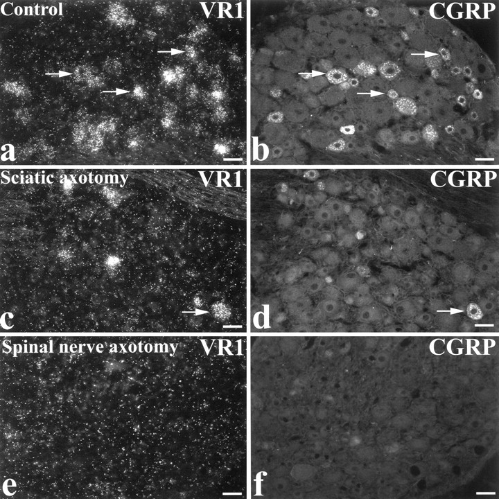
- Fig. 6.
Expression of VR1 mRNA decreases in the DRG after axotomy. Combined in situ hybridization for VR1 mRNA and CGRP immunofluorescence was performed on sections from the contralateral control (a, b), ipsilateral sciatic nerve-axotomized (c, d), and spinal nerve-axotomized (e, f) dorsal root ganglia. In the control DRG, there is robust labeling for both VR1 mRNA and CGRP. Many cells coexpress both products (a, b, arrows). Ipsilateral to the sciatic nerve axotomy, there is a dramatic reduction in the number of cells labeled for VR1 or CGRP. Many of the remaining VR1-labeled cells are CGRP-immunoreactive. Arrows inc and d point to one example. After spinal nerve axotomy, both VR1 mRNA and CGRP expression is reduced throughout the ganglion. Scale bars, 50 μm.


