

Article Figures & Data
Figures
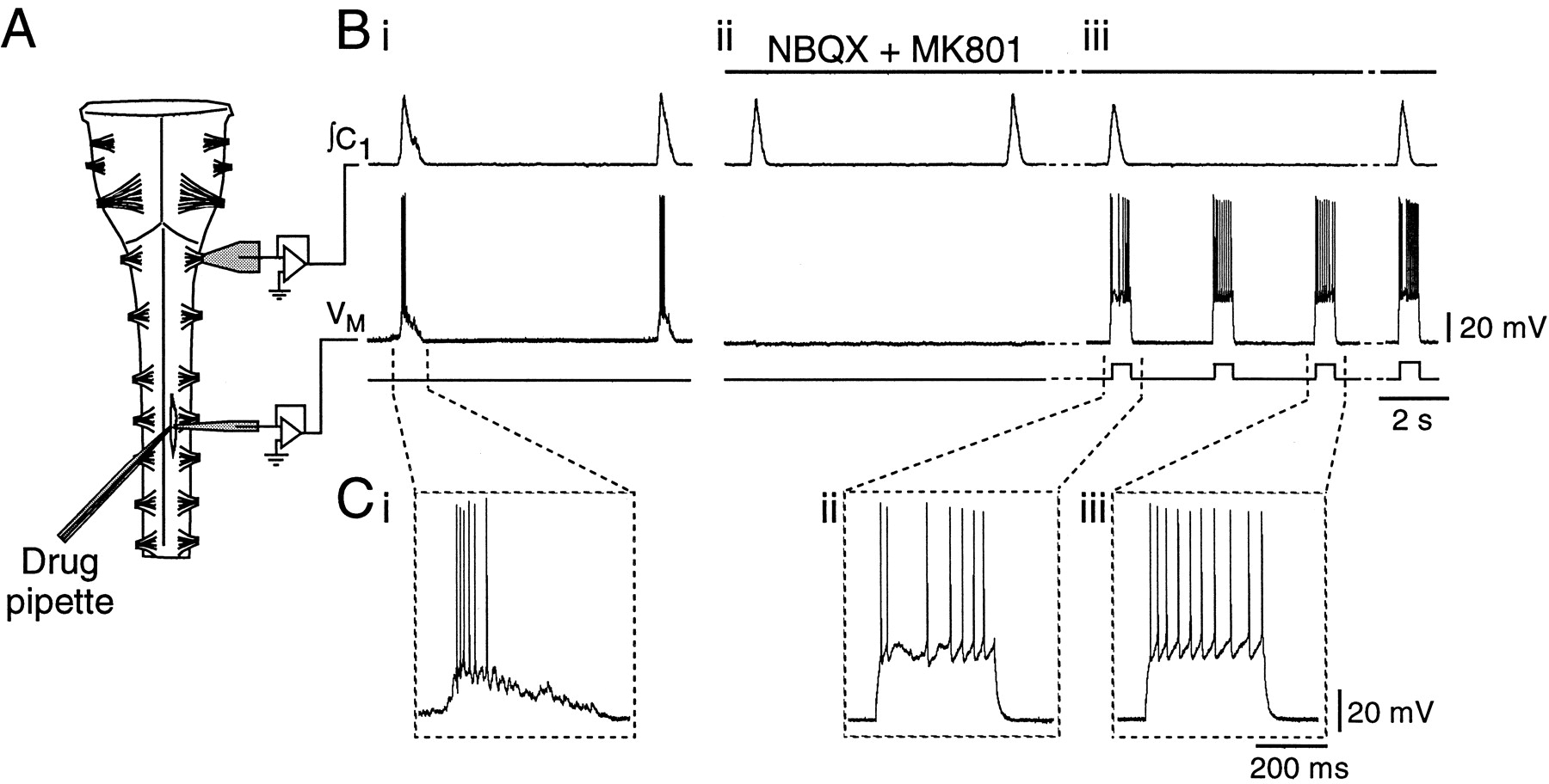
- Fig. 1.
Experimental set-up and protocol for comparison of PMN firing behavior during inspiratory and expiratory phases.A, Schematic of the rhythmically active brainstem–spinal cord preparation showing the configuration of the suction electrode for recording first cervical nerve (C1) activity, the whole-cell recording electrode at the level of the fourth cervical nerve for recording PMN activity, and the triple-barrel drug ejection pipette. B, Rectified, integrated recording of C1 output (∫C1, top trace) showing population inspiratory activity and whole-cell current-clamp record of a PMN (VM, middle trace) (i) under control conditions with inspiratory synaptic potential present, (ii) after complete blockade of the PMN excitatory inspiratory input with NBQX and MK801, and (iii) during the repetitive firing protocol in the continued presence of NBQX and MK801. After block of excitatory inspiratory drive to the PMN (Bii), population inspiratory activity on ∫C1 was used to trigger injection of square-wave current pulses during inspiration (Biii). Responses of PMNs to current pulses injected during inspiratory and expiratory periods were then compared. C, Expanded versions of voltage traces (VM) inB, showing the firing responses of the PMN to (i) endogenous inspiratory synaptic input, (ii) a 380 pA pulse delivered during inspiration, and (iii) the same amplitude current pulse delivered during expiration.
- Fig. 2.
PMN excitability is reduced in inspiration relative to expiration. A, Responses of one PMN to four levels of current steps presented during inspiration and at two times during expiration after block of excitatory inspiratory synaptic input. Responses to pulses presented during the inspiratory phase are shown in the left-hand traces(INSPIRATION); responses to pulses presented during the expiratory phase are shown in the middle(EXPIRATION, Pulse 1) and right-hand traces (EXPIRATION, Pulse 2). Pulse amplitude is indicated to the left of each triplet. B, Plot of total pulse firing frequency versus current step number calculated from responses to pulses presented during inspiration (Insp) and at two times during expiration (Exp 1, Exp 2) (n = 14). C, Plot of instantaneous firing frequency versus current step number calculated from responses to the fourth, sixth, and eighth current steps during inspiration (Insp) and at two times during expiration (Exp 1, Exp 2) (n = 14).D, Plot of percentage reduction in inspiratory-phase action potential (AP) output (number of APs relative to expiratory phase discharge) as a function of current step number.E, Plots of interspike interval (ISI) duration versus interval number for repetitive firing in one PMN. Firing was elicited by the sixth (left-hand trace) and eighth (right-hand trace) current steps delivered during inspiration (■) and by the same current levels at two times during the expiratory phase (Exp 1, ▴; Exp 2, ●). Compare with Figure 4C.
- Fig. 3.
PMNs receive hyperpolarizing input during inspiration. A, Current-clamp recordings from one PMN illustrating the magnitude of the inspiratory hyperpolarization at five different membrane potentials (shown at left of traces). Traces represent averages of five inspiratory cycles. B, Time course of the excitatory (top trace) and inhibitory inspiratory potentials (middle trace) relative to the inspiratory burst recorded from C1 (bottom trace). Traces represent averages of 10 inspiratory cycles. C, The high degree of overlap between these inspiratory excitatory and inhibitory synaptic inputs is demonstrated by inverting and amplifying (8.7 times) the inhibitory input (gray trace) and superimposing it on the excitatory input (thin black trace).
- Fig. 4.
The hyperpolarizing inspiratory inhibition of PMNs and the reduction in PMN firing during inspiration are blocked by bicuculline. A, The same cell as in Figure3A at −56 mV. The membrane potential of a PMN during the inspiratory phase, before (CONTROL) and after local application of 200 μm bicuculline (BICUCULLINE). Traces are averages of eight inspiratory cycles. B, Bicuculline (200 μm) blocked the reduction in spike output during inspiration relative to expiration (same cell as in Fig. 2A). C, Bicuculline also blocked the increases in inspiratory interspike interval (same cell and current steps as in Fig.2E).
- Fig. 5.
GABA reduces PMN excitability. A, Current-clamp recording from a PMN before (CONTROL), during (GABA), and after (WASH) local application of GABA (1 mm) over the C4 phrenic motoneuron pool. During GABA application, endogenous excitatory synaptic drive did not elicit action potentials. B, The same PMN as in A, showing that injected current pulses that elicited firing in control did not bring the cell to threshold during GABA application. C, Plot of membrane potential versus injected current for one PMN, showing a decrease in slope of theV–I relationship in the presence of GABA (1 mm, ■) or muscimol (0.01 mm, ▵) relative to control (●), indicating a reduction in cellRN. D, Application of GABA (1 mm) also reduced RN after block of synaptic transmission with 0.8 μm TTX (different cell than in C).
- Fig. 6.
A, Sixty second application of GABA (1 mm) over the PMN pool nearly abolished ∫C4 population inspiratory activity in a P1 rat. B, Pooled data showing the dose-dependent reduction in ∫C4 burst amplitude (expressed as percentage of control amplitude) produced by 10, 30, and 60 sec applications of 1 mm GABA over the C4 motoneuron pool (n = 5; mean ± SE).
- Fig. 7.
Bicuculline increases C4 inspiratory output without changing RN or firing responsiveness of PMNs during expiration. A, Rectified, integrated C4 nerve recording from a P1 rat showing increased burst amplitude in response to a 60 sec application of bicuculline (1 mm) over the PMN pool (bar under trace). B, Time course of the effects of bicuculline on C4 inspiratory burst amplitude expressed as a percentage of control (n = 5; mean ± SE). C, Bicuculline (0.2–1.0 mm) had no significant effect onRN of PMNs measured during expiration (n = 10). D, Current-clamp recording from a PMN showing repetitive firing during expiration elicited by current steps injected under control conditions and during local application of bicuculline (1 mm) over the C4 motoneuron pool. E, Plot of firing frequency versus injected current during expiration for a PMN in control (●) and bicuculline (1 mm, ■).
- Fig. 8.
GABA-mediated gain modulation of PMN inspiratory activity. A, Superimposed voltage profiles of the integrated C4 nerve inspiratory burst envelopes (averaged from 10 cycles) for one preparation before (Control) and after local application of bicuculline (1 mm) over the C4 PMN pool. Bicuculline increased C4 burst amplitude and the slope of dV/dt, but did not change burst duration. B, Measurements of voltage, taken at 25 msec intervals through the decrementing phase (region enclosed in box) of the control and bicuculline C4 burst envelopes shown in Awere used to generate a plot of voltage in bicuculline (V(bic)) versus control (V(con)). Linear regression provided estimates of the slope (1.55) and intercept of this relationship and indicated a 1.55-fold increase in gain of the population PMN inspiratory input–output relationship. The line of identity was produced by plotting V(con) versusV(con).
- Fig. 9.
Renshaw cells do not mediate the bicuculline-sensitive inspiratory inhibition. A, Rectified, integrated record of C4 inspiratory activity from a P1 rat brainstem–spinal cord showing no change in nerve burst amplitude after a 60 sec application of 1 mm strychnine (bar under trace) to the C4 PMN pool. B, Pooled data confirm that strychnine applied to the C4 PMN pool (60 sec starting att = 0, indicated by bar) had no effect on C4 inspiratory output (n = 6; mean ± SE). C, Addition of mecamylamine (50 μm) to the medium perfusing the spinal cord (split-bath configuration) did not block the potentiation of ∫C4 inspiratory burst amplitude induced by local application of bicuculline (1 mm, 60 sec) (n = 4; error bars indicate SE). D, Potentiation of C4 burst amplitude induced by local application of bicuculline (60 sec, 1 mm) over the PMN pool (Control) was not affected by addition of mephenesin (1 mm) to the spinal cord bath (n = 5; error bars indicate SE).
- Fig. 10.
The inspiratory-related inhibition does not arise from structures rostral to the pre-Bötzinger Complex.A, Sagittal view of the brainstem showing levels of transection (dashed lines labeled 1through 6) taken serially, in relation to organization of ventrolateral brainstem respiratory nuclei.B, Time course of the effects on ∫C4 inspiratory burst amplitude (in volts) and burst frequency (F) of locally applying bicuculline (60 sec, 1 mm) over the C4 PMN pool after transection of the brainstem at levels 1(Bi) and 5 (Bii). Individual data points represent amplitude and frequency of single inspiratory cycles. The line in the top panel ofBi represents the moving average of the C4 burst amplitude response to bicuculline applied after transection at level1. This moving average is included in Biito illustrate that the effects of bicuculline were unaffected with progressively more caudal transections up to level 5. Removal of the rostral component of the pre-Bötzinger Complex with transection at level 6 disrupted both respiratory rhythm and C4 burst amplitude (Biii), precluding further analysis of responses to bicuculline. BötC, Bötzinger Complex; VII, facial nucleus;XII, hypoglossal nucleus; cNA, compact division of nucleus ambiguus; LRN, lateral reticular nucleus; NA, nucleus ambiguus;Pre-BötC, pre-Bötzinger Complex;rVRG, rostral–ventral respiratory group).


