

Article Figures & Data
Figures
- Fig. 1.
Sequential analysis of signaling events in rat pineal gland in vitro. Explanted rat pineal glands were stimulated with NE (10−7m) for 30 min or 6 hr. b–d, f,h, Semiquantitative analyses of SUMDENS values as analyzed from immunoblots (n = 3–7).e, RT-PCR analysis of RNA extracts using AA-NAT–specific primers. Equal loading was confirmed by coamplification of template cDNA with GAPDH primers (Pfeffer et al., 1998). g, Melatonin values analyzed from the culture medium by ELISA. Co, control; M, molecular weight markers. **p < 0.001 versus control.
- Fig. 2.
Twenty-four hour NE stimulation of explanted pineal glands. Top, Representative immunoblots for CREB, pCREB, and ICER in protein extracts from NE-stimulated rat pineal glands (10−6m). Blots were run simultaneously with extracts from the same pineal gland. Protein sizes are indicated on the left. Bottom, Images from the top analyzed by semiquantitative densitometry. The obtained SUMDENS values were corrected for CREB signals. For comparison, values were normalized so that the peak SUMDENS values for pCREB (squares with dashed line) and ICER (triangles with solid line) equal 100%.
- Fig. 3.
Diurnal rhythms in pCREB, AA-NAT, and ICER. Representative immunoblots for CREB, pCREB, AA-NAT, and ICER in rat pineal glands taken ex vivo at the indicated time points are shown. The two bands in the ICER immunoblots likely represent the isoforms ICER and ICERγ (Molina et al., 1993; Foulkes et al., 1996;Razavi et al., 1998). In addition, autoradiographic images from a representative in situ hybridization with an antisense AA-NAT cDNA ribonucleotide probe and ICER immunocytochemical images of sections from rat pineal glands are shown. Animals were maintained under 12:12 hr light/dark conditions with lights off at ZT12. Scale bars: AA-NAT in situ hybridization, 200 μm; ICER immunocytochemistry, 20 μm.
- Fig. 4.
Phosphorylation of CREB in rat pineal gland is a circadian event. a, The phosphorylation of CREB occurs under conditions of 12:12 hr light/dark (LD) shortly after the onset of the dark period (lights off at ZT12), as shown with immunocytochemical images from sections of rat pineal glands takenex vivo at the indicated time points. Scale bar, 50 μm. b, The onset of CREB phosphorylation persists under constant darkness (DD). For comparison of CREB with pCREB, SUMDENS values are normalized from immunoblot analyses (n = 3) so that peak values equal 100%. Theopen bar (11.5) represents a time point during subjective daytime; the closed bars represent time points at subjective night. c, A 30 min exposure of animals to light during subjective nighttime (ZT18) leads to the expected decline of AA-NAT protein levels (rightmost column) (see also Gastel et al., 1998); however, CREB, pCREB, and ICER levels remain unaltered. n.d., Not detectable.
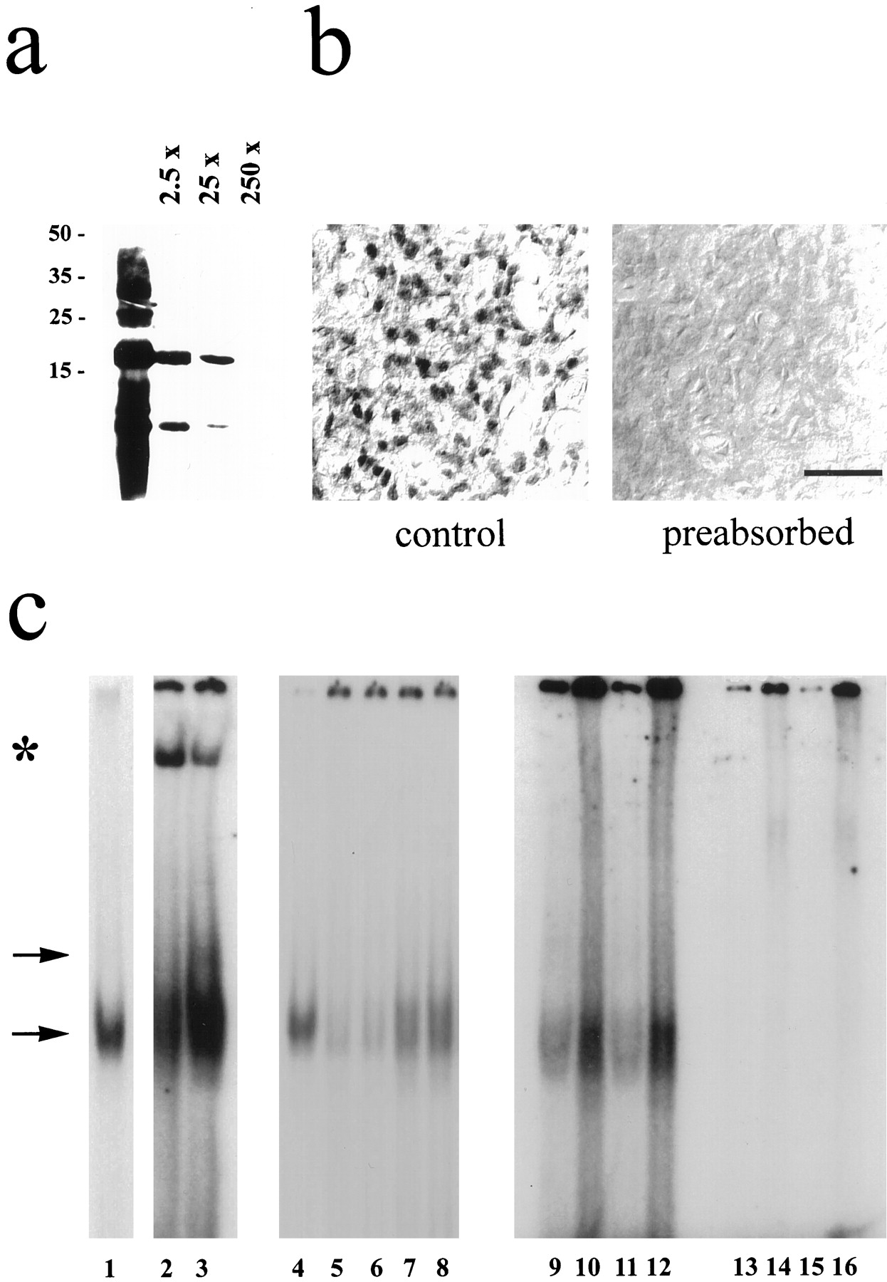
- Fig. 5.
Characterization of the ICER antibody.a, b, ICER immunoreaction could be abolished by preincubation of the antibody with the antigenic peptide in a dose-dependent manner in immunoblots (a) (2.5- to 250-fold excess of ICER protein) and immunohistochemical preparations (b) (250-fold excess of ICER protein). Similar results were obtained in two additional experiments. Scale bar, 20 μm. c, Gel mobility shift analyses with nuclear extracts obtained from rat pineal glands incubated with a labeled AA-NAT CRE always revealed a specific band of retarded mobility (lower arrow) that comigrated with bacterially generated ICER-IIγ (compare lanes 1, 4 with lanes 2, 3, 7–12). An additional ICER-specific signal of unknown quality is indicated by the upper arrow. Coincubation with the ICER antibody generated an additional low mobility complex (lanes 2, 3; indicated by a star). Excess of unlabeled AA-NAT CRE (100×) suppressed specific binding (comparelanes 5, 6 with lanes 7, 8). Notably, the labeled AA-NAT CRE (lanes 9–12) has a higher affinity for nuclear extracts as compared with that of a labeled somatostatin-CRE (lanes 13–16) (lanes 9, 11, 13, 15, 1 μg of nuclear extract; lanes 10, 12, 14, 16, 10 μg of nuclear extract). Similar results were obtained from four separate nuclear extract preparations.
- Fig. 6.
Silencing of ICER in rat pinealocytes primarily diminishes inducibility in ICER immunoreactivity. Top panels, NE induces nuclear pCREB (a) and ICER (b) immunoreactivity in isolated pinealocytes (compare columns 1 and 2).Bottom panels, The SUMDENS values from semiquantitative image analyses (n = 12) corrected for the total area covered by the cells [corrSUMDENS (Wicht et al., 1999)] are shown. Similar results were obtained with the β1-adrenergic agonist isoproterenol (data not shown).Column 1, Unstimulated pinealocytes (control).a, The NE-induced increase in pCREB immunoreactivity in pinealocyte preparations (compare columns 1 and2) is independent of transfected DNA (comparecolumns 2 and 3–5). b, Silencing ICER (column 5, pICERas) decreases NE-induced ICER immunoreactivity, as compared with that of NE-stimulated pinealocytes that were untransfected (column 2) or transfected with control DNA (column 3, pRcCMV) or with pICERs (column 4).
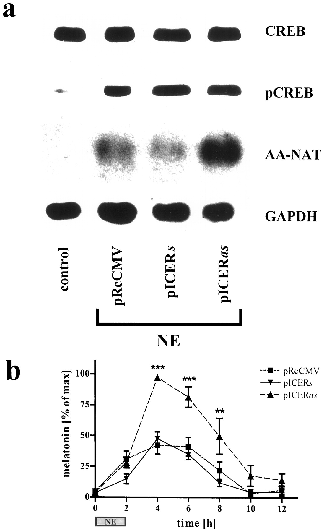
- Fig. 7.
NE-induced AA-NAT mRNA levels and melatonin synthesis are disinhibited in pICERas-transfected pinealocytes. a, CREB and pCREB immunoblots from unstimulated (control) and stimulated (NE, 10−6m; 5 hr) pinealocytes transfected with control DNA (pRcCMV), pICERs, or pICERas are shown. The amount of pCREB protein is highly induced in transfected and NE-stimulated cells (columns 2–4), as compared with control. Semiquantitative analysis of autoradiographic images from Northern blots probed for AA-NAT mRNA and corrected for the GAPDH signal revealed a fivefold superinduction in pICERas-transfected cells (column 4) as compared with controls (columns 2, 3). b, NE-induced (10−6m; 2 hr; n = 7) melatonin synthesis in pinealocytes is primarily increased in amplitude by silencing ICER. For equalizing differences between experiments in the absolute amounts of net melatonin synthesis, data are normalized so that the peak values equal 100%. **p < 0.01; ***p< 0.001 versus the basal value at 0 hr.
- Fig. 8.
Diurnal rhythms in elements determining melatonin synthesis in rat pineal gland. a, SUMDENS values for CREB (squares with dashed line) and pCREB (circles with solid line) from immunoblot analyses. For comparison, values were normalized so that the peak SUMDENS values for pCREB and CREB equal 100%. The semiquantitative analysis for AA-NAT mRNA (triangles with small dashed line) from in situ hybridization was corrected against the background signal and hybridization signal obtained with the sense AA-NAT probe. b, SUMDENS values for AA-NAT protein (squares with solid line) and melatonin synthesis (circles withdashed line). For comparison, values were normalized so that the peak SUMDENS values for AA-NAT and maximum melatonin values equal 100%. c, Semiquantitative analysis of ICER mRNA (triangle with dashed line) as revealed by in situ hybridization. The values were corrected against the background signal and hybridization signal obtained with the sense ICER probe. SUMDENS values for ICER protein (circles with solid line) were normalized so that peak SUMDENS values equal 100%.


