

Article Figures & Data
Figures
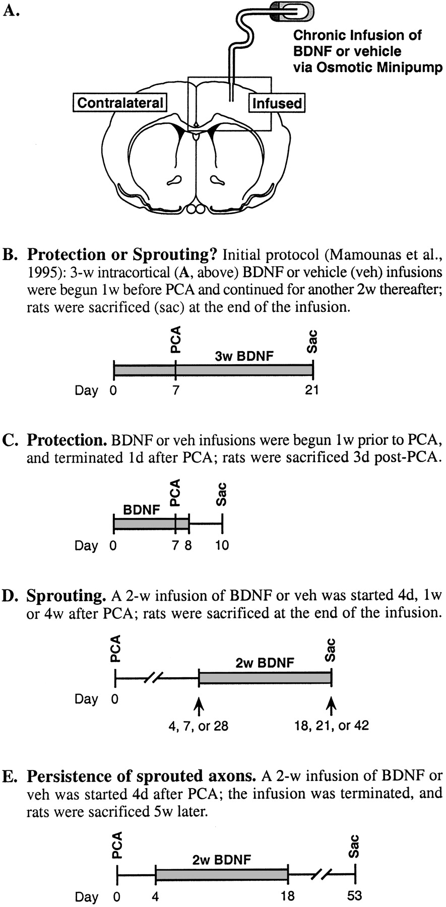
- Fig. 1.
Method of BDNF infusion and experimental paradigms used to characterize the neurotrophic effects of BDNF on 5-HT axons in rat cortex. A, Cannula placement in frontoparietal cortex; rectangle overlaying the infused cortex depicts the location and orientation of subsequent photomicrographs.B–E, Treatment paradigms; the temporal parameters of BDNF (0.1–36 μg/d) and PCA (10 mg/kg, s.c., one time) delivery were manipulated to determine whether BDNF protects 5-HT neurons from axonal injury by PCA or augments their regenerative sprouting after injury.
- Fig. 2.
Effects of vehicle or BDNF (12 μg/d) infusions on serotonergic axons in cortex of PCA-lesioned rats. Dark-field photomicrographs depict bright 5-HT-immunoreactive axons on a dark background (coronal sections). A, Protection or sprouting? (protocol, Fig. 1B); 5-HT neurons were exposed to BDNF for a considerable duration both before and after PCA administration, yielding a dense plexus of 5-HT axons at the BDNF infusion site in cortex (cannula tract in center).B, Intact; the normal 5-HT innervation in cortex of non-PCA-lesioned, non-infused rats. C, D,Protection (protocol, Fig. 1C); few 5-HT axons are spared when BDNF (C) infusions are limited to the 1 week period before PCA administration, and animals are killed 3 d after PCA (D, vehicle). E,F, Sprouting (protocol, Fig. 1D); robust sprouting of prelesioned 5-HT axons is found when 2 week BDNF (E) but not vehicle (F) infusions are started 4 d after PCA. G,H, Persistence of sprouted axons (protocol, Fig.1E); once eliciting the 5-HT sprouting response with BDNF (G), the sprouted fibers persist for at least 5 weeks after terminating the BDNF infusion (H, vehicle). Scale bar, 1 mm.
- Fig. 3.
The density of serotonin axons (SERT-immunoreactive) in frontoparietal cortex of PCA-lesioned animals and expressed as a percentage of the normal density found in the homologous cortex of intact animals. A, The normally slow reinnervation of cortex by 5-HT axons after PCA administration, as determined in the contralateral cortex of vehicle- and BDNF-infused animals (the two measures were not significantly different at any time point, and were thus collapsed for presentation). B,Effects of vehicle or BDNF infusions (12 μg/d) on PCA-lesioned 5-HT axons (measured locally at the infusion site), as investigated in the following treatment paradigms: Protection (protocol, Fig.1C; leftmost set of histograms); Sprouting (protocol, Fig. 1D) where 2 week BDNF infusions were initiated at 4 d or 4 weeks after PCA administration (center two sets of histograms, respectively); and Persistence of sprouted axons (protocol, Fig. 1E) at 5 weeks after terminating the BDNF infusion (rightmost set of histograms). Gray histogram bars, The contralateral cortex of vehicle- and BDNF-infused animals (the two values were not significantly different in any treatment paradigm, and were thus pooled); white bars,vehicle infusion in the ipsilateral (right) cortex; black bars, BDNF infusion. In all treatment paradigms, the 5-HT axon density was higher in the BDNF-infused cortex relative to the vehicle-infused and contralateral cortex (ANOVA followed by the Newman–Keuls multiple range test, p < 0.05), whereas the control conditions did not differ significantly (p > 0.05).
- Fig. 4.
The dose–response profile for BDNF (0.4–36 μg/d) to stimulate 5-HT axon sprouting in cortex (protocol, Fig.1D, 2 week BDNF infusions were started 4 d after PCA). A, C, E, G, 5-HT-immunoreactive axons in cortex (dark-field photomicrographs). B, D, F, H, BDNF diffusion as determined by BDNF immunocytochemistry in adjacent sections (bright-field photomicrographs). A, B, BDNF, 0.4 μg/d; C, D, BDNF, 1 μg/d; E, F,BDNF, 4 μg/d; G, H, 36 μg/d of BDNF. The effects of vehicle infusion and the 12 μg/d dose of BDNF on 5-HT axons (using this same treatment protocol) are shown in Figure 2, Fand E, respectively. Scale bar, 1 mm.
- Fig. 5.
The density of serotonin axons (SERT-immunoreactive) after intracortical infusion of vehicle or 0.1, 0.4, 1, 4, 12, or 36 μg/d of BDNF (protocol, Fig.1D, 2 week infusions were started 4 d after PCA); measured in the area of BDNF-positive immunoreactivity (determined in adjacent sections) and expressed as a percentage of the normal density in intact animals. Closed squares,Vehicle or BDNF infusions in the ipsilateral (right) cortex;open squares, the contralateral (noninfused) side of cortex. ANOVA followed by the Bonferonni post hoc test revealed that 5-HT axon density was significantly higher after 0.4–12 μg/d of BDNF relative to vehicle; the 12 and 36 μg/d doses of BDNF resulted in a lower density than the 4 μg/d dose (F(6,24) = 15; p < 0.0001).
- Fig. 6.
The serotonergic innervation after infusion of vehicle or BDNF (4 μg/d) for 3, 7, or 14 d in cortex of previously PCA-lesioned animals. A, SERT axon density was measured at the BDNF infusion site and expressed as a percentage of the density in the contralateral cortex (noninfused but PCA-lesioned). Treatment paradigms are described in Materials and Methods (Experimental paradigm). *p < 0.05, relative to the vehicle-infused and contralateral cortex (ANOVA followed by the Newman–Keuls multiple range test). B, Dark-field photomicrograph of 5-HT-immunoreactive axons after a 1 week BDNF infusion (4 μg/d; initiated at 7 d after PCA administration) in cortex. The effects of vehicle and 2 week BDNF (4 μg/d) infusions on 5-HT axons (using a similar treatment protocol) are shown in Figures2F and 4E, respectively. Scale bar, 0.5 mm.
- Fig. 7.
The serotonergic innervation in dorsal hippocampus after local infusion of vehicle or BDNF (4 μg/d) in PCA-lesioned animals (protocol, Fig. 1D, 2 week infusions were started 1 week after PCA). A–D, F, H,5-HT-immunoreactive axons in hippocampus (dark-field photomicrographs; coronal sections); E, G, I, area of BDNF diffusion as determined by BDNF immunocytochemistry in adjacent sections (bright-field photomicrographs). A, The normal 5-HT innervation in hippocampus of intact rats. B, Vehicle infusion in the PCA-lesioned hippocampus (note cannula tract at center). C, The extent of 5-HT denervation normally seen 3 weeks after PCA administration in the contralateral hippocampus.D, E, Sprouting of 5-HT axons in the dentate gyrus (D) after local infusion of BDNF (E). F, G, Sprouting of 5-HT axons in CA1 and the dentate gyrus (F) in response to BDNF infusion (G). H, I, Sprouting of 5-HT axons in CA3 (H) after infusion of BDNF (I); note the higher magnification inH and I than in A–G. Scale bars: A–G (shown in G), 1 mm;H, I (shown in I), 0.5 mm.
- Fig. 8.
The densities of serotonergic (SERT), catecholaminergic (TH), and cholinergic (AChE) axons after intracortical infusion of vehicle or BDNF (4 μg/d) for 18 d in intact (non-PCA-lesioned) rats. Values were measured within the area of BDNF-positive immunoreactivity in cortex and expressed as a percentage of the normal density found in the contralateral cortex. White histogram bars,vehicle infusions; black bars, BDNF infusions. *p < 0.05, relative to the vehicle-infused and contralateral cortex (ANOVA followed by the Newman–Keuls multiple range test).
- Fig. 9.
The dose–response profile for BDNF infusions to stimulate Trk activity (as reflected by Trk tyrosine autophosphorylation) in cortex. BDNF (0.1–12 μg/d) or vehicle was chronically infused into cortex for 24 hr before assaying Trk proteins at the BDNF infusion site. A, Immunoblots of Trk tyrosine autophosphorylation (Trk P-tyr) and total full-length (gp145) TrkB protein after intracortical BDNF infusion in intact (left) or PCA-lesioned (right; 10 mg/kg, s.c., administered 1 week before starting the intracortical infusion) animals. Top set of immunoblots, To assay the levels of Trk P-tyr, Trk family proteins were immunoprecipitated (IP) with anti-panTrk (Trk) antibody, and Western-blotted (WB) with anti-phosphotyrosine (P-tyr) antibody (4G10; Upstate Biotechnology);middle, above immunoblots were reprobed with TrkB antibody to measure total levels of catalytic (full-length; gp145) TrkB protein; bottom, to assess the levels of exogenously delivered BDNF protein, an aliquot of the same lysate (used in each case above) was Western-blotted with anti-BDNF antibody.B, Quantitation of Trk P-tyr (top; expressed as -fold induction over vehicle infusion) and total full-length (gp145) TrkB protein (bottom; expressed as a percentage of vehicle). Open circles, Vehicle or BDNF infusions in cortex; closed circles, homologous cortex from naive (i.e., noninfused) animals.
- Fig. 10.
Cortical Trk tyrosine autophosphorylation after local BDNF administration. A, Three hours after a single injection of vehicle or BDNF (4 or 12 μg). B, Chronic infusion of vehicle or BDNF (4 μg/d) for 7 d. Trk P-tyr, Tyrosine-phosphorylated Trk; gp145 TrkB, total full-length (catalytic) TrkB protein; gp95 TrkB, truncated isoform of the TrkB protein; BDNF, exogenously delivered BDNF. Levels are quantified in Table 1.


Tables
- Table 1.
Effects of chronic 7 d infusion of BDNF (4 μg/d) or vehicle in rat frontoparietal cortex on TrkB protein and mRNA expression
TrkB Protein/mRNA Levels Vehicle BDNF Student's t test (two-tailed) Trk tyrosine phosphorylation 1.0 ± 0.4 3.3 ± 0.2 t12 = 4.8;p < 0.0005 (Trk P-tyr; fold induction over vehicle) Full-length TrkB protein 100 ± 3 66 ± 7 t12 = 4.5; p < 0.001 (gp145 TrkB; percent vehicle) Truncated TrkB protein 100 ± 5 78 ± 9 t6 = 2.1; p < 0.08, NS (gp95 TrkB; percent vehicle) TrkB/GAPDH mRNA 100 ± 5 88 ± 6 t8 = 1.6; p = 0.15, NS (percent vehicle) At the end of a 7 d intracortical infusion of BDNF (4 μg/d) or vehicle in intact animals, the infused cortex was microdissected, and levels of Trk protein were assayed by immunoblotting (data shown in Fig. 10). TrkB mRNA was analyzed by RNAse protection assay (described in Materials and Methods) using a cDNA probe that recognizes all TrkB isoforms; a GAPDH probe was included in the same reaction mixture to assess relative levels of RNA present in each hybridization (data calculated as the ratio of TrkB to GAPDH). Values shown are means ± SEM for 4–7 rats per group. NS, Not significant at p < 0.05.


