

Article Figures & Data
Figures
- Fig. 1.
Morphological characteristics of PV neurons viewed in conventionally oriented coronal sections. a, A montage of confocal laser-scanning microscopic images. Somata of PV neurons are located mainly in and around the pyramidal cell layer (P) and the granule cell layer (G). Their axon terminals also are distributed in the same layers, causing band-like staining in lower magnification.b, PV neurons in the pyramidal cell layer of the CA1 region give rise to dendrites spanning all hippocampal layers.A, Alveus; O, stratum oriens;R, stratum radiatum; Lm, stratum lacunosum-moleculare. c, d, Double immunostaining with antibodies against PV (c) and GAD (d). Note multiple PV-containing GAD-immunoreactive (ir) boutons (arrows) abutting on the PV-ir soma. Arrowheads indicate similarly double-labeled boutons, some of which may abut on unstained somata of pyramidal neurons. Scale bars: a, 0.5 mm; b, 50 μm; c, d, 5 μm.
- Fig. 2.
CLSM images in a section cut tangentially along the border between the alveus and the stratum oriens of the CA1 region.a, PV-ir dendrites running two-dimensionally within a single 40-μm-thick section form an extremely dense network. Some dendrites appear to be bundled together (open arrows), whereas others cross one another with a fairly wide angle. Four PV-ir somata are of the horizontal type (see Results). b, Stereo pair of the enlargement of the framed area ina. PV-ir dendrites make multiple contacts with one another, only three of which are indicated by arrows.c–e, Single confocal optical images of the three contact sites designated in b, suggesting a direct contact between these dendrites. Scale bars: a, 50 μm;b–e, 1 μm.
- Fig. 3.
Electron micrographs showing the dendrodendritic gap junctions. a, Two PV-ir dendrites (D) receiving multiple presynaptic terminals (asterisks) make direct contact with each other. b, Enlargement of the contact site ina. A gap junction is formed between the two PV-ir dendrites. Note the close apposition of the plasma membranes of the two contacting cells as demarcated by arrows. A synaptic bouton (asterisk) forming a synapse of asymmetrical type is located in close vicinity to the gap junction. c, Electron micrograph of a specimen without immunoreaction, demonstrating a profile similar to a. The contacting dendrites (D) receive presynaptic terminals (asterisks). d, Enlargement of the contact site in c with the same magnification as inb, g, and i to facilitate comparison. Plasma membranes of the two cells are closely apposed, as demarcated by arrows. Note a layer of cytoplasmic semidense material (arrowheads) undercoating either side of the junction, which is characteristic of neuronal gap junctions.e, Further enlargement of the contact site ind, showing a narrow central gap, 2.7 nm wide, between the outer leaflets of the apposed unit membranes. f–i, Other examples of gap junctions formed between PV-ir dendrites (D). The contact sites in f andh are enlarged in g and i, respectively, with the same magnification as in b andd. Scale bars: a, c,f, h, 1 μm; b,d, e, g, i, 0.1 μm.
- Fig. 4.
Electron micrographs showing two novel forms of dendrodendritic contacts between PV neurons. a, Two PV-ir dendrites (D) receiving multiple presynaptic terminals (asterisks) make direct contact with each other (arrow). Inset, Enlargement of the contact site demonstrating synaptic vesicles, widening of the synaptic cleft, and less prominent thickening of the postsynaptic density; the last feature is consistent to conventional symmetrical synapses, including those formed by PV-ir axon terminals.b,c, Serial ultrathin sections with different magnifications demonstrating that two PV-ir dendrites (D), receiving multiple presynaptic terminals (data not shown in these sections), establish a mixed type of synapse between each other. Synaptic vesicles (Sv) accumulate in one side of the contact as in a, but in this case the gap junction can be identified in line with the chemical synapse inc. Note a layer of cytoplasmic semidense material (arrows) undercoating gap junction as in Fig.3d, g, i. Scale bars:a, b, 1 μm; Inset ina, c, 0.1 μm.
- Fig. 5.
Comparison of the reconstructed dendritic arborization of PV neurons between the vertical type (left) and the horizontal type (right), viewed in two directions (top, top view;down, front view) with the use of a computer-assisted neuron tracing system. The top and front views are of the same cell in each type and are aligned.
- Fig. 6.
Top. CLSM images showing several morphological aspects of PV-ir neurons viewed in tangential (a, c–e) and coronal (b) sections. a, Triple-immunostained section at the border between the stratum oriens and the alveus. PV-ir dendrites (green) receive numerous synaptophysin (red)-positive and GAD (blue)-negative boutons (arrows). The mutual contact sites in dendrites are shown byarrowheads. b, A PV neuron of the horizontal type (arrow) located at the border between the stratum oriens (O) and the alveus (A) gives rise to long dendrites (arrowheads) along the border. The stratum oriens contains many obliquely running dendrites originating from PV-ir somata located in and around the stratum pyramidale (P).c, Dendrites of horizontal PV neurons extend in all directions from their somata along the horizontal plane.d,e, Enlargement of the PV-ir neuron, indicated by an arrow in c, double-immunostained with antibodies against PV (green) and GAD (red). CLSM images in d and e were taken at different depths from the section surface. Note numerous PV-containing GABAergic boutons (arrows) abutting on the soma and proximal dendrites. Scale bars: a, 5 μm; b,c, 50 μm; d, e, 10 μm.
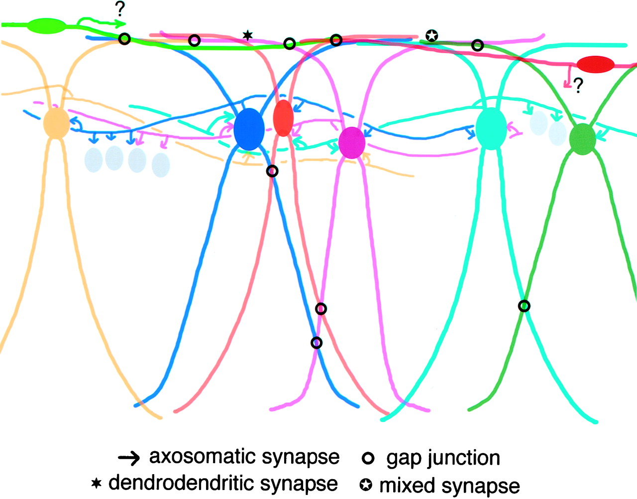
- Fig. 7.
Bottom. A schematic drawing representing the dual networks of hippocampal PV neurons connected by chemical and electrical synapses. PV neurons are shown in color, whereas pyramidal cell somata are depicted as gray ovals. Frequent occurrence of gap junctions between horizontally oriented dendrites (top of the figure) was confirmed by quantitative electron microscopic analysis, but it remains unknown whether both or either of the two neuronal types (vertical and horizontal) actually forms gap junctions there. See Discussion for details.


