

Article Figures & Data
Figures
- Fig. 1.
Estrogen produces both negative and positive feedback on gonadotropin secretion. Representative composite hormone profile based on serial blood samples taken from ovariectomized female guinea pigs. Filled circles represent plasma LH concentrations determined by radioimmunoassay at various time points before and after E2 (25 μg) administration.
- Fig. 2.
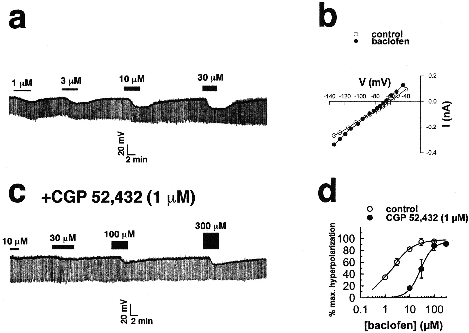
Stimulation of GABAB receptors inhibits POA neurons by activating a K+ conductance.a, Successively increasing doses of baclofen (1, 3, 10, and 30 μm) hyperpolarized this POA neuron (resting Vm = −55 mV) at 8.5, 11, 16, and 18 mV, respectively. b, An I/Vplot derived from a POA neuron just before (control; open circles) and near the end of the application of a maximal concentration of baclofen (100 μm; filled circles) is shown. The reversal potential for the baclofen response was −94 mV, and a Δg of 0.67 nS between −60 and −80 mV and a Δg of 1.63 nS between −100 and −130 mV were also observed. c, Dose–response relationship from the cell shown in a is then generated in the presence of CGP 52,432 (1 μm). Successively increasing doses of baclofen (10, 30, 100, and 300 μm) elicited hyperpolarizations of 2.5, 4.5, 11, and 12.5 mV, respectively. d, Composite baclofen dose–response curves in the absence (open circles) and presence (filled circles) of CGP 52,432 (1 μm) are shown. Cells were perfused with successively higher concentrations of baclofen (1, 3, 10, 30, 100, and 300 μm; 4–7 min/dose; n = 2–10).Symbols represent means, and vertical lines are 2 SEMs of the baclofen-induced hyperpolarization normalized to the ΔVmax. Before CGP 52,432, the mean baclofen EC50 value was 2.3 ± 0.5 μm, whereas in the presence of CGP 52,432, the EC50 was shifted to 33.0 ± 10.0 μm. The estimatedKi for CGP 52,432 was 64.0 nm. BAC, Baclofen.
- Fig. 3.
Estrogen attenuates the efficacy of GABAB receptor-mediated neurotransmission in the POA 24 hr after its administration. a, Successively increasing doses of baclofen (1, 3, and 10 μm) hyperpolarized this POA neuron from a vehicle-treated animal (resting Vm = −45 mV) 10, 22.5, and 25.5 mV, respectively. The upward deflection represents the return of low-threshold spikes and/or action potentials (truncated) seen during the later stages of drug washout. b, Successively increasing doses of baclofen (3, 10, 30, and 100 μm) hyperpolarized this POA neuron (resting Vm = −50 mV) from an EB-treated (25 μg; 24 hr) animal by 1.5, 2.5, 3, and 4 mV, respectively. c, Composite dose–response curves from recordings of POA neurons obtained from vehicle- and EB-treated animals are shown. Cells were perfused with successively higher concentrations of baclofen (1, 3, 10, and 30 μm; 4–7 min/dose; n = 2–10). Symbolsrepresent means, and vertical lines are 1 SEM of the hyperpolarizations elicited by a given concentration of baclofen. The ΔVmax obtained via logistic fit for POA neurons from vehicle-treated animals was 13.5 mV, whereas that obtained for POA neurons from EB-treated animals was 7.5 mV. *, Hyperpolarizations obtained with 10 and 30 μm baclofen that are significantly different (multifactorial ANOVA and LSD;p < 0.05) from those obtained with 1 or 3 μm baclofen are shown. #, Hyperpolarizations of POA neurons obtained from EB-treated animals are significantly lower (multifactorial ANOVA and LSD; p < 0.05) than those obtained from vehicle-treated animals at all doses tested.d, Composite bar graph illustrates the baclofen-induced Δg in POA neurons from vehicle- and EB-treated animals (n = 5–8). Columns represent means, and vertical lines are 1 SEM of the baclofen-induced Δg estimated by linear regression between −60 and −80 mV and between −100 and −130 mV. *, Values of Δg obtained in POA neurons from EB-treated animals that are significantly different (multifactorial ANOVA and LSD;p < 0.05) from those obtained from vehicle-treated controls are shown.
- Fig. 4.
Distribution of GAD65 in the guinea pig hypothalamus. a, b, Dark-field photomicrographs that illustrate the distribution of GAD65 in the rostral (a) and caudal (b) POA.c, d, Dark-field photomicrographs of coronal sections through the MBH from rostral to caudal illustrating the distribution of GAD65 mRNA. AC, Anterior commissure;BST, bed nucleus of the stria terminalis;fx, fornix; LS, lateral septum;LSv, lateral septum (ventral part); ME, median eminence; MS, medial septum; OC, optic chiasm; PSCH, suprachiasmatic preoptic nucleus;PVN, paraventricular nucleus; VMH, ventromedial nucleus of the hypothalamus; 3V, third ventricle.
- Fig. 5.
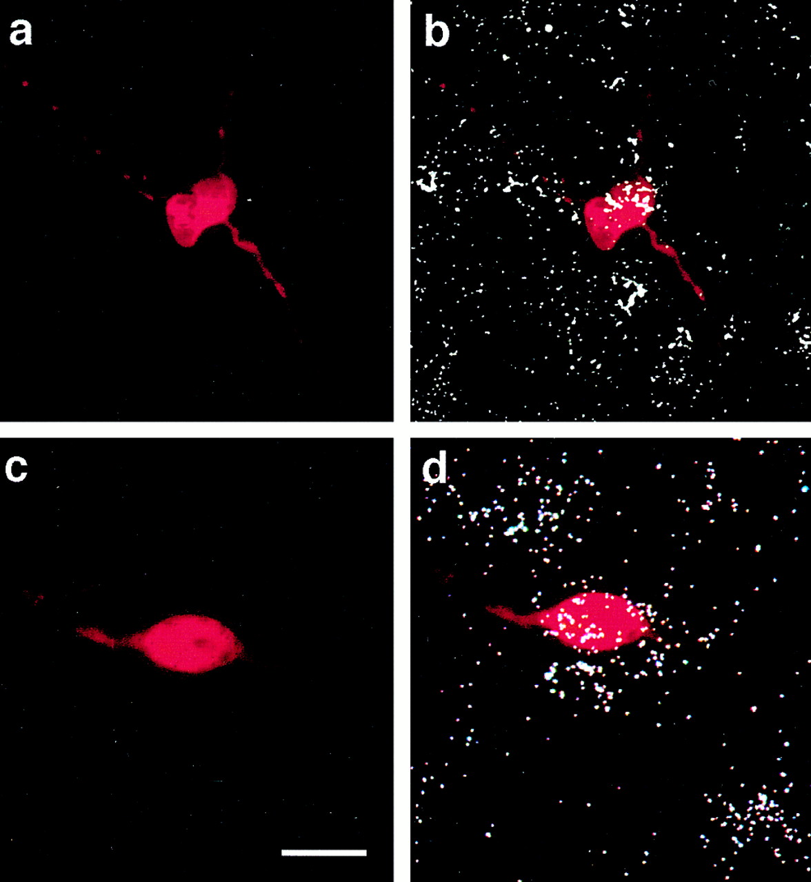
GABAergic POA neurons are identified using combined histofluorescence and in situ hybridization for GAD65 after electrophysiological recording.a, Photomicrograph of the biocytin-streptavidin-Texas Red fluorescent labeling of a recorded cell from a vehicle-treated animal. b, An overlay of the fluorescent labeling ina and the hybridization signal that clearly illustrates the double labeling for GAD65. c, Photomicrograph of the biocytin-streptavidin-Texas Red fluorescent labeling of a recorded POA neuron from an EB-treated (25 μg; 24 hr) animal. d, An overlay of the fluorescent labeling inc and the hybridization signal illustrating the double labeling for GAD65. Scale bar, ∼15 μm (for all photomicrographs).
- Fig. 6.
Estrogen produces a subsequent decrease in the POA expression of GAD67 and GAD65 42 hr after its administration. a, Distribution and quantitative analysis using a sensitive ribonuclease protection assay of GAD65 and GAD67 mRNA in the POA and MBH obtained from female guinea pigs. *, Denotes the levels of GAD67 that are significantly greater (Student'st test; p < 0.05) than those obtained for GAD65. b, A representative ribonuclease protection assay of total RNA (3 μg/lane) from vehicle- and EB-treated (25 μg; 42 hr) animals illustrating the levels of GAD67 mRNA detected in the POA and the rostral (r) and caudal (c) MBH. Sense RNA (125–4000 fg) was used to construct a standard curve.c, Quantitative analysis of GAD65(left) and GAD67 (right) mRNA in hypothalamic brain tissue obtained from vehicle- and EB-treated animals. Bands were normalized to their corresponding cyclophilin band and quantified from their respective sense mRNA standard curves.Columns represent means, and vertical lines are 2 SEMs of the EB-induced percent change in the GAD65 and GAD67 levels with respect to those observed in vehicle-treated animals. *, Denotes a significant change (paired t test; p < 0.05) in the level of GAD65 caused by EB relative to that observed in the POA of vehicle-treated controls. **, Denotes a significant change (paired t test; p < 0.01) in the level of GAD67 caused by EB relative to that observed in the POA of vehicle-treated controls. DP, Digested probe;UP, undigested probe.
- Fig. 7.
Schematic representation illustrating the biphasic central feedback actions of estrogen on the mammalian female reproductive axis. a, During negative feedback, estrogen uncouples postsynaptic GABAB receptors from their effector K+ channels in GABAergic POA neurons. This leads to a decreased autoinhibition and an increased inhibitory tone onto GnRH neurons, thereby reducing GnRH release and ultimately LH secretion.b, During positive feedback, estrogen decreases POA GAD expression, thereby diminishing intraneuronal transmitter levels and the release of GABA. This reduces inhibitory GABAergic tone onto GnRH neurons, which facilitates excitatory (e.g., glutamatergic, noradrenergic) inputs in generating the preovulatory LH surge.


