

Article Figures & Data
Figures
- Fig. 1.
Development of midline glia and the corpus callosum. GFAP-positive cells are present by E14 in mouse cortex (arrow in A). These glia send long radial-glial-like processes toward the midline, coalescing into a wedge-shaped structure by E15 (arrow inB). By E17, a second midline glial population arises in the indusium griseum (arrowhead in C), and the glial wedge is still present (arrow inC). At P0, both the indusium griseum glia (arrowhead in D) and the glial wedge (arrow in D) are present, but the glial wedge remains confined to the ventricular zone of the corticoseptal boundary. Staining of the GFAP-lacZ (nuclear-targeted) mice with an antibody to β-galactosidase shows that the cell bodies of the glial wedge remain within the ventricular–subventricular zone (arrow in E). F, Double-labeling of the glial wedge (arrow) and the indusium griseum glia (arrowhead) with GFAP antibody (green) and the callosal axons with DiI (red) show that the callosal axons do not enter these glial structures but pass between them. Scale bars: (inD) A, B, 150 μm;C, D, 240 μm; (inF) E, 100 μm; F, 200 μm.
- Fig. 2.
The glial wedge suppresses and repels E17 cortical axons. A, Schematic of the experimental paradigm. Cortical explants (Ctx) and glial wedge explants (GW) were dissected from coronal sections (350 μm in thickness) of E17 mouse brain. Cortical explants were cocultured with either glial wedge explants (D–F) or with other cortical explants (B, C) in three-dimensional collagen gels. After 3 d, the cultures were fixed, and DiI was injected into the cortical explant. When cortex was paired with cortex as a control, the cortical axons projected symmetrically from the explant (B), growing into the target cortical explant (arrow in C; C is a higher power view of B). When cortex was paired with glial wedge, the length of axons on the proximal side facing the glial wedge was greatly diminished (D–F), with many axons turning away (arrow in F) or stopping at the edge of the glial wedge explant (D andE are two different examples; F is a higher power view of E). G, Quantification of axonal length (mean axon length was derived by measuring the length of the 10 longest axons on each side of the explant) and number on the proximal side versus the other three sides of the explants. Statistically significant results are labeled (*). Scale bar (in F): B,D, F, 300 μm; C,F, 150 μm.
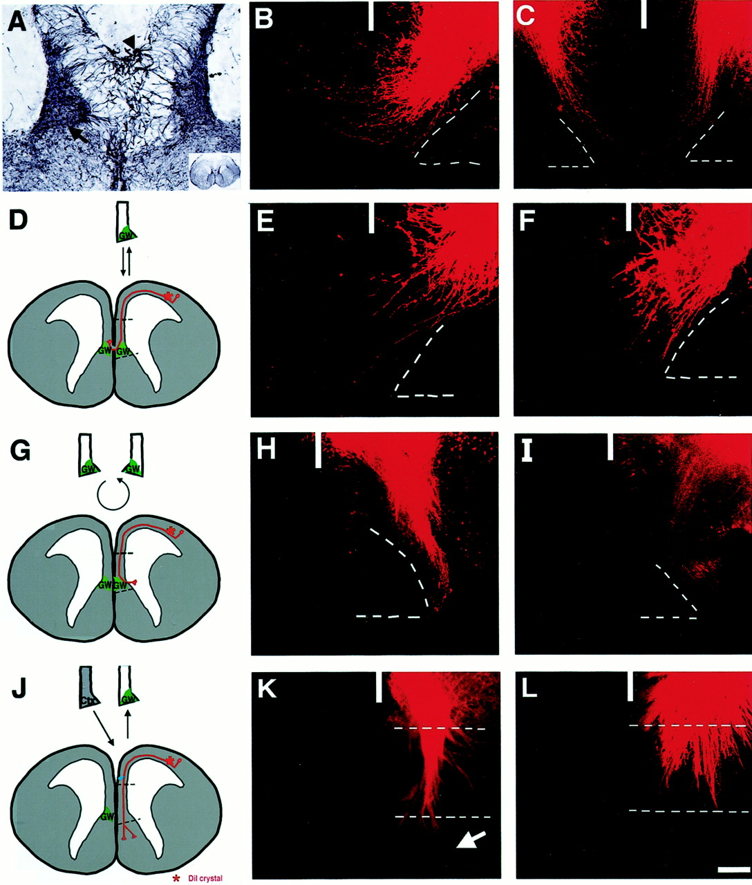
- Fig. 3.
The glial wedge directs the growth of callosal axons in situ. A, The glial wedge forms in organotypic slices. E17 mouse brains were sectioned at 350 μm, grown for 3 d in culture, fixed, resectioned at 50 μm, and stained with a GFAP antibody. Both the glial wedge (arrow) and the indusium griseum glia (arrowhead) maintain their in vivomorphology after 3 d in vitro. B,C, The corpus callosum forms in organotypic slices.B and C represent two examples of uncut control slices, cultured for 3 d and fixed, and a crystal of DiI was added to label the callosal axons. In C, DiI crystals were placed on both sides of the midline to show that cortical axons from both hemispheres still cross in the same organotypic slice (in all other slices, DiI was added to only one hemisphere).D–L, Replacement or reorientation of the midline results in axonal misrouting. D–F, The corpus callosum forms normally in sham-operated slices (D shows the experimental paradigm, and E and F are two examples). G–I, Reorienting the glial wedge by 180° causes the axons to turn away from the midline (Gshows the experimental paradigm, and H andI are two examples). J–L, The glial wedge is required for axons to turn toward the midline. When the glial wedge–indusium griseum region is replaced on one side by a piece of cortex, cortical axons fail to turn and instead grow straight through the graft, in many cases entering the septum (arrow inK; J shows the experimental paradigm, andK and L are two examples). Thewhite broken lines in B,C, E, F, H, and I represent the position of the glial wedge; inK and L, they represent the edges of the cortical graft. The solid white line inB, C, E, F,H, I, K, andL represent the position of the midline. Scale bar (inL): A, 120 μm; C,E, F, K, L, 200 μm; B, H, I, 100 μm.
- Fig. 4.
During callosal axon targeting, the glial wedge and the indusim griseum express slit-2. In situ hybridizations using slit-2 antisense (A, C) probes show that slit-2is expressed within the ventricular zone of the septum and corticoseptal boundary. The control slit-2 sense probe shows no specific labeling (B). slit-2 is expressed in both the glial wedge (arrow inC) and the indusium griseum glia (arrowhead in C; C is a higher power view of A). D,E, Double-labeling of anti-β-galactosidase (driven by the GFAP promoter and targeted to the cytoplasm) immunohistochemistry and slit-2in situ hybridization shows that cells within the glial wedge express both slit-2 and the lacZ transgene (E is a higher power view ofD). In wild-type embryos, glia within the glial wedge (arrow in F) and the indusium griseum (arrowhead in F) are double-labeled with slit-2 (purple) and GFAP (brown). F is a higher power view of the boxed region in the inset. Examples of double-labeled cells are shown in the indusium griseum (arrows in G) and the glia wedge (arrows in H; G andH are higher power views of F). All sections are from E17 mouse brains. Scale bar (inC): A, B,D, 950 μm; C, 220; E, 110 μm; F, 170 μm; G, 25 μm;H, 20 μm.
- Fig. 5.
The slit-2 receptors robo-1and robo-2 are expressed in the neocortex during callosal axon targeting. In situ hybridization using antisense probes against robo-1 (A, C) or robo-2 (D, F) show that both receptors are expressed within the cortical plate at E17 (C and F are higher power views of theboxed regions in A and D, respectively). Control sense probes for either robo-1(B) or robo-2(E) show no specific labeling. Scale bar (inF): A, B,D, E, 900 μm; C,F, 80 μm.
- Fig. 6.
Slit-2 suppresses and repels E17 cortical axons. Cortical explants (Ctx) derived from E17 mouse brains were cocultured with 293T cells transfected with either a control (vector alone) construct (A, D;D is a higher power view of A) or a Slit-2 expression construct (B, C,E, F; 4 different examples). Explants were cultured for 3 d and fixed, and then the cortical explant was injected with DiI to label axonal outgrowth. Cortical explants display a symmetrical growth when cocultured with control transfected cells (A), even growing into the transfected cell block (arrow in D). However, when cocultured with Slit-2-expressing cells, cortical axon outgrowth was severely suppressed in the proximal side facing the cell block (B, C), with some axons turning away (arrow in E) and refusing to enter the cell block (arrow in F).G, Quantification of axonal length (mean axon length was derived by measuring the length of the 10 longest axons on each side of the explant) and number on the proximal side versus the other three sides of the explants. Statistically significant results are labeled (*). Scale bar (in C): A–C, 300 μm;D–F, 150 μm.
- Fig. 7.
Model of axon guidance across the corpus callosum by midline glial populations. The glial wedge (GW) and the indusium griseum glia (IG) express Slit-2, repelling axons away from these structures. Because slit-2 acts as a short-range guidance cue, callosal axons (shown in red; expressing theslit-2 receptors robo-1 and robo-2) are not inhibited from approaching the midline but turn when they encounter the glial wedge. Callosal axons may then use positive guidance signals from the glial sling (GS) to cross the midline and may then be deflected dorsally by the glial wedge in the opposite hemisphere. LV, Lateral ventricle.


Tables
Experiment Mean ± SE length of axons (μm) Mean ± SE number of axons Total number of explants/total number of experiments Proximal side Other 3 sides combined Proximal side Other 3 sides combined Cortex vs cortex 364 ± 24 341 ± 27 61 ± 7 52 ± 7 29 /7 Cortex vs glial wedge 209 ± 15* 311 ± 14 33 ± 4* 41 ± 3 44 /7 Cortex vs control transfected cells 428 ± 21 451 ± 15 89 ± 8 89 ± 7 38 /4 Cortex vs Slit-2-transfected cells 234 ± 19* 393 ± 22 22 ± 3* 56 ± 4 51 /4 ↵* p < 0.05 indicates significant difference; Student's t test.


