

Article Figures & Data
Figures
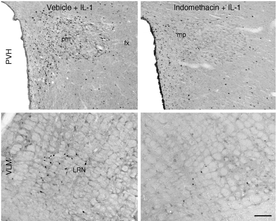
- Fig. 1.
Central prostaglandin synthesis blockade disrupts systemic IL-1-induced activation of the paraventricular nucleus and its aminergic afferents. Bright-field photomicrographs show IL-1-induced Fos-ir expression in the paraventricular nucleus (PVH, top) and the C1 region of the ventrolateral medulla (VLM, bottom) in rats pretreated by intracerebroventricular injection of vehicle (left) or indomethacin (10 μg/5 μl) (right). As reported previously, intravenous IL-1 (1.87 μg/kg) evokes a robust Fos response within the PVH and C1 regions. However, pretreatment with central infusion of indomethacin, a nonselective inhibitor of COX activity, results in a marked diminution of IL-1 effects at the levels of both medulla and hypothalamus. This finding supports the view that induced synthesis of prostaglandins within the brain is required for the activation of HPA control systems in response to systemic (intravenous) IL-1. Scale bar, 100 μm.
- Fig. 2.
Effects of central indomethacin on IL-1-induced Fos expression in hypothalamus and ventrolateral medulla. Mean ± SEM number of Fos-ir neurons in the ventrolateral medulla (VLM, left) and PVH (right) and in rats pretreated centrally (ICV) with 10 μg indomethacin (Indo) or vehicle (Veh), and systemically (IV) with 1.87 μg/kg IL-1. Indomethacin treatment results in a marked diminution of IL-1 effects at the levels of both medulla and hypothalamus. Neither of the groups pretreated with indomethacin had counts in either region that were significantly different from those in the Veh/Veh control group. *p < 0. 01 compared with Veh/Veh group.+ p < 0.05 compared with Veh/IL-1 group (n = 5 per group).
- Fig. 3.
Basal and IL-1-stimulated COX-2 mRNA expression. Dark-field photomicrographs of sections from rats killed 1 hr after intravenous injection of vehicle (top row) or 1.87 μg/kg IL-1 (bottom row), at the levels of the preoptic area (left), paraventricular nucleus (middle), and medulla (right). In vehicle-treated rats, COX-2 mRNA is evident throughout the isocortex, hippocampal formation, and the area postrema. Some signal is also clearly evident within the meninges (men) and a few blood vessels (bv). IL-1 treatment does not appear to alter neuronal expression of COX-2 mRNA, although expression by cells associated with the vasculature and the meninges is clearly increased throughout the brain. Scale bar, 100 μm. ac, Anterior commissure; och, optic chiasm; men, meninges; OT, olfactory tubercule; CP, caudate putamen; Pir, piriform cortex; iso, isocortex;DG, dentate gyrus; CA3, field CA3 of ammon's horn; NLOT, nucleus of lateral olfactory tract;AP, area postrema; cc, central canal;SNV, spinal nucleus of trigeminal.
- Fig. 4.
Immunoreactive COX-2 expression in the brain. Bright-field images of COX-2-ir cells in the isocortex and meninges (top) and cells associated with the vasculature in the forebrain (middle) and medulla (bottom), from rats killed 4 hr after vehicle (left) or IL-1 injection (right). In agreement with findings at the mRNA level, constitutive COX-2-ir is seen within some cortical neurons and, at lower levels, in the meninges and perivascular regions. At 4 hr after IL-1 (1.87 μg/kg) treatment, a clear increase in the number and staining intensity of COX-2-positive cells is seen within the meninges and perivascular regions, but not in neurons. Scale bar, 100 μm.
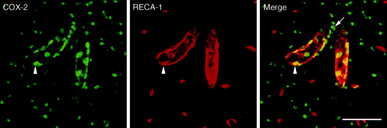
- Fig. 5.
IL-1-induced COX-2 expression in perivascular cells. SCLM images showing dual immunostaining for COX-2 (green) and a marker for perivascular cells/macrophages (ED2, red,top and middle panels) or endothelial cells (RECA-1, red, bottom panels) in blood vessels in the medulla (top andbottom panels) or forebrain (middle panel). Results of dual immunolabeling of material from IL-1-challenged rats revealed that very nearly all COX-2-ir cells also stained positively for the ED2 antigen, suggesting that they may be considered perivascular cells as defined by Graeber et al. (1989). COX-2-ir was never found to colocalize with the endothelial marker RECA-1 (bottom panel), suggesting that endothelial cells do not manifest substantial COX-2 expression in this paradigm. The yellow color in the top right and middle two panels is from merged confocal images and represents a positive signal for both COX-2-ir and ED2-ir. The arrow indicates a single COX-2-positive, ED2-negative cell. The open arrow indicates a COX-2-positive cell immediately adjacent to a RECA-1-positive vessel. Scale bars, 50 μm. POA, Pre-optic area.
- Fig. 6.
Fine structure of ED2-ir and COX-2-ir perivascular cells. Electron micrographs showing pre-embedding immunolabeling for ED2 (top panel) and COX-2 (bottom panel) near blood vessels in the medulla of a rat treated with IL-1 (1.87 μg/kg). The ED2 antiserum recognizes a surface antigen, and the reaction product (black arrows) outlines a cell in a perivascular location, adjacent to a smooth muscle cell (SM). The arrowheads indicate reaction product associated with membrane-bound vacuoles. COX-2 immunoreactivity is distributed diffusely within the cytoplasm (CY) of a cell of similar morphology and location. It also appears to be enclosed within a basal lamina, consistent again with the perivascular cell designation.EC, Endothelial cell; bl, basal lamina;L, lysosome; N, nucleus;v, vacuole; bv, blood vessel. Scale bars, 1 μm.
- Fig. 7.
Dual localization of ED2-ir and IL-1R1 mRNA. Bright-field photomicrographs of sections from untreated rats, showing the distribution of ED2-ir perivascular cells (brown) and IL-1R1 mRNA (black grains) around blood vessels in the ventrolateral medulla. Black arrows indicate cells expressing both markers. Not all receptor-bearing cells are ED2 positive (open arrows), suggesting that other cell types associated with the vasculature (i.e., endothelial or other vasculature-associated cell types) may exhibit IL-1 sensitivity, and not all ED2-positive cells appear to express the receptor (black arrowheads), suggesting some heterogeneity among ED2-positive perivascular cells. Scale bar, 50 μm.
- Fig. 8.
Immunoreactive COX-2 expression in vasculature-associated cells in response to IL-1 versus LPS. Bright-field images of COX-2-ir cells associated with the vasculature in the forebrain (top) and medulla (bottom) from rats killed 4 hr after intravenous injection of IL-1 (1.87 μg, left) or LPS (100 μg/kg,right). As with IL-1, at 4 hr after LPS treatment, a clear increase in the number and staining intensity of COX-2-positive cells is seen within perivascular regions throughout the brain. However, the predominant cell types manifesting enzyme expression after each treatment are morphologically distinct. COX-2-positive polygonal/multipolar cells (open arrows) are seen in response to each treatment and exclusively in material from IL-1-treated animals. COX-2-positive round cells (arrowheads) are evident only in rats treated with LPS. Scale bar, 100 μm.
- Fig. 9.
LPS-induced COX-2 expression in vasculature-associated cells. SCLM images show dual immunostaining for COX-2 (green, left) and RECA-1, a marker for endothelial cells (red,middle), in blood vessels in the forebrain. Results of dual immunolabeling of material from rats challenged with 100 μg/kg LPS revealed that many round COX-2-ir cells coexpress the endothelial marker RECA-1 (right panel). Another population of COX-2-ir cells, polygonal or multipolar in form, stained positively for the ED2 antigen (data not shown) in LPS-treated rats. Theyellow color in the merged image (right) represents a positive signal for both markers and is consistent with a perinuclear distribution of COX-2-ir in activated endothelial cells.Arrowhead indicates a COX-2- and RECA-1-positive cell.Arrow indicates a multipolar COX-2-positive cell that did not express RECA-1. Scale bar, 50 μm.
- Fig. 10.
Fine structure of LPS-sensitive vasculature-associated cells. Electron micrographs showing pre-embedding immunoperoxidase labeling for COX-2 in vasculature-associated cells in the forebrain of a rat treated with 100 μg/kg LPS. COX-2-ir is distributed diffusely within the cytoplasm of a cell (top panel, dotted line) that is not an integral component of the vascular wall, is segregated from the brain parenchyma by a basal lamina, and displays morphological features similar to ED2-positive perivascular cells. Thebottom panels show examples of COX-2-ir within endothelial cells. Note the perinuclear distribution of the reaction product, consistent with the light-level appearance of COX-2-ir in this cell type. Arrows indicate positive labeling for COX-2.N, Nucleus; bl, basal lamina;EC, endothelial cell; bv, blood vessel. Scale bar, 1 μm.
- Fig. 11.
Vascular COX-2-ir induction as a function of IL-1 dose. Bright-field images show blood vessels stained for COX-2 (top row) or the PVN labeled for Fos-ir (bottom row), from rats given vehicle (left panels), 1.87 μg/kg (middle panels), or 30 μg/kg IL-1 (right panels). To provide an index of the strength of the stimulus, Fos-ir induction in the PVH seen in response to the same treatments is shown (bottom row). In vehicle-treated rats, few to no COX-2-ir cells are found in association with blood vessels (top left), and Fos expression is not detected within the PVH (bottom left). As documented previously, 1.87 μg/kg doses of IL-1 stimulate COX-2-expression within polygonal or multipolar cells presumed to conform to ED2-positive perivascular cells (top, middle, open arrows); moderate Fos-ir induction is localized principally to the medial parvocellular (mp) part of the PVH, with lesser involvement of the dorsal parvocellular (dp)and posterior magnocellular (pm) subdivisions (bottom, middle). The 30 μg/kg IL-1 dose produces more robust Fos induction in the PVH (bottom, right), comparable to that seen in response to 2 μg/kg LPS. Nevertheless, only elements exhibiting perivascular cell morphology manifest COX-2-ir in response to the higher IL-1 dose (top, right). Scale bar, 100 μm.
- Fig. 12.
Strength and locus of COX-2 induction as a function of LPS dose. Bright-field images show vessels stained for COX-2 (top row) from rats given 0.1 μg/kg (left), 2 μg/kg (middle), or 100 μg/kg (right) LPS. In rats treated with 0.1 μg/kg LPS (left), only cells exhibiting perivascular cell morphology manifest COX-2-ir (left, open arrows). Even this low dose provokes significant activation of neurons within the PVH, especially its CRF-rich medial parvocellular (mp) subdivision. In response to the 2 μg/kg LPS dose (middle), both polygonal/multipolar (open arrows) and round-shaped cells (closed arrowheads) exhibit COX-2-ir, suggesting involvement of both perivascular and endothelial cells. Fos induction under this condition is marginally increased, with greater involvement of the dorsal parvocellular (dp) and posterior magnocellular (pm) aspects of the PVH. The 100 μg/kg LPS dose (right) also provokes COX-2-ir expression in both polygonal/multipolar (open arrows) and round (arrowheads) cells, whose number and staining intensity are enhanced. Fos induction in the PVH is most robust under this condition and distributed uniformly throughout all subregions of the nucleus. Scale bar, 100 μm.


