

Article Figures & Data
Figures
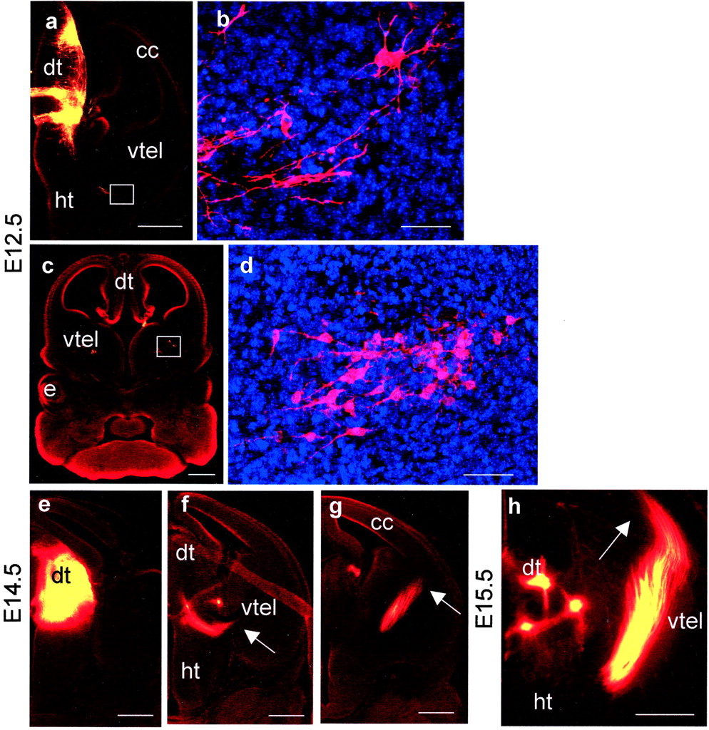
- Fig. 1.
Labeling of thalamic afferents and efferents in control embryos by DiI placement in the dorsal thalamus at E12.5 (a–d), E14.5 (e–g), and E15.5 (h). a, A caudal E12.5 section showing the injection site and axons growing laterally at the diencephalic/telencephalic boundary (white box).b, A higher magnification of the area boxed in a showing retrogradely labeled cell bodies marking the lateral limit of DiI diffusion. c, A more rostral E12.5 section with higher magnification (d) of the area boxed inc showing retrogradely labeled cell bodies in the medial part of the ventral telencephalon. e–g, Caudal to rostral series of sections showing injection site and the trajectory of the thalamocortical tract at E14.5. Arrowsmark the lateral limit of the tract in each section. h, By E15.5 the tract has reached the cerebral cortex (marked witharrow). All sections were cut in the coronal plane.e, Eye; cc, cerebral cortex;dt, dorsal thalamus; ht, hypothalamus;vtel, ventral telencephalon. Scale bars:a, c, e–h, 500 μm; b, d, 50 μm.
- Fig. 2.
a,f–i, Labeling inPax6−/− embryos after DiI placement in the dorsal thalamus at E12.5 (a) and E14.5 (f–i).a, An E12.5 rostral section, at a level similar to that in Figure 1c, showing that no retrogradely labeled cells were seen in the ventral telencephalon. Boxed areaindicates region of ventral telencephalon shown inb–e. b–e, Mash1 immunohistochemistry in E12.5 Pax6+/+(c) and Pax6−/−(e) ventral telencephalon. In both cases cells expressing Mash1 protein (green) are distributed in the ventricular zone (filled yellow arrows) and not in the central region. b, d, Phase-contrast images corresponding to fluorescent images inc and e. E14.5 caudal (f) and rostral (g) sections showing injection site and the absence of DiI labeling in the ventral telencephalon. h, Thalamic axons descending through the thalamus (indicated by arrow); boxed area in h is shown at higher magnification ini illustrating an axon tipped with a growth cone (arrow in i). dt*,Pax6−/− correlate of thePax6+/+ dorsal thalamus;ht, hypothalamus; vtel, ventral telencephalon. Pax6−/− embryos lack eyes altogether. All sections were cut in the coronal plane. Scale bars: a,f–h, 500 μm;b–e, 50 μm; i, 10 μm.
- Fig. 3.
E12.5Pax6−/−↔Pax6+/+ chimeras show an autonomous requirement for Pax6 in the dorsal thalamus but not in the medial part of the ventral telencephalon. a, d, Low magnification of horizontal sections of chimeras like those used to examine the contribution of cells to the medial part of the ventral telencephalon (a) and the dorsal thalamus (d) (red boxes indicate location and orientation of higher magnification fields). b,c, e–g, High magnification of portions of typical fields used for quantification:c, f, g, inPax6−/−,Tg+ ↔Pax6+/+ chimeras; b,e, inPax6+/+,Tg+↔Pax6+/+ chimeras. In these images medial is to the right, and rostral is at the top. The Tg signals appear asbrown spots in the nuclei (here stainedblue). In the medial part of the ventral telencephalon, bothPax6+/+,Tg+ (b) andPax6−/−,Tg+ (c) cells intermingle equally well withPax6+/+, Tg− cells. In the dorsal thalamus, althoughPax6+/+,Tg+ (e) cells are well mixed with thePax6+/+,Tg− cells,Pax6−/−,Tg+ (f, g) cells do not intermingle with their wild-type counterparts and instead form stripes highly enriched for Pax6−/− cells (red arrows in f, g) separated by regions consisting almost exclusively ofPax6+/+ cells (red arrowheads in f). In some cases the larger congregations ofPax6−/− cells seem to have caused the ventricular surface of the dorsal thalamus to buckle inward (red arrow in g).e, Eye; cc, cerebral cortex;dt, dorsal thalamus; vtel, ventral telencephalon. Scale bars: a, d, 500 μm; b, c,e–g, 50 μm.
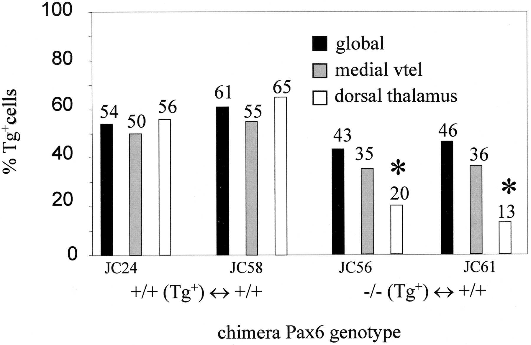
- Fig. 4.
Histogram showing quantification of chimerism inPax6−/−↔Pax6+/+ andPax6+/+ ↔Pax6+/+ chimeras. Global chimerism and chimerism for the medial part of the ventral telencephalon and the dorsal thalamus are shown for each chimera, with thenumbers above each bar indicating the percentages of Tg+ cells. Primary Tg+ signal/nuclei counts for the medial ventral telencephalon and the dorsal thalamus were as follows:Pax6+/+,Tg+ ↔Pax6+/+ chimeras: JC24, medial vtel = 2168/3721, dt = 2613/4390; JC58: medial vtel = 2135/3325, dt = 2427/3465;Pax6−/−,Tg+ ↔Pax6+/+ chimeras: JC56, medial vtel = 1412/3484, dt = 692/3179; JC61: medial vtel = 1513/3591, dt = 511/3633. These primary counts were divided by the tissue-specific correction factors (1.16 for the medial ventral telencephalon, 1.07 for the dorsal thalamus) to give the corrected hybridization index, which gives a true estimate of the percentage of Tg+ cells in the tissue for comparison with the global Tg+ contribution (see Materials and Methods). Large variation between tissue-specific and global Tg+ contribution, indicating a requirement for Pax6, is seen only in the dorsal thalamus ofPax6−/− ↔Pax6+/+ chimeras (marked with ∗), where Pax6−/− cells are also abnormally distributed (Fig. 3). vtel, Ventral telencephalon.
- Fig. 5.
DiI labeling inFoxg1−/− embryos at E12.5 (a–d), E14.5 (e–g), and E15.5 (h–j).a–h, DiI injections into dorsal thalamus. a, A caudal E12.5 section showing the injection site and absence of DiI labeling from telencephalic structures (the Foxg1−/− telencephalon lacks recognizable ventral structures so is markedcc*). b, A higher magnification of thetop boxed area in a showing that the very few axons which approach the telencephalon are disorganized.c, A higher magnification of the bottom area boxed in a showing that labeled axons grow ventrally and medially rather than turning laterally into the telencephalon. d, A more rostral E12.5 section showing that no retrogradely labeled cells or axons can be detected in the rostral telencephalon. e–g, Caudal to rostral series of sections at E14.5 show the injection site and the trajectory of thalamic efferents. These do not penetrate the telencephalon; arrows mark lateral limit of the tract in each section. h, By E15.5 the tract has not penetrated the telencephalon but has continued ventrally within the thalamus toward the hypothalamus (marked with arrow).i, j, DiI injection into telencephalic structures at E15.5. i, Although no substantial tract leaves the telencephalon inFoxg1−/− embryos, higher magnification of the area boxed in jshows that a few disorganized axons are able to cross the telencephalic/diencephalic boundary. All sections were cut in the coronal plane. cc* and e* denoteFoxg1−/− correlates ofFoxg1+/+ cerebral cortex and eye;dt, dorsal thalamus; ht, hypothalamus. Scale bars: a, d,e–h, 500 μm; b,c, 50 μm.
- Fig. 6.
DiI labeling of axons from the optic cup in Foxg1−/−(f–j) and control (a–e) embryos at E15.5.a–e, In control embryos, a rostral to caudal series shows the tract leaving the optic cup (a), growing over the lateral surface of the hypothalamus (b), and reaching the lateral aspect of the dorsal thalamus (c). Boxed areas in b and c are shown at higher magnification in d and e. d, Axons do not penetrate into telencephalic structures.e, Axons form a smooth tract running in a dorsoventral direction in the lateral dorsal thalamus.f–j, InFoxg1−/− embryos, a rostral to caudal series shows the tract leaving the optic cup (f), growing over the lateral surface of the hypothalamus (g) (unfilled yellow arrow in g shows axons penetrating the telencephalon), and reaching the lateral aspect of the dorsal thalamus (h). Boxed areas ing and h are shown at higher magnification in i and j. i, Some axons leave the main tract and penetrate into telencephalic structures (unfilled yellow arrow). j, Axons form a smooth tract running in a dorsoventral plane in the lateral dorsal thalamus. The main tract is indicated by yellow arrows in a–c andf–h. Note that short labeled fibers, marked with yellow arrowheads, sprouting medially from the main tract into the diencephalon are seen in both control (d, e) andFoxg1−/− (i, j) embryos. cc* ande* denoteFoxg1−/− correlates ofFoxg1+/+ cerebral cortex and eye;dt, dorsal thalamus; ht, hypothalamus;vtel, ventral telencephalon. All sections were cut in the coronal plane. Scale bars: a–c,f–h, 500 μm; d,e, i, j, 50 μm.


