

Article Figures & Data
Figures
- Figure 1.
Short-term depression at the calyx of Held. A, Transmission is not saturated because raised calcium increases EPSC amplitude. Initial EPSC amplitude is plotted with [Ca2+]oof 1 (n = 4), 2 (n = 12), and 4 mm (n = 4). Inset shows the effect of changing calcium concentration on the amplitude of a single EPSC; traces are means of 10 sweeps at each calcium concentration. B, Short-term depression generated by a 1 sec train at 50 Hz (left) with the first seven EPSCs expanded (right); top trace, control; bottom trace, during perfusion with 50 μm CTZ. Dotted lines indicate measured EPSC amplitude (on the right); note the apparent summation in the presence of CTZ. Stimulus artifacts have been blanked for clarity. C, Average EPSC amplitude (n = 4 cells) in the presence (gray squares) and absence (black diamonds) of 50 μm CTZ. D, Normalized EPSC amplitude expressed as a percentage of initial EPSC amplitude in the presence (gray squares) and absence (black diamonds) of 50 μm CTZ.
- Figure 2.
γ-DGG reduces the EPSC amplitude, unmasks facilitation, and reduces short-term depression. Ai, Effect of γ-DGG on the amplitude of a single EPSC. Traces are mean of 20 EPSCs in the absence (control) and presence of either 2 or 4 mm γ-DGG. Aii, EPSC amplitudes in the presence ofγ-DGG normalized to the peak EPSC in control conditions. B–D, Effect ofγ-DGG on the EPSC amplitude during a 50 Hz train. Control (B),γ-DGG (C), and normalized data (D) from two different neurons are shown in i and ii. Bi, Control. After an initial large EPSC, there is rapid depression to a low steady-state level (< 10% of initial EPSC amplitude), which is maintained throughout the train. Bii, Control. In this neuron, the EPSC declines slowly, and the relative steady-state amplitude is greater than that for i, being 50% of initial EPSC amplitude. C, Bath application of 4 mm γ-DGG reduces EPSC amplitude by 85% in both neurons (Ci, Cii). Di, The trace shown in Ci is scaled to the amplitude of the first EPSC in the control train (Bi). The amplitude of the steady-state EPSCs is higher with respect to the first EPSC. ii, The trace shown in Cii is scaled to the amplitude of the first EPSC in the control train (Bii). The EPSC now facilitates from the second to the eighth stimulus and then depresses, but to a higher steady-state amplitude relative to control.
- Figure 3.
γ-DGG had no effect on short-term depression during 10 Hz trains but has a marked effect during 50 Hz trains. Average EPSC amplitudes during the train are shown for four cells with EPSC trains at a frequency of 10 and 50 Hz, under control conditions (black diamonds) and in the presence of 4 mm γ-DGG (gray squares). A, Average EPSC amplitude during a 10 Hz train. B, Average EPSC amplitude during a 50 Hz train. C, Normalized EPSC amplitude during a 10 Hz train. Data with and withoutγ-DGG are superimposed and show a 60% depression at 1 sec. D, Normalized EPSC amplitude during a 50 Hz train. At this frequency,γ-DGG unmasks facilitation and reduces depression from 78 to 50% at 1 sec.
- Figure 10.
Kynurenate and γ-DGG block desensitization at physiological temperature and stimulus frequency (37°C; 200 Hz train). Average data are shown for four cells, under control conditions (black diamonds) and in the presence of either 4 mm γ-DGG or 2 mm KYN (graysquares). A, Normalized data for control EPSC trains and trains in the presence of KYN. B, Normalized data for control trains and with γ-DGG. C, Bar graph showing that γ-DGG and KYN block initial EPSC amplitude to the same extent. D, KYN and γ-DGG decrease the amount of depression observed during a train. The graph shows the amplitude of the final EPSC expressed relative to the amplitude of the initial EPSC of the train. Asterisks indicate statistical significance (Student's t test; p < 0.05).
- Figure 4.
A computational model of transmission accounts for the frequency-dependent effect of γ-DGG by a simple competitive antagonist mechanism. A, The model of synaptic transmission at the calyx of Held: 500 active zones with initially five vesicles per active zone. Release probability is 0.17 per vesicle. See Materials and Methods for rate constants. B–E, Simulated EPSC short-term depression. Data are expressed as a mean ± SEM of 10 individual runs in a manner similar to the experimental data shown in Figure 3. Black traces are in control conditions; light gray traces are in the presence of 4 mm γ-DGG. Dark gray traces are with AMPA receptor desensitization disabled (Rd1 and Rd2 set to 0) in the presence of 4 mmγ-DGG. B and C are simulated EPSC amplitudes for trains at frequencies of 10 and 50 Hz. D and E are normalized EPSC amplitudes for trains at 10 and 50 Hz, respectively.
- Figure 5.
Competitive antagonists with slow kinetics do not suppress desensitization. A, Changing the kinetics of the competitive antagonist has a marked effect on the efficacy of relief from desensitization. The unblocking rate constant is plotted against the amount of relief from desensitization. Unblocking rates of an antagonist were changed from 0.000006 mm-msec -1 to 60,000 mm-msec -1 for a mechanism with a single blocked state. The ratio of blocking and unblocking rates was kept constant (Rbl /Rub = 11.67 mm). Data are mean ± SEM of 10 runs at 50 Hz at each unblocking rate. The bell-shaped distribution shows that there is an optimum rate that provides maximal relief from desensitization. γ-DGG has rate constants that provide 93% relief of desensitization. In contrast, NBQX has kinetics that provides no relief (arrows). B, C, Antagonists with slow kinetics fail to protect from desensitization in the model. Normalized data in the presence (gray squares) and absence (black diamonds) of 100 nm NBQX. Average data are shown as mean ± SEM of 10 separate runs for 10 and 50 Hz trains.
- Figure 6.
Experimentally, NBQX reduces EPSC amplitude but has no effect on the rate of short-term depression. A, Raw data: Top trace, control; bottom trace, 100 nm NBQX. The first 200 msec of a 50 Hz train of 1 sec duration is shown. NBQX blocks EPSC amplitude by 60%. B, C, Normalized EPSC amplitudes (n = 4) in the presence (gray squares) and absence (black diamonds) of 100 nm NBQX. Average data are mean ± SEM for four cells in both conditions. Both γ-DGG and NBQX reduce EPSC amplitude, but in contrast to γ-DGG, NBQX has no effect on short-term depression during low- or high-frequency trains.
- Figure 7.
Use of CTZ and γ-DGG together provide no relief from desensitization. Average data are shown for four cells in the same format as Figure 3 with EPSC trains at 10 and 50 Hz in the presence of 50 μm CTZ, before (black diamonds) and during (gray squares) application of 4 mm γ-DGG. A, Average data for a 10 Hz train. B, Average data for a 50 Hz train. C, Normalized data for the 10 Hz train. D, Normalized data during the 50 Hz train. In the presence of CTZ, γ-DGG has no effect on the steady-state EPSC amplitude measured at the end of a 1 sec train.
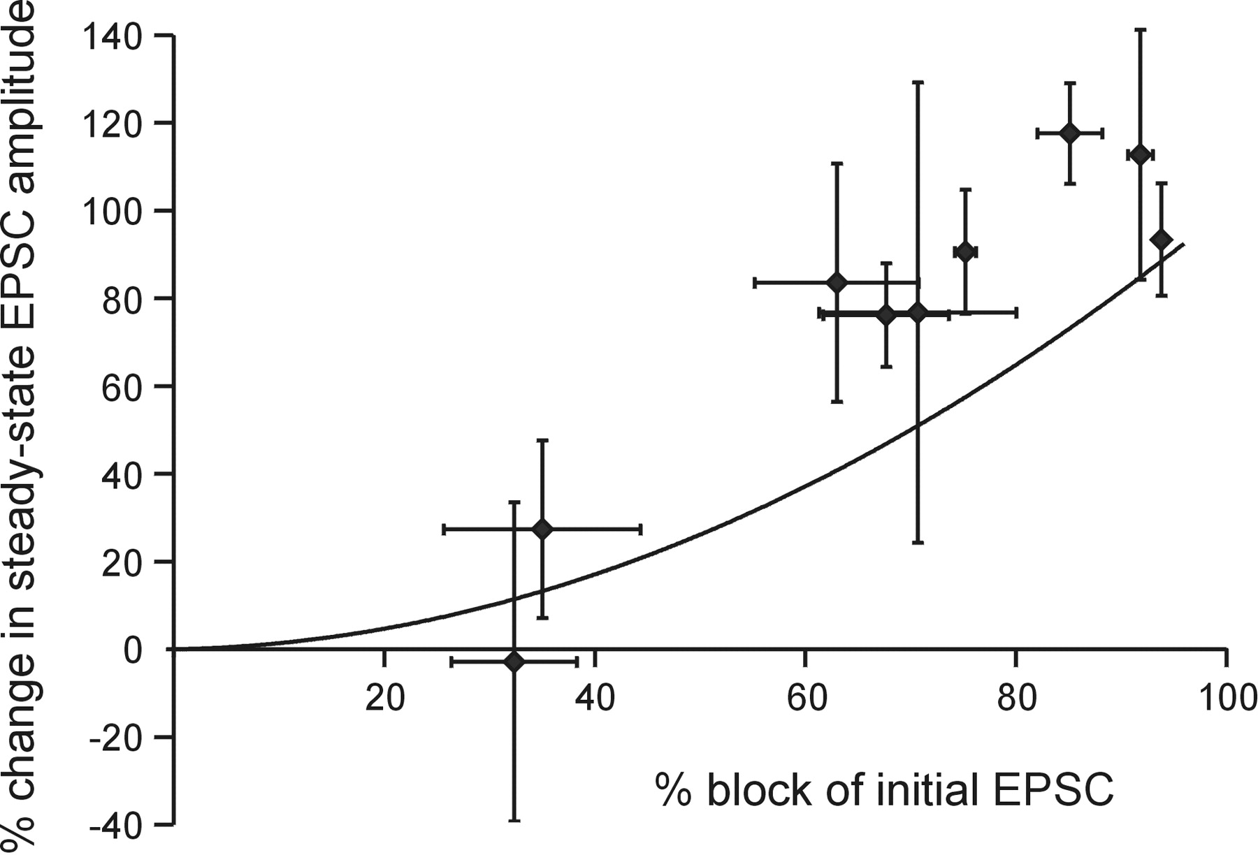
- Figure 8.
Magnitude of EPSC block by γ-DGG correlates with block of desensitization. The change in the steady-state EPSC amplitudes on application of γ-DGG (an index to the extent of desensitization) is plotted against the percentage block of the initial EPSC by γ-DGG. Data are plotted as one cell per data point using a stimulus frequency of 50 Hz. Line is a polynomial fit to data (r2 = 0.99) modeled at different concentrations of γ-DGG producing between 20 and 90% block of initial EPSC amplitude (0.25 and 4 mm γ-DGG).
- Figure 9.
Estimation of the readily releasable pool size from cumulative EPSC amplitude after block of desensitization. Data are mean of four cells with EPSC trains at a frequency of 50 Hz, under control conditions (black diamonds) and in the presence (gray squares) of 4 mm γ-DGG. A, γ-DGG reduces EPSC amplitude and hence the cumulative EPSC amplitude during a 50 Hz train. B, Scaling the γ-DGG trace relative to the amplitude of the initial EPSC corrects for the EPSC block, permitting estimation of the readily releasable pool size in the absence of desensitization. Linear regression fits were constructed by fitting a straight line through the data points obtained during the last half of the train (0.5–1 sec) in both cases. The y-axis intercept of the regression line gives the estimate of the pool size, suggesting that under control conditions, the pool size has been underestimated by approximately half. γ-DGG also increased the gradient of the regression line, from 44 ± 5 to 79 ± 4.5 nA/sec.


Tables
Control 4 mm γ-DGG Frequency (Hz) EPSClast/EPSC1 EPSC2/EPSC1 EPSClast/EPSC1 EPSC2/EPSC1 10 38 ± 4.9 89 ± 4.5 43 ± 5.1 82 ± 5.1 20 25 ± 3.8 89 ± 4.9 62 ± 10 117 ± 30 50 22 ± 3.8 70 ± 9.2 49 ± 8.7 112 ± 9.7 100 11 ± 2.4 58 ± 11 24 ± 4.8 84 ± 20 Data are mean ± SEM of four different cells at all frequencies before and during bath application of 4 mm γ-DGG. EPSClast/EPSC1 shows the extent of depression during the train; EPSC2/EPSC1 shows paired-pulse facilitation/depression. γ-DGG increases the EPSClast/EPSC1 and the paired-pulse ratio at all frequencies >10 Hz.


