

Article Figures & Data
Figures
- Figure 1.
Vestibular primary afferent mossy fiber and climbing fiber projections to uvula-nodulus. Sequences in activation are indicated by solid lines for excitatory pathways and dashed lines for inhibitory pathways, listed numerically. 1, Roll-tilt onto the left side increases primary afferent discharge; 2, primary afferent mossy fibers project to ipsilateral Psol, Y-group, and granule cell layer of nodulus; 3, Psol projects to ipsilateral β-nucleus and dmcc; 4, climbing fibers from β-nucleus and dmcc project to contralateral nodulus; 5, vestibular nuclei project bilaterally to Y-group; 6, Y-group projects to contralateral dorsal cap, β-nucleus, and dmcc; Amb, nucleus ambiguus; β, β-nucleus; cf, climbing fiber; Cu, cuneate nucleus; dc, dorsal cap; DVN, LVN, MVN and SVN, descending, lateral, medial, and superior vestibular nucleus, respectively; Fl, flocculus; Gc, granule cell; icp, inferior cerebellar peduncle; LRN, lateral reticular nucleus; mf, mossy fiber; LCN, MCN and IntP, lateral, medial and interpositus cerebellar nucleus, respectively; NPH, nucleus prepositus hypoglossi; Nsol, nucleus solitarius; Pc, Purkinje neuron; pf, parallel fiber; PFl, paraflocculus; PO, principal olive; Psol, parasolitary nucleus; sol, tractus solitarius; SpV, spinal trigeminal nucleus; spV, spinal trigeminal tract; X, dorsal motor nucleus of the vagus; XII, hypoglossal nucleus; VI, abducens nucleus; Y, Y-group: 8n, auditory-vestibular nerve.
- Figure 2.
Topographic distribution of CFR optimal planes within uvula-nodulus. Sinusoidal vestibular stimulation was used to classify optimal response planes of CFRs. A, B, Figurines illustrate optimal (A1) and null (B1) CFR response planes of a Purkinje cell recorded in the left uvula. Stimulation in the optimal plane (A2) evoked increased CFRs and decreased SSs when the rabbit was rotated onto its left side. When stimulated in the null plane (B2), neither CFRs nor SSs were modulated. C, Sagittal view of the rabbit cerebellum. The shaded area (folia 9d and 10) indicates the region of the uvula-nodulus receiving vestibular primary afferent projection. D, Folia 9-10 are represented as a two-dimensional topographic sheet. Optimal planes for CFRs recorded from 205 Purkinje cells are represented on this surface. Cells with optimal planes corresponding to stimulation in the plane of the ipsilateral posterior semicircular canal (LPC) are illustrated as circles. Cells with optimal planes corresponding to stimulation in the plane of the ipsilateral anterior semicircular canal (LAC) are illustrated as squares. Open symbols indicate cells in which the optimal plane was determined only for sinusoidal stimulation. Filled symbols indicate cells that were tested for static sensitivity and were positive. Black diamonds indicate cells that responded only to horizontal optokinetic stimulation (LHOK) of the ipsilateral eye in the posterior-anterior direction. The more lateral aspects of folia 9-10 were unexplored.
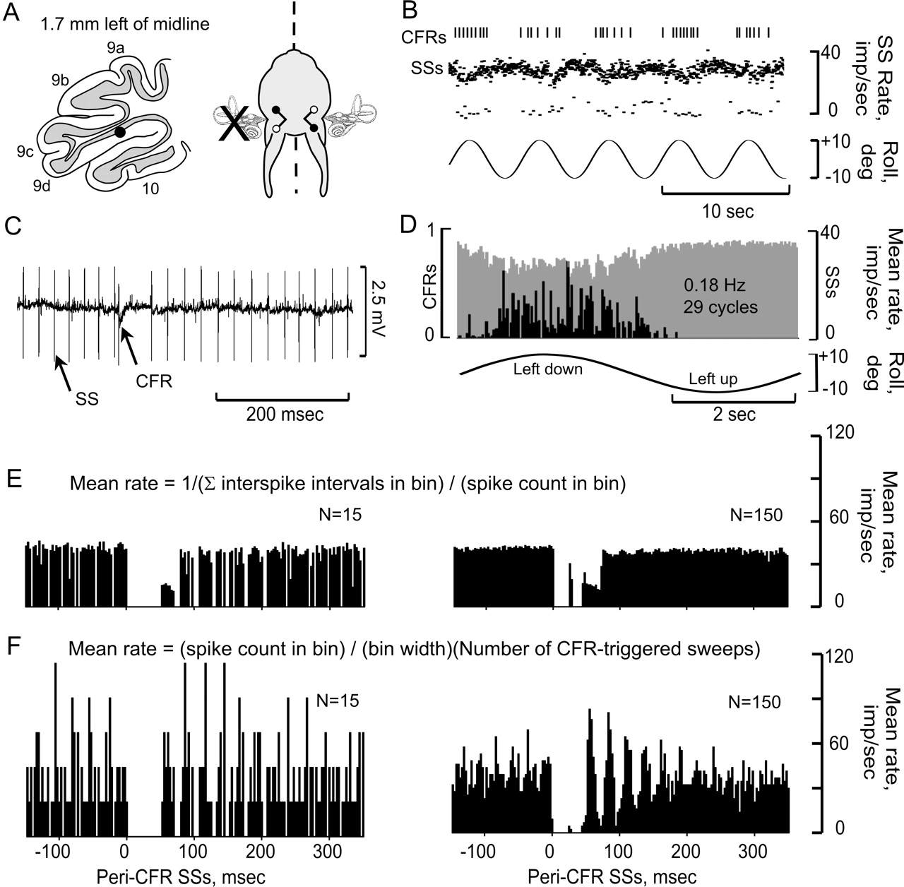
- Figure 3.
CFRs and SSs are modulated in a Purkinje cell ipsilateral to UL. A, Purkinje cell was located 1.7 mm lateral to the midline and ipsilateral to the UL. The optimal plane for modulating CFRs was orthogonal to the longitudinal axis. B, CFRs and SSs, modulated by sinusoidal stimulation at 0.2 Hz, are displayed as vertical lines (CFRs) and instantaneous frequency dot displays (SSs). C, Action potentials are illustrated at a faster time base. D, Peristimulus histograms reveal antiphasic modulation of CFRs (black bars) and SSs (gray bars). E, F, Peri-CFR-triggered SS histograms were constructed using two different methods. In E, Mean rate = 1/(∑ interspike intervals in bin)/(spike count in bin). In F, Mean rate = (spike count in bin)/(bin width)(total CFR-triggered sweeps). Both methods sorted the SSs into 180 bins. The histograms in E and F show different totals of CFR-triggered sweeps, 15 and 150. Bin width, 2.78 msec.
- Figure 4.
Modulation of CFRs and SSs ipsilateral to UL and frequency-independent CFR-associated SS pause. A, Antiphasically modulated CFRs and SSs were recorded from a nodular Purkinje cell, 1.35 to the left of the midline. B, CFRs and SSs were modulated optimally during sinusoidal stimulation in the plane of the intact contralateral posterior semicircular canal. C, Modulation of CFRs and SSs was antiphasic. D-F, Peristimulus histograms were constructed at three different stimulus frequencies (0.05, 0.20, and 0.5 Hz). CFRs are depicted as black, and SSs as gray. G-I, Peri-CFR-triggered SS histograms for each stimulus frequency show that the duration of the CFR-associated pause in SSs was independent of stimulus frequency.
- Figure 5.
Vestibular modulation of CFRs and SSs in Purkinje cell ipsilateral to UL. A, Vestibularly modulated CFRs and SSs were recorded from a Purkinje cell in folia 9d, 0.75 mm to the left of the midline after a left UL. B, C, CFRs and SSs were modulated antiphasically during stimulation in an optimal plane, corresponding to the intact right anterior semicircular canal. D-F, Peristimulus histograms for three stimulus frequencies (0.01, 0.10, and 0.20 Hz), show the antiphasic occurrences of CFRs and SSs. G-I, Peri-CFR SS histograms indicate a CFR-associated pause of 10 msec.
- Figure 6.
Vestibular modulation of CFRs and SSs in Purkinje cell contralateral to UL. A,CFRs and SSs were recorded from a Purkinje cell in the right nodulus after a left UL. B, The optimal plane of vestibular stimulation corresponded to the intact right anterior semicircular canal. CFRs increased and SSs decreased when the rabbit was rotated onto its right side during sinusoidal (C, D) or static (F, G) roll-tilt. Peri-CFR-triggered SS histograms recorded during sinusoidal (E) and static roll-tilt (H) vestibular stimulation show a SS pause, lasting ∼125 and 111 msec, respectively.
- Figure 7.
UL reduces the depth of modulation of SSs associated with CFRs in the contralateral uvula-nodulus. The phase and Dm of CFRs and SSs are plotted in polar coordinates for 28 Purkinje cells in the ipsilateral (A1) and 21 Purkinje cells in the contralateral (A2) uvula-nodulus after a left UL. Phase was measured relative to ipsilateral side-down head position. Vestibular stimulation was in the optimal plane (0.1-0.2 Hz). B1,2, Peak SS modulation was plotted against peak CFR modulation. The slope of this function was reduced in Purkinje cells recorded in the contralateral (right) uvula-nodulus. The dotted lines indicate the 95% confidence intervals for each fitted regression line. The slope of the regression line fitting the data for Purkinje cells ipsilateral to the UL was statistically greater than that for data from contralateral Purkinje cells.
- Figure 8.
Localization of microlesions of caudal β-nucleus of inferior olive. Electrolytic microlesions, made in the caudal right β-nucleus caused reduced modulation of CFRs and SSs recorded in the contralateral uvula-nodulus. A, Vestibularly modulated responses of a neuron recorded in the right caudal β-nucleus were used to guide the placement of the microlesion. The optimal plane corresponded to the right anterior semicircular canal. B, Microlesions for five rabbits are illustrated in six transverse sections listed in rostral to caudal order. The distance from the caudal pole of the inferior olive to each section is indicated by the number in upper right corner. The size and location of microlesions in the caudal β-nucleus are indicated with different colors. Each of five rabbits is identified by number at the level of the transverse section for which its microlesion was largest: R239, blue; R248, green; R269, gray; R270, yellow; R271, red. The locations of 18 β-nucleus neurons, characterized by their optimal planes, are also indicated. Ten of these neurons (filled circles) had optimal planes aligned with the ipsilateral posterior semicircular canal. Eight neurons (open circles) had optimal planes aligned with the ipsilateral anterior semicircular canal. Neurons with optimal planes aligned with the ipsilateral posterior semicircular canal were located rostral to the section at 925 μm. Those with optimal planes aligned with the ipsilateral anterior semicircular canal were located caudal to the section at 1200 μm. C1, Photomicrograph of a transverse section, stained with neutral red, shows the maximal extent of the microlesion (R248) made in the right caudal β-nucleus. C2, The, microlesion is illustrated schematically at a level 925 μm rostral to the caudal pole of the inferior olive. β, β-Nucleus; Cu, external cuneate nucleus; DAO, dorsal accessory olive; DVN, descending vestibular nucleus; dc, dorsal cap of Kooy; icp, inferior cerebellar peduncle; MAO, medial accessory olive; MVN, medial vestibular nucleus; Nsol, nucleus solitarius; Psol, parasolitary nucleus; Pyr, pyramidal tract; sol, tractus solitarius; SpV, spinal trigeminal nucleus; spV, spinal trigeminal tract; vlo, ventrolateral outgrowth; X, dorsal motor nucleus of the vagus; XII, hypoglossal nucleus; XII n, hypoglossal nerve.
- Figure 9.
Microlesions of the caudal right β-nucleus reduce the depth of modulation of contralateral CFRs and SSs. A, Optimal planes for CFRs and depth of modulation for CFRs and SSs were measured in Purkinje cells in uvula-nodulus after microlesions were made in the right β-nucleus of five rabbits. For Purkinje cells in the ipsilateral (right) uvula-nodulus, CFRs increased and SSs decreased during rotation of the rabbit onto its right side. Conversely, in the contralateral (left) uvula-nodulus, CFRs increased and SSs decreased during rotation onto the left side. A key for categorizing responses is located in the upper right corner. Shading in the left half of each symbol characterizes the Dm of the CFR. Shading in the right half characterizes the Dm for the SS. Circles indicate Purkinje cells with optimal responses in the plane of the ipsilateral posterior-contralateral anterior semicircular canals. Squares indicate Purkinje cells with optimal responses in plane of the ipsilateral anterior-contralateral posterior semicircular canals. B, Polar plot compares Dm for CFRs and SSs recorded from medial zones in the ipsilateral (green) and contralateral (red) uvula-nodulus after microlesions of the right β-nucleus. C, Polar plot compares Dm for all CFRs and SSs recorded in the ipsilateral and contralateral uvula-nodulus after microlesions of the right β-nucleus. The vector representing Dm for contralateral SSs in both B and C was reduced relative to that for ipsilateral SSs.


