

Article Figures & Data
Figures
- Figure 1.
Localizing aversive and attractive memory formation. After Pavlovian training with either sugar or electric shock reinforcement, the same olfactory cues elicit attraction (PI > 0) or repulsion (PI < 0) in the 3 min memory test. A, Wild-type Canton-S acquires a strong aversive memory after a single trial of electric shock reinforcement, whereas at least two training trials of sugar reward are required for a significant positive memory score (p < 0.05). However, a total of four training trials does not result in an additional increase in performance (p > 0.05). Therefore, in all of the following experiments on sugar learning, two training trials are used. In sugar (B) and electric shock learning (C), mutant rut2080 flies (including rut;UAS-rut+ and rut;247-Gal4 flies) show only ∼40% of wild-type memory scores [p < 0.001, compared with rut rescued flies (rut;UAS-rut+;247-Gal4) or wild-type in A]. Expression of the rut+ cDNA in ∼700 Kenyon cells of the 247-Gal4 driver line (rut-rescue) is sufficient to restore performance of sugar and electric shock memory to wild-type levels (p > 0.05). D, Restrictive temperature throughout the experiment (black columns) completely abolishes sugar memory in flies expressing the UAS-shits1 transgene exclusively in the Kenyon cells of the MBs (247-Gal4/UAS-shits1; p < 0.001). No temperature-dependent decrease is found in genetic control flies heterozygous for each of the transgenes alone (247-Gal4/+ and UAS-shits1/+; p > 0.05). At the permissive temperature, all genotypes show normal memory (p > 0.05). E, Electric shock memory is strongly reduced at the restrictive temperature in flies expressing the Shi-transgene in the Kenyon cells (247-Gal4/UAS-shits1) compared with genetic controls at either permissive or restrictive temperature (p < 0.001). Data are the means ± SEM of at least six experiments.
- Figure 2.
Sugar memory formation is independent of synaptic output from Kenyon cells during acquisition. Synaptic output from the Kenyon cells is selectively blocked during either acquisition or retrieval of memory in 247-Gal4/UAS-shits1 flies. A, When 247-Gal4/UAS-shits1 flies are trained at the permissive temperature (26°C) and tested 60 min later at the restrictive temperature (34°C), performance is decreased compared with the heterozygous control groups UAS-shits1/+ and 247-Gal4/+ (p < 0.001). B, In contrast, when 247-Gal4/UAS-shits1 flies are trained at the restrictive and tested at the permissive temperature, memory is not affected compared with the genetic controls (p > 0.05). Temperature is shifted to 34°C 15 min before the training. Means and SEMs of six experiments are shown. The same temperature regimen has been applied to electric shock learning with very similar results (data not shown), confirming previous results (Dubnau et al., 2001; McGuire et al., 2001; Schwaerzel et al., 2002).
- Figure 3.
Octopamine is necessary for the acquisition of sugar memory. A, Flies lacking octopamine caused by a mutation in the mutant TβHM18 show normal electric shock memory (3 min memory; p > 0.05 compared with control lines described in Materials and Methods; note that electric shock memory of the TβH mutant and control lines is slightly lower than that of our CS wild-type). B, In contrast, no sugar memory is detected in TβH mutant flies (p < 0.001). C, Mutant TβHM18 flies with a heat-shock inducible TβH+ cDNA (TβH; hs1) show normal sugar memory after heat shock (HS+) (p < 0.05). Neither the hs1-construct alone (TβH; hs1 - HS-) nor the heat shock itself have a significant effect compared with the TβHM18 mutant (p > 0.05) D, Mutant TβHM18 flies show normal sugar memory after feeding on octopamine (10 mg/ml) for 1 or 18 hr before the experiment (p < 0.001). E, Octopamine is required during acquisition. If TβHM18 flies are fed octopamine for 1 hr starting right after the training, no memory is detected, although the feeding itself does not abolish 1 hr memory in the control flies (p < 0.001). Data are means and SEMs of six experiments (except for 12 experiments on the TβHM18 mutant in A and B).
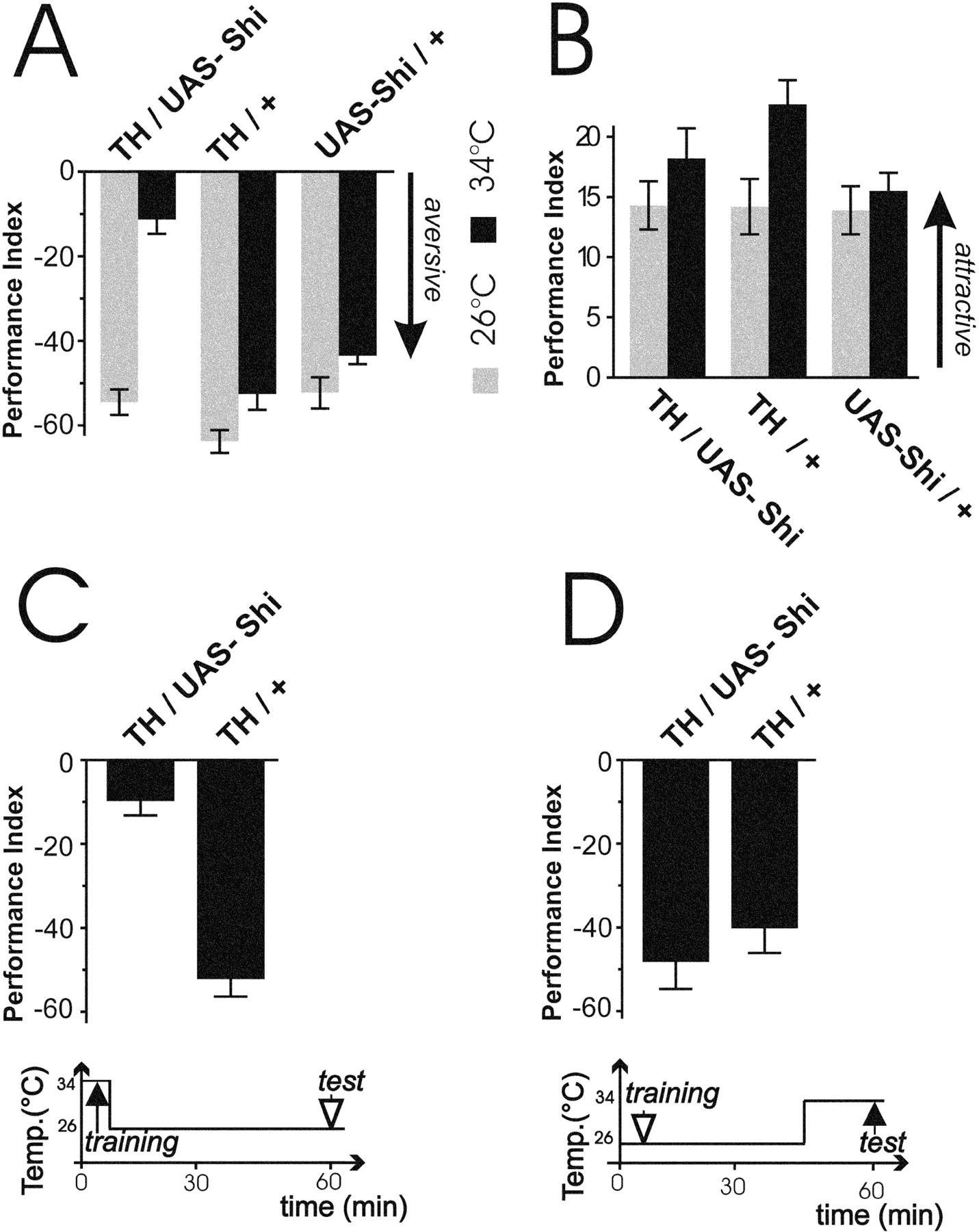
- Figure 4.
DA is necessary for acquisition but not retrieval of electric shock memory. A, Blockade of transmission from (putatively) dopaminergic neurons in TH-Gal4/UAS-shits1 flies at 34°C severely disturbs electric shock 3 min memory (p < 0.001). This temperature-dependent decrease in performance is absent in the genetic controls (TH-Gal4/+ and UAS-shits1/+) (p> 0.05). B, In contrast, no temperature-dependent decrease in sugar memory is observed in any of the groups (TH-Gal4/UAS-shits1, TH-Gal4/+, UAS-shits1/+) (p > 0.05, except for TH-Gal4/+). Here, performance is significantly increased at the restrictive temperature: p < 0.05). C, One hour memory in electric shock learning is strongly decreased if transmission in dopaminergic neurons is blocked during acquisition only (p < 0.001). Flies are transferred to a restrictive temperature 15 min before the experiment. D, Memory is not affected if the neurons are blocked only during retrieval (p > 0.05). Data are means and SEMs of six experiments.
- Figure 5.
Alternative representations of olfactory memory traces. In the MBs, modulatory neurons representing specific USs (e.g., electric shock or sugar) have synaptic input to Kenyon cells representing the fly's odor space (all perceivable odors). Each modulatory neuron is the functional companion of an MB output neuron (CR neuron), which can mediate a conditioned response. A CR neuron will be recruited to respond to a particular odorant if the companion US and the odorant coincide. A, If the odor space is represented in the MBs several times in parallel (i.e., by separate sets of Kenyon cells), each set could be connected to just one US-CR pair (schematized as circles). B, If the odor space is represented in the MBs only once, the Kenyon cells would have to be connected to several US-CR pairs. C, In this case, different memory traces would be stored in the same set of Kenyon cells at different occasions using the same molecular mechanism independently at different locations along the axon.


Tables
Odorant avoidance Shock avoidance EA IAA Sugar reactivity Genotype 1:36 1:6 1:36 1:6 Rut2080 68.6 ± 3.1 13.8 ± 5.9 16.9 ± 5.3 9.4 ± 3.4 76.6 ± 3.9 78.0 ± 6.0 Rut2080;USA-rut+ 76.6 ± 3.0 8.5 ± 4.7 23.4 ± 5.6 14.8 ± 5.8 70.6 ± 1.7 81.0 ± 5.7 Rut2080;247 72.6 ± 3.0 15.3 ± 4.0 36.8 ± 8.2 4.8 ± 1.7 64.2 ± 5.1 74.5 ± 6.3 Rut-rescue 71.7 ± 3.8 7.2 ± 3.0 35.0 ± 7.0 16.9 ± 6.9 72.9 ± 5.6 85.8 ± 4.0 247/UAS-shi(26°C) 76.2 ± 2.9 −3.0 ± 4.0 36.0 ± 4.8 11.5 ± 4.6 60.8 ± 2.2 83.2 ± 7.9 247/UAS-shi(34°C) 78.2 ± 2.1 −8.1 ± 8.2 41.7 ± 6.7 16.3 ± 2.0 44.6 ± 8.4 72.3 ± 7.9 TβH+ control ND ND ND ND ND 81.8 ± 5.1 TβHM18 ND ND ND ND ND 80.8 ± 5.6 TH/UAS-shi (26°C) 79.0 ± 4.4 ND ND ND ND ND TH/UAS-shi (34°C) 77.0 ± 6.7 ND ND NDg ND ND Electric shock, sugar, and olfactory sensitivities of experimental and control animals. Odors were tested at the normal (dilution 1:36) and a sixfold higher concentration. Mutant TβHM18 flies were tested only for sugar sensitivity and TH/UAS-shits1 flies only for sensitivity to electric shock, because they had normal memory scores in the alternative learning assays. No significant differences (p > 0.05) in any of the assays were detected between experimental and control flies. For each experiment, the means of six (and, in the case of sugar, the means of at least 20 experiments, except for 40 experiments on the TβHM18 mutant) are shown. Errors are SEMs. ND, Not determined.
HTML Page - index.htslp
Files in this Data Supplement:
- Supplemental Figure 1 - Putative dopaminergic (DA) neurons innervating the mushroom body (MB) neuropil. TH-Gal4 / UAS-tau flies (Ito et al., 1997) were double stained with anti-TAU and the general neuropil marker antibody nc82 (curtsey E. Buchner, W�rzburg). Complete3D data sets of adult brains were obtained by CLM microscopy and reconstructed using Amira and Standardbrain visualization software (Rein et al., 2002). (a) Transparent MBs (light blue, surface view) and tau-expressing fibers (yellow) within the MB neuropil. MBs were labeled manually, tau expressing fibers were detected by a threshold criterion (see Rein et al., 2002 for further details). In (b) a stack of 10 and in (c) of 30 virtual sections are compiled. MB neuropils are highlighted by white dotted contour lines. (b) The calyx is innervated only marginally by few non-branching fibers [arrows in (a) and (b)]. (c) In the heel [double arrow in (a) and (c)] and α-lobe [arrowhead in (a) and (c)] profusely arborizing fibers are found. Scale bar: 50 micro-meters.


