

Article Figures & Data
Figures
- Fig. 1.
Interneuron loss in the dentate gyrus of a pilocarpine-induced epileptic rat. Nissl-staining (a,b), GAD65 mRNA in situ hybridization (c, d), and somatostatin (e, f) and parvalbumin immunocytochemistry (g, h) demonstrate the loss of interneurons in adjacent sections from an epileptic rat (b, d, f,h) compared with a control (a,c, e, g). GAD65 mRNA expression appears more intense in surviving interneurons in the epileptic rat (d) compared with the control (c). The surviving somatostatin-immunoreactive somata and axons in the outer molecular layer (arrowheads) are labeled more intensely in the epileptic rat (f) compared with the control (e). Interneuron profiles (GAD65 mRNA-, somatostatin-, and parvalbumin-positive) were counted within the borders of the dentate gyrus that are demonstrated in a. Nissl-stained hilar neurons were counted only within the hilus (h). m, Molecular layer;g, granule cell layer; CA3, CA3 pyramidal cell layer. Scale bar, 250 μm.
- Fig. 2.
Analysis of interneuron loss in the dentate gyrus of pilocarpine-induced epileptic rats and in rats 3–7 d after status epilepticus. a, Parvalbumin- and somatostatin-immunoreactive and GAD65 mRNA-positive interneuron profiles were counted in the most dorsal (Fig. 1) and the most ventral slice prepared from each rat. Slices between were used for electrophysiology. The number of interneuron profiles per dentate gyrus for each rat was calculated by averaging the values from the most dorsal and the most ventral slice. The averages of each experimental group are plotted in this graph. Epileptic and 3–7 d post-status epilepticus rats had fewer interneurons than did controls (*p < 0.0001; unpaired t test). Parvalbumin-positive interneurons could not be analyzed in 3–7 d post-status epilepticus rats (see Results). Error bars indicate SEM. b, The number of somatostatin-immunoreactive interneuron profiles per dentate gyrus was correlated with the number of Nissl-stained hilar neurons from the same slice (r = 0.86). Analysis of only the data from epileptic and 3–7 d post-status epilepticus rats also revealed a significant correlation (p < 0.005; Spearman Rho test).
- Fig. 3.
Granule cells in epileptic rats are hyperexcitable and less inhibited. Sharp electrode, current-clamp recordings of responses evoked by outer molecular layer stimulation reveal one action potential in the granule cell from a control rat (a) and five action potentials in the granule cell from an epileptic rat (b1). b2, Lower stimulation intensity revealed a prolonged depolarization in the granule cell from an epileptic rat. c, Granule cells in epileptic rats discharged more action potentials than controls (p < 0.0001; unpaired ttest). d, Granule cells from epileptic (and control) rats did not discharge bursts of action potentials in response to injected current steps. e–h, Representative examples of IPSPs evoked by molecular layer stimulation and recorded at a range of holding currents. Evoked potentials were analyzed at latencies of 20 and 150 msec, which are near the peaks of the early and late IPSPs, respectively. IPSPs recorded in normal ACSF in control (e) and epileptic rats (f). Action potentials evoked in the epileptic tissue are clipped.f, The reversal potential at the 20 msec latency in the epileptic rat was more depolarized (arrow).g, h, Monosynaptic IPSPs recorded in the presence of CNQX/d-APV. Reversal potentials were similar in the epileptic rat (h) and control (g) (Table 1).
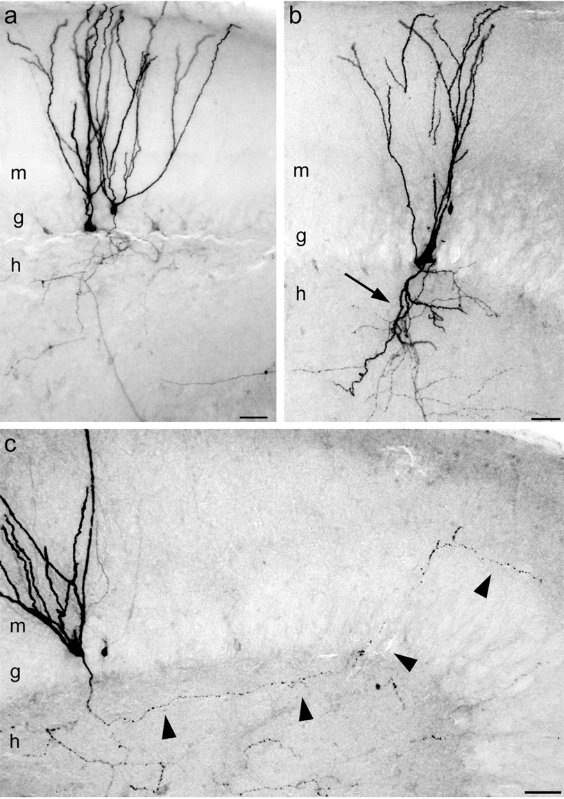
- Fig. 4.
Morphological evidence for changes in the excitatory circuitry of granule cells in epileptic rats.a, Biocytin-labeled granule cells in a control rat extended dendrites into the molecular layer (m) and axons into the hilus (h). b, In an epileptic rat, one of two labeled granule cells extended a basal dendrite into the hilus (arrow). c, In an epileptic rat, an axon collateral (arrowheads) projected from the hilus, through the granule cell layer (g), and into the molecular layer. Scale bars, 50 μm.
- Fig. 5.
Reduced paired-pulse depression of monosynaptic GABAA receptor-mediated IPSCs recorded with whole-cell voltage clamp in granule cells from epileptic rats. IPSCs were evoked by outer molecular layer stimulation. a, Superimposed pairs of evoked IPSCs at interstimulus intervals of 30, 70, 100, 200, and 500 msec (average of 4 trials). Horizontal barsindicate the average peak of the first response. b, Time course of paired-pulse responses in controls (n = 13) and epileptic animals (n = 10). Error bars indicate SEM. *p < 0.05; **p< 0.01; ***p < 0.001 (unpaired ttest).
- Fig. 6.
Fewer spontaneous IPSCs recorded in normal ACSF in epileptic (a) and 3–7 d post-status epilepticus rats (b) compared with their respective controls.a, b, Middle andbottom traces show time-expanded view of regions indicated by bars under top traces.c, Frequency, charge transfer, amplitude, and 10–90% rise time of spontaneous IPSCs in each group. Error bars indicate SEM. *p < 0.05; ***p < 0.001 (unpaired t test).
- Fig. 7.
Fewer miniature IPSCs recorded in CNQX/d-APV/TTX in epileptic (a) and 3–7 d post-status epilepticus rats (b) compared with their respective controls. a, b,Middle and bottom traces show time-expanded view of regions indicated by bars undertop traces. c, Frequency, amplitude, and 10–90% rise time of miniature IPSCs in each group. Error bars indicate SEM. *p < 0.05; **p< 0.01; ***p < 0.001 (unpaired ttest).
- Fig. 8.
Two types of GABAA receptor-mediated mIPSCs in granule cells based on 10–90% rise times. a, In a granule cell from a control rat, minimal stimulation of outer molecular layer evoked slower rise time and smaller amplitude IPSCs compared with minimal stimulation of the granule cell layer.b, Paired whole-cell patch-clamp recording from a putative basket cell and a granule cell. Action potentials evoked by a short-duration current pulse injected into the basket cell generated fast-rising and large-amplitude unitary IPSCs in the granule cell. Traces were aligned at the peak time of the action potential.ML, Molecular layer; GCL, granule cell layer; S, stimulation electrode; R, recording electrode. c, Responses of a putative basket cell (same as one shown in b) display nonadapting repetitive firing to a small (left side) and a larger (right side) current step. d, Consecutive mIPSCs in a granule cell from an epileptic rat arranged in columns from the top trace of the left panel to thebottom trace of the right panel.e, Distribution of 10–90% rise times of mIPSCs obtained from the same cell as shown in d. The distribution was fit with two Gaussians (dotted lines). The thick line shows the summation of these curves.
- Fig. 9.
Histograms of average 10–90% rise times (a) and mIPSC amplitudes (b). a, Both fast- and slow-rising mIPSCs were less frequent in epileptic and 3–7 d post-status epilepticus rats. b, Both low- and high-amplitude mIPSCs were less frequent in epileptic and 3–7 d post-status epilepticus rats. Error bars indicate SEM.


Tables
- Table 1.
Granule cell intrinsic physiology and evoked responses recorded with current-clamp and sharp intracellular electrodes
Control n (cells) Epileptic n(cells) Input resistance (MΩ) 102 ± 8 37 110 ± 11 26 Resting membrane potential (mV) −77 ± 1 37 −76 ± 2 26 Normal ACSF Stimulation intensity (mA) 0.34 ± 0.04 33 0.35 ± 0.05 25 20 msec latency Conductance (nS) 26.5 ± 2.2 33 19.7 ± 3.9 25 Reversal potential (mV) −60 ± 1 33 −51 ± 21-165 25 150 msec latency Conductance (nS) 9.8 ± 0.8 33 7.5 ± 1.0 25 Reversal potential (mV) −91 ± 1 33 −91 ± 2 25 CNQX/d-APV Stimulation intensity (mA) 2.88 ± 1.20 8 5.28 ± 1.24 9 20 msec latency Conductance (nS) 44.1 ± 15.3 8 10.2 ± 1.7* 9 Reversal potential (mV) −71 ± 2 8 −67 ± 1 9 150 msec latency Conductance (nS) 16.1 ± 3.3 8 5.2 ± 1.01-160 9 Reversal potential (mV) −90 ± 2 8 −92 ± 3 9 Stimulation of the outer molecular layer was set at an intensity that evoked a maximum amplitude IPSP at a 150 msec latency. Stimulation intensity was reset after switching to CNQX/d-APV. Synaptic responses were measured at latencies of 20 and 150 msec, which are near the peaks of the early and late IPSPs, respectively. Values represent mean ± SEM.
↵* p < 0.03;
↵F1-160 p < 0.005;
↵F1-165 p< 0.001; unpaired t test.
Adult control Epileptic Young control 3–7 d post-status Number of cells 12 16 15 30 Fast-rising mIPSCs 10–90% rise time (msec) 0.89 ± 0.06 0.86 ± 0.05 0.93 ± 0.07 0.84 ± 0.04 Proportion 0.29 ± 0.03 0.30 ± 0.01 0.27 ± 0.02 0.33 ± 0.03 Amplitude (pA) 10.9 ± 1.12-159 19.8 ± 3.72-167,* 10.7 ± 0.92-159 17.1 ± 1.32-159,2-160 Slow-rising mIPSCs 10–90% rise time (msec) 2.11 ± 0.08 2.17 ± 0.11 2.02 ± 0.14 2.05 ± 0.09 Proportion 0.71 ± 0.03 0.70 ± 0.01 0.73 ± 0.02 0.67 ± 0.03 Amplitude (pA) 8.8 ± 0.7 9.9 ± 0.5 9.2 ± 0.7 11.3 ± 0.6* Histograms of mIPSC rise times from individual cells were fit with two Gaussian curves (Fig. 8e). From the two Gaussian curves, the average rise times of fast-rising and slow-rising events were calculated. Those results were compiled for cells from animals in all four experimental groups, and the values in this Table represent the means ± SEM. There were no significant differences in the average rise times or proportion of fast- versus slow-rising events between epileptic, 3–7 d post-status epilepticus, and control rats (t test). Average amplitudes of the fastest fast-rising (10–90% rise time less than the median minus 1 SD) and the slowest slow-rising (10–90% rise time more than the median plus 1 SD) were calculated and are shown in this Table. Within each experimental group, the average amplitudes of the fastest fast-rising mIPSCs were larger than the average amplitudes of the slowest slow-rising mIPSCs (
↵F2-159 p < 0.05;
↵F2-167 p < 0.001; paired ttest). The average amplitude of the fastest fast-rising mIPSCs in epileptic rats was 182% of that of adult controls (
↵* p < 0.05; unpaired t test). Similarly, the average amplitude of the fastest fast-rising mIPSCs in 3–7 d post-status epilepticus rats was 160% of that of young controls (
↵F2-160 p < 0.001; unpaired t test). The average amplitude of the slowest slow-rising mIPSCs in 3–7 d post-status epilepticus rats was 123% of that of young controls (*p < 0.05; unpaired ttest).


