

Article Figures & Data
Figures
- Figure 1.
Nuclear delineation in the posterior thalamus. Adjacent frontal sections of a rhesus monkey thalamus at the level of the posterior pole of the VP complex, Nissl stained, histochemically stained for CO, and immunocytochemically stained for parvalbumin or calbindin. Arrowheads indicate the thin fiber lamina that demarcates the VPM nucleus, including its small-celled region and the VMb nucleus. The arrows indicate profiles of the same blood vessels. Note the close matching of the CO staining with the immunostaining for parvalbumin, in which the rod domain of larger Nissl-stained cells in VPM is delineated. The VMb and the small-celled regions are clearly identifiable by their smaller cells (Nissl) and weaker staining for CO. The asterisk in the section stained for CO marks an area corresponding to the densest calbindin immunostaining well within the medial tip of VPM. This area is also characterized by dense CO staining and dense parvalbumin immunostaining, locating it well inside the boundaries of the VPM nucleus. Note that for a correct identification of the boundaries of VPM and adjacent nuclei, it is essential to take all forms of staining into account. (For a complete series of sections through the posterior thalamus stained with these and other markers, see http://neuroscience.ucdavis.edu/Jones/ThalamusImages/Monkey/.) R, Reticular nucleus; s, small-celled zone of VPM. Scale bar, 1 mm.
- Figure 2.
A, Serial plottings of the cSpV showing the extent and localization of the injection of tracer in the three animals (RM75, RM76, RM77). The tracer injections primarily involved the superficial-most laminas I and II in all three cases, with minor spread into lamina III. The rostrocaudal extent of the injections ranged between 1 mm (RM77) and 1.5 mm (RM76). B, Drawings of cross sections through the brainstem at selected medullary, pontine, and midbrain levels, showing the location of labeled ascending fibers (in RM77). 4, Fourth ventricle; cp, cerebral peduncle; Cu, cuneate nucleus; DR, dorsal raphe nucleus; DV, dorsal motor nucleus of the vagus; eCu, external cuneate nucleus; mlf, medial longitudinal fasciculus; Gr, gracile nucleus; IO, inferior olivary complex; LR, lateral reticular nucleus; mcp, middle cerebellar peduncle; ml, medial lemniscus; PN, pontine nuclei; py, pyramidal tract; SA, sylvian aqueduct; SC, superior colliculus; SpV, spinal trigeminal nucleus; STN, solitary tract nucleus; V, principal trigeminal nucleus; VNC, vestibular nuclear complex; VR, ventral raphe nucleus; xbc, decussation of the brachium conjunctivum; XII, hypoglossal nucleus; xpy, pyramidal decussation.
- Figure 3.
Localization of an injection in frontal sections of the cSpV nucleus. A, Nissl-stained section showing two tracks left by the injecting-recording micropipette reaching the marginal zone (lamina I) and the superficial part of lamina II. B, Section adjacent to A, immunostained for calbindin. The discontinuity in the calbindin-rich superficial laminas of the cSpV is caused by the injection of tracer and indicates the involvement of laminas I and II, primarily in its external portion. Asterisks in A and B mark the same blood vessel. C-E, Dual-channel laser confocal micrographs showing the site of injection of the fluorescent tracer (D, E, green) in relation to the calbindin immunostaining (C-E, red). Laminas I and II and the superficial part of lamina III are clearly identified by the dense calbindin immunolabeling of cell bodies and neuropil. The merged image in E shows the deposit of tracer concentrated in lamina I and superficial lamina II, with less involvement of inner lamina II and minor involvement of lamina III. Scale bars: A, B, 300 μm; C-E, 200 μm.
- Figure 4.
A, D, Drawings from MD2 plottings of serial frontal sections of the posterior thalamus contralateral to the injection site, arranged from posterior (A) to anterior (D) in a representative case (RM76) and showing the terminations of labeled sVTT fibers (red). The labeled fibers arising from cells in the contralateral cSpV do not terminate in a single focal region but are widely distributed across a number of nuclei in the posterior thalamus. Note that the densest cluster of terminations in C, although forming a well defined zone, is well inside the boundaries of VPM, as delineated in Figure 1. Other substantial clusters of sVTT fiber ramifications are found in Po and caudal VPL; many scattered labeled fibers are also present in CM, VPI, VMb, and Pla. CL, Central lateral nucleus; LG, lateral geniculate nucleus; LP, lateral posterior nucleus; MD, mediodorsal nucleus; MGd, dorsal medial geniculate nucleus; PF, parafascicular nucleus; Pi, inferior pulvinar nucleus; R, reticular nucleus; s, small-celled zone of VPM nucleus; thi, habenulopeduncular tract. Scale bar, 1 mm.
- Figure 5.
Frontal sections through the medial tip of VPM stained for CO with superimposed plottings of the principal clusters of labeled sVTT fiber terminations. The patches of dense CO staining correspond to the rods of VPM as seen in Figure 1, and the labeled terminations lie inside the boundaries of VPM, lateral to its medial tip, in which the dense CO staining corresponds to the region of densest calbindin immunostaining (Fig. 1). Scale bar, 1 mm.
- Figure 6.
Dual-channel laser confocal micrographs from frontal sections showing the distribution of labeled sVTT terminations (green) in relation to the region of densest calbindin immunostaining (red) in the medial tip of VP. The labeled fibers terminate close to the region of calbindin-rich neuropil but rarely coextensive with it. A, C, D, The principal clusters of terminations lie lateral to the calbindin-rich medial tip of VPM. B, Some clusters of terminations are located in VPM areas characterized by calbindin-immunopositive cells of the matrix but lacking the dense calbindin-immunopositive fiber plexus at the medial tip of VPM. Scale bar, 400 μm.
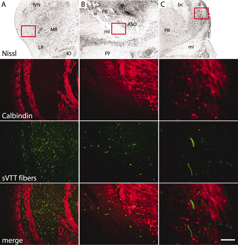
- Figure 7.
Labeled sVTT fibers and calbindin immunostaining. Frontal sections at different levels of the ascending sVTT pathway. The red boxes on the images of the Nissl-stained sections localize the areas depicted in the corresponding laser confocal micrographs. A, In the ipsilateral medulla rostral to the injection site, labeled fibers (green) of the trigeminothalamic tract run between bundles of calbindin-immunopositive fibers (red) that do not contain any fluorescent tracer. B, At the level of the pontine nuclei, labeled sVTT fibers run along the dorsal aspect of the medial lemniscus (calbindin immunonegative) and ventral to the pontine reticular formation, which is rich in a calbindin-immunopositive fiber plexus; calbindin-positive cells of the accessory superior olivary nucleus (ASO) are also shown. C, More rostrally in the brainstem, labeled sVTT fibers run dorsolateral to the medial lemniscus and ventromedial to the lateral lemniscus (ll). None of the labeled fibers are calbindin positive. bc, Brachium conjunctivum; IO, inferior olive; IVN, inferior vestibular nucleus; LR, lateral reticular nucleus; ml, medial lemniscus; MR, medullary reticular formation; PR, pontine reticular formation; py, pyramidal tract; SO, superior olivary nucleus; vi, abducens nerve.
- Figure 8.
Antibody specificity. Right, Western blot of total protein lysate from adult rhesus monkey thalamus. A, B, Both the commercial monoclonal (A) and polyclonal (B) anti-calbindin antibodies recognized a single band of ∼28 kDa. Left, Dual-channel laser confocal micrographs of a rhesus monkey brain section immunostained with both anti-calbindin antibodies; the immunostaining obtained with the polyclonal (A) and the commercial monoclonal antibody (B) are merged in C, where almost complete overlap is evident. Note that the polyclonal antiserum immunostained a slightly broader area of neuropil (C, red) than the monoclonal antibody.


