

Article Figures & Data
Figures
- Figure 1.
EtOH increases GABAergic transmission to granule cells. A, Simplified representation of the basic cerebellar cortical circuit. For clarity, we show only the components of the circuit that are relevant to this study. Mossy fibers, which come from the spinal cord and brain stem, provide excitatory input to granule and Golgi cells. Granule cells receive inhibitory input from Golgi cells. Granule cells provide excitatory input to Purkinje cells and Golgi cells via the parallel fibers. B, Sample traces of GABAergic currents recorded from a cerebellar granule neuron before (control; Ctrl) and during application of 50 mm EtOH and after washout (W/O). Note that EtOH induced a reversible increase in sIPSCs frequency, an inward current shift, and an increase in background noise. GABAergic currents were blocked by bicuculline (20 μm). Calibration: 40 pA, 10 sec. C, Expanded time scale sIPSC traces illustrating that these events can be clearly distinguished from background noise. Calibration: 50 pA, 200 msec. D, Frequency histogram corresponding to the recording shown in B, illustrating the effect of EtOH on sIPSC frequency as a function of time. E, Cumulative probability frequency histograms corresponding to the recording shown in B. F, Same as in E but for amplitude. G, Average sIPSC traces obtained from the recording shown in B. Calibration: 10 pA, 5 msec. Note that EtOH does not affect the amplitude or decay of these currents.
- Figure 2.
Effect of increasing concentrations of EtOH on sIPSCs and tonic current. Left, Summary of the effect of 10 mm (n = 5), 20 mm (n = 6), 35 mm (n = 6), 50 mm (n = 9), 75 mm (n = 6), and 100 mm (n = 9) EtOH on sIPSC frequency and amplitude. Note the lack of an effect of EtOH on amplitude even at a concentration of 100 mm. Right, Summary of the effect of 10 mm (n = 6), 20 mm (n = 7), 35 mm (n = 7), 50 mm (n = 14), 75 mm (n = 8), and 100 mm (n = 6) EtOH on the tonic current noise variance. *p < 0.05; **p < 0.005; ***p < 0.0005, by one-sample t test versus theoretical mean of zero.
- Figure 3.
EtOH does not affect evoked IPSCs recorded in cerebellar granule cells. A, Sample traces of IPSCs evoked in a cerebellar granule cell by Golgi cell stimulation under control conditions, in the presence of 50 mm EtOH and in the presence of 100 μm furosemide. The arrows indicate the peak, early phase (30 msec), and spillover phase (200 msec). Calibration: 50 pA, 25 msec. B, Summary graph illustrating the lack of an effect of 50 mm EtOH on sIPSC peak amplitude, early decay phase, and spillover phase (n = 11). As expected, furosemide inhibited both decay phases, given that these are mediated byα6 subunit-containing receptors (n = 10); the effect of furosemide on the early phase did not reach statistical significance because of high variability, but there was a clear inhibitory effect in 9 of 10 cells tested. *p < 0.01; by one-sample t test versus theoretical mean of 100.
- Figure 4.
The EtOH-induced increase of GABAergic transmission to a cerebellar granule cell is blocked by TTX. A, Sample GABAergic current traces from a cerebellar granule neuron. Application of TTX dramatically reduced IPSC frequency and also reduced both the tonic current amplitude and noise variance. Note that in the presence of TTX, application of 50 mm EtOH did not affect mIPSC frequency or amplitude and did not change tonic current amplitude or current noise. All of the currents were blocked by the GABAA receptor antagonist bicuculline (20 μm). Calibration: 25 pA, 10 sec. B, Summary of the effect of TTX on sIPSC frequency and amplitude (n = 10). C, Summary of the effect of TTX on the amplitude of the tonic current (I Bic; n = 16) and noise variance (n = 21). D, Summary of the effect of 50 and 100 mm EtOH in the presence of TTX on the amplitude of the tonic current (I Bic; n = 10–11), tonic current noise (n = 11–14), and mIPSC frequency and amplitude (n = 4–6). *p < 0.03; **p < 0.008; ***p < 0.0005, by one-sample t test versus theoretical mean of zero.
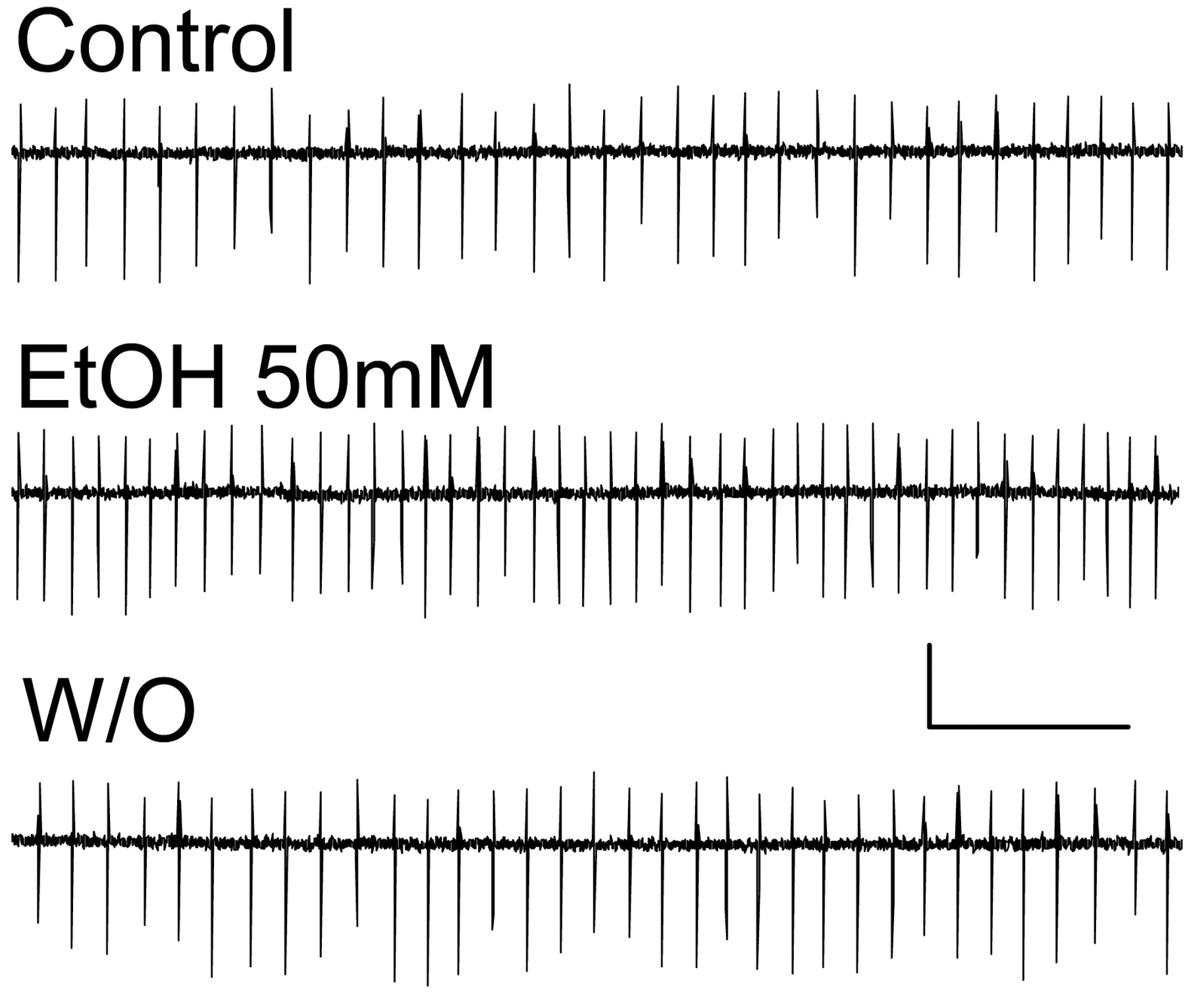
- Figure 5.
EtOH increases the spontaneous firing of Golgi cells. Sample traces of loose-patch cell-attached recordings illustrating that application of 50 mm EtOH induces a reversible increase in the frequency of spontaneous action potentials. Calibration: 40 pA, 400 msec. EtOH increased the firing frequency by 25 ± 8% with respect to control (p < 0.01 by one-sample t test vs a theoretical mean of zero; n = 13).
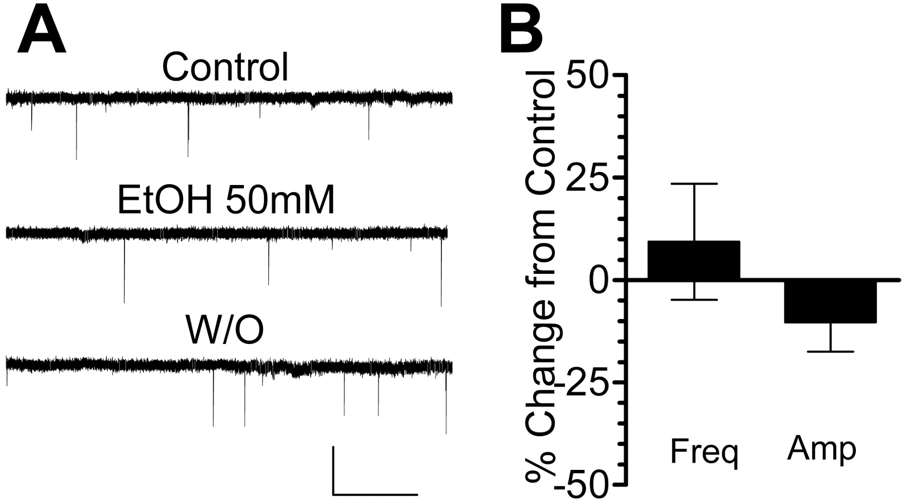
- Figure 6.
EtOH does not affect glutamatergic transmission at mossy fiber-to-granule cell synapses. A, Sample traces of sEPSC recordings from cerebellar granule neurons obtained in the presence of bicuculline (20 μm) showing that application of 50 mm EtOH did not significantly affect the frequency or amplitude of these events. Calibration: 40 pA, 5 sec. B, Summary of the effect of 50 mm EtOH on the frequency and amplitude of sEPSCs (n = 5).


