

Article Figures & Data
Figures
- Figure 1.
The nrc mutation is a stop codon in synj1, and Synj1 is expressed in brain and retina. A, Positional cloning defines the critical genetic region for the nrc mutation. The number between genetic markers represents the number of recombinants identified. B, Schematic of Synj1 domains, SacI (Sac1), 5′-phosphatase (5′Ptase), and proline-rich protein binding domain (pppp). The site of the nrc mutation is shown as a stop on the domain schematic. DNA sequence wave data of the mutation site reveals C to T nonsense mutation in nrc larvae. The full-length protein sequence of zebrafish Synj1 is shown with the mutation site demarcated in red. The border between phosphatase domains is marked by a bold line. The green rectangle demarcates the putative clathrin-binding domain. The putative amphiphysin-binding domain is outlined by a blue rectangle, and the endophilin-binding domain is demarcated by a red rectangle. C, Antisense, but not sense, RNA in situ hybridization reveals synj1 in WT 6 dpf larval brain. The arrowheads point to the brain region. D, Two-week-old mouse retinal section immunohistochemistry with anti-Synj1 proline-rich domain antibody localizes Synj1 to the OPL and IPL. Scale bar, 50 μm. E, Dendrogram shows the amino acid conservation between zebrafish Synj1 and other previously published Synjs. Zebrafish (Zf) Synj1 is more similar to human and rat Synj1 than to human (Hum) and rat Synj2. Dros, Drosophila. The dendrogram was generated using Vector Nti version 9.0 (Informax, Bethesda, MD). The GenBank accession number for zebrafish synaptojanin is AY736013.
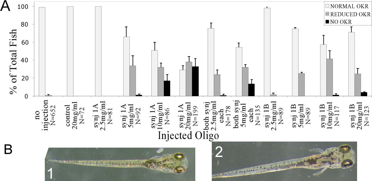
- Figure 2.
synj1 morpholinos phenocopy the nrc visual behavior defect. A, Percentage of total larvae with no OKR, normal OKR, or reduced OKR plotted for each injection scenario. B, The overall morphology of control (20 mg/ml morpholino, robust OKR) (1) injected and synj1-MO-A (20 mg/ml, no OKR) (2)-injected larvae are grossly indistinguishable. Representative 5 dpf larvae are shown.
- Figure 3.
Retina of morpholino-injected larvae. A, Light microscopy of retinas from control (20 mg/ml morpholino, robust OKR) injected and synj1-MO-A (20 mg/ml, no OKR)-injected 5 dpf larvae. The retina is grossly normal. Photoreceptors (PR), secondary neurons (2°), and ganglion cells (GCL) appear properly positioned in the laminated retina. The OPL and IPL are present. However, the OPL in synj1 morpholino-injected larvae appears thinner than in control-injected larvae. B, Electron micrographs of photoreceptor outer segments from control (20 mg/ml morpholino, robust OKR)-injected and synj1-MO-A (20 mg/ml, no OKR)-injected 5 dpf larvae. Both have formed outer segments with nicely stacked discs. Scale bar, 1 μm.
- Figure 4.
synj1 morpholinos phenocopy the nrc ultrastructural retinal defects of the OPL. EM of the OPL of control (20 mg/ml morpholino, robust OKR)-injected and synj1-MO-A (20 mg/ml, noOKR)-injected 5 dpf larvae. Ribbon structures (R) are unanchored in the retina of MO-A larvae but not in controls. Matrix-rich areas (*) devoid of organelles are also seen in MO-A-injected larvae, consistent with nrc findings in this study (Fig. 6). Representative synapses are shown. Scale bar, 1 μm.
- Figure 5.
Phosphatase function and Synj1 protein in nrc larvae. A, B, Free phosphate release from PI(4)P and PI(4,5)P2 substrates is reduced in extracts from nrc mutants. Free phosphate-generated per mg of cytosolic protein from 6 dpf zebrafish larval extracts is plotted for WT and nrc mutants. A, PI(4)P was used as substrate to test the activity of the SacI phosphoinositide phosphatase domain. B, PI(4,5)P2 was used as substrate to test the activity of the inositol 5′-phosphatase domain. Error bars represent SDs. Standard two-tailed t tests show that the differences between mutant and wild type are statistically significant [PI(4)P, p = 0.03; PI(4,5)P2, p = 0.005]. C, Immunoblotting of 6 dpf zebrafish brain extracts reveals reduced levels of ∼150 kDa Synj1 (S) in nrc brain extracts. Five brains are loaded per lane. The ∼65 and ∼30 kDa proteins do not correspond to expected sizes of isoforms or SacI truncated products of synaptojanin and appear to be nonspecific. D, Endophilin overlay of larval zebrafish brain extracts. The 150 kDa protein (Synj1) that binds to GST-endophilin is not present in nrc 6 dpf larval brains. However, the GST-endophilin binding to the ∼95 kDa protein (predicted size of dynamin) is observed in both WT and nrc brains from 6 dpf larvae. Lanes 1 and 2 are independent brain samples. Five brains are loaded per lane. S, Zebrafish Synj1; D, putative zebrafish dynamin with the expected electrophoretic mobility.
- Figure 6.
Actin localization. Fluorescence micrographs showing polymerized actin in the photoreceptor layer of 6 dpf light-adapted nrc retinas, as revealed by staining with phalloidin. The phalloidin staining is more uneven in nrc than in wild type. Punctate regions of phalloidin staining are seen throughout the nrc OPL. No obvious differences in phalloidin staining were seen in the IPL (data not shown). All nrc larvae examined showed similar staining.
- Figure 7.
Vesicles at nrc photoreceptor synapses vesicles. A, WT cone synapse in the OPL, showing the even distribution of vesicles and typical ribbon (R) organization. The ribbon is anchored to the plasma membrane via the arciform density (A). Secondary neurons properly invaginate and juxtapose against the anchored ribbon. B, nrc photoreceptor synapse, demonstrating that vesicles are fewer and unevenly distributed. Organelle-free areas (asterisk) occupied by a dense cytomatrix are present among vesicle clusters. The ribbon (R) is unanchored, and increased endosomes (E) are present throughout the cytoplasm. Insets, Vesicles surrounded by a coat. nrc photoreceptor synapses showing a cluster of floating ribbons (C) and peculiar rows of vesicles (D), like beads on a string. Scale bars: A, B, 1 μm; C, D, 200 nm. E, Quantification of vesicles in 18 WT and 20 nrc light-adapted cone photoreceptor synapses is shown. The difference in overall vesicle number per synapse section is statistically significant (p = 6 × 10⁻⁹). Similarly, the difference in vesicle number per micrometer of plasma membrane is statistically significant (p = 6 × 10⁻⁹). Error bars report the SEM. Vesicles numbers were reduced to nearly the same extent in nrc dark-adapted photoreceptor synapses (24 wild type and 25 nrc; data not shown) as in light-adapted synapses. The difference in vesicle number in nrc versus WT dark-adapted synapses is also statistically significant (p = 2 × 10 vesicle number per micrometer of plasma membrane).


