

Article Figures & Data
Figures
- Figure 1.
IPSC regulation by mGluR on second-order medial NTS neurons in brainstem slices. After identifying second-order NTS neurons by their minimal (<150 μsec) synaptic jitter to ST activation (data not shown; see Materials and Methods), ST-evoked postsynaptic responses were blocked by addition of ionotropic Glu antagonists NBQX (20 μm) and AP-5 (100 μm). Typically, second-order medial NTS neurons appeared spindle shaped, with two major processes exiting the poles of the long axis of the cell (top right micrograph). After ionotropic GluR blockade, large-amplitude, long-duration spontaneous GABAergic IPSCs were prominent (Control traces, left and right). ST activation consisted of trains of five shocks (T2) or 10 shocks (T3) with intershock intervals of 20 msec (i.e., 50 Hz), delivered as short bursts that were repeated every 0.5 sec as depicted at the top left; horizontal bars mark periods of ST stimulation. Note that IPSCs were inward because of the high Cl- (50 mm) internal solution and holding potentials of -60 mV. Left, In decrease-type neurons, ST stimulation decreased the sIPSC rate in a frequency-dependent manner; bottom, 20 consecutive original traces superimposed for each condition. The lowest trace shows that T3 stimulation nearly eliminated IPSCs. Bin plots indicate that sIPSC rates rapidly rebound to near control levels within 10-20 sec after the halt of stimulation. Right, In increase-type neurons, ST stimulation strongly increased the sIPSC rates in a frequency-dependent manner; bottom, 10 consecutive original traces superimposed for each condition. Bin plots indicate that sIPSC rates increase more rapidly for T3 than T2. For clarity, stimulus artifacts have been truncated in some of the original traces.
- Figure 2.
Subtype-specific mGluR receptor antagonists block the effects of ST activation on sIPSCs. In brainstem slices (see Fig. 1 for conditions), ST stimulation modulated the frequency of sIPSCs in subtype-representative second-order NTS neurons. A, Decrease-type neuron. T2 and T3 (horizontal bars) depressed sIPSCs. Modest concentrations of combined group II (LY 341495; 20 nm) and group III (MSOP; 200 μm) mGluR antagonists reversibly blocked the highest intensity (T3) of ST-induced depression of GABA release, and this block was rapidly reversed by return to control perfusate. B, Increase-type neuron. High-frequency ST volley (T3) abruptly increased sIPSC frequency that rapidly returned to control after cessation of ST stimulation. Group I mGluR antagonism (LY 367385; 160 μm) strongly attenuated T3 ST stimulation-induced increases in GABA release. Blockade reversed rapidly with control perfusion so that T2 and T3 volleys produced frequency-graded increases in sIPSC frequency.
- Figure 3.
Summary of average frequency dependence of ST stimulation on sIPSC frequency and specific mGluR antagonism in subtypes of NTS second-order neurons. The frequency of sIPSCs has been normalized to control (1.0; broken horizontal line). Each graph is broken to indicate that different numbers of neurons were subjected to the ST stimulation protocols than the antagonist protocols. *Significant difference from control (p < 0.05). Error bars represent mean ± SEM. A, Decrease-type neurons (n = 6). On average, T2 depressed sIPSC rates by ∼40% and T3 by more than half (p < 0.05), with full recovery after cessation of ST stimulation. Combined group II LY 341495 (20 nm) and group III MSOP (200 μm) antagonism had no effect (p > 0.9) on basal sIPSC rates but reversibly blocked T3 ST stimulation-induced depression of GABA release. B, Increase-type neurons (n = 10). ST stimulation increased sIPSC rates in a graded manner, but note that T1 (2 shocks every 0.5 sec) significantly enhanced sIPSC frequency (p < 0.05); this ST stimulation rate had no effect on decrease-type neurons. These effects were fully reversible (recovery). Group I antagonist LY 367385 (160 μm) had no effect on basal sIPSC rates (p > 0.7) but greatly reduced T3-induced increases in IPSC frequency (p < 0.02).
- Figure 4.
Isolated NTS neurons were dispersed from the medial region of NTS for recording of spontaneous synaptic currents. A, Spindle-shaped neurons were selected for recording because they closely resembled second-order NTS neurons found in slices. B, All of the neurons had spontaneous synaptic currents in control conditions (top single-sweep trace). IPSCs were isolated for study by blocking EPSCs with NBQX (20 μm; middle trace). Note that under our recording conditions (internal Cl- 50 mm) and a holding potential of -60 mV, IPSCs were inward. Combined antagonist block (lowest trace) with bicuculline (100 μm) and NBQX eliminated all of the synaptic events. C, Clear kinetic differences also distinguished IPSCs from EPSCs in this same neuron. Expanded mean traces illustrate the prolonged time course of IPSCs compared with the rapid time course of EPSCs (amplitudes normalized to 100%). The IPSC and EPSC traces are averages of 69 and 80 synaptic currents, respectively. All of the traces and the photo are from the same NTS neuron.
- Figure 5.
Exogenous Glu applied to dispersed NTS neurons either depressed or increased the spontaneous release of GABA (sIPSCs) in different NTS neurons. In all of the experiments, ionotropic Glu responses were blocked with NBQX (20 μm) and AP-5 (100 μm). IPSC detection counts in all of the cases were collected in 10 sec bins, and points are means ± SEM. A, Decrease-type NTS neurons (n = 10). Glu application (1 mm; horizontal bar) rapidly decreased sIPSC frequency in decrease-type neurons, and this effect was rapidly reversed after switching back to control solution. Bottom, Representative samples of sIPSCs recorded from a single neuron during control, Glu, and wash with control solution. B, Increase-type NTS neurons (n = 5). Glu increased the sIPSC frequency averaged at each time bin. Bottom, Representative samples from a single neuron during control, Glu, and wash with control solution.
- Figure 6.
Glutamate acts via presynaptic mechanisms at both increase- and decrease-type NTS neurons. Broken line indicates control sIPSC levels normalized to 1.0. *Significant difference from control (p < 0.05). A, The sIPSC rate in decrease-type NTS neurons (n = 22) was reversibly reduced by application of exogenous Glu (1 mm). Amplitudes of sIPSCs were unaltered in all three of the conditions. B, In increase-type neurons (n = 6), Glu increased the mean frequency of spontaneous GABAergic IPSCs by nearly fivefold. Glu did not alter sIPSC mean amplitudes in increase-type neurons.
- Figure 7.
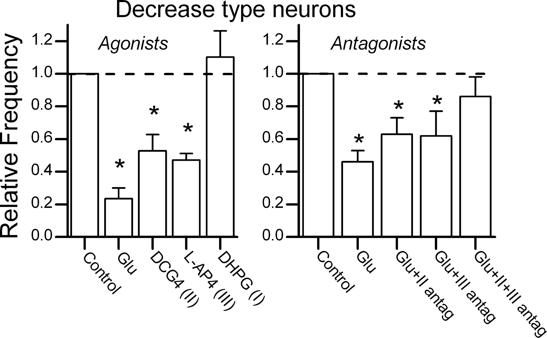
Selective group II and group III mGluR drug actions on decrease-type NTS neurons. Bins of sIPSCs were counted over intervals of 10 sec. Left, Glu (1 mm) substantially depressed sIPSC rates in decrease-type neurons (n = 12; p < 0.0001). The group II mGluR-selective agonist DCG4 (10 μm; n = 6) or the group III mGluR-selective agonist l-AP-4 (10 μm; n = 8) mimicked the actions of Glu in these neurons (p < 0.02). Not all of the neurons were tested for all of the agents. Four of these neurons responded similarly to Glu as well as DCG4 and l-AP-4 (within-neuron tests). The group I mGluR-selective agonist DHPG (10 μm; n = 6) failed to alter sIPSCs (p = 0.58). Five of these DHPG-insensitive neurons were sensitive to l-AP-4, and three of these DHPG-insensitive neurons were sensitive to l-AP-4 and DCG4. Right, In a different set of tests of neurons (n = 12), Glu (1 mm) depressed sIPSC rate by more than half (p < 0.001). Combined antagonists for group II (LY 341495; 10 nm) and group III (MSOP; 200 μm,) blocked the Glu effects (n = 7; p > 0.3). In five of the neurons tested with II-III combined, either group II antagonist (n = 4) or group III antagonist (n = 4) was additionally tested separately. These single subtype-selective antagonists failed to eliminate Glu responses (p < 0.05). Three of the seven neurons subjected to II-III combined drugs were tested separately for both antagonists as well. The data suggest that both group II and group III mGluR receptors are present on GABA terminals of decrease-type neurons, but these neurons are devoid of group I mGluR receptors.
- Figure 8.
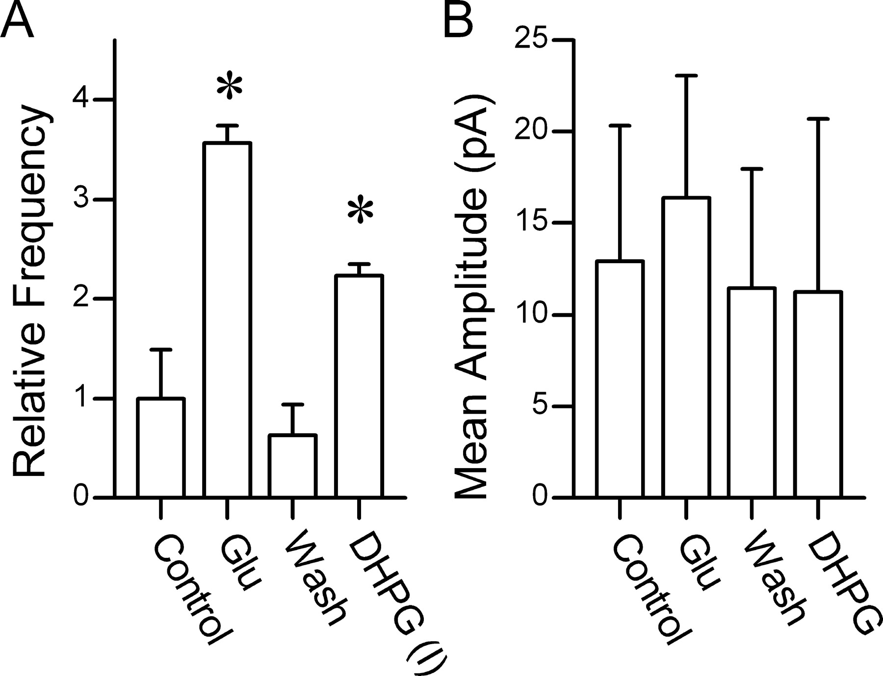
In increase-type NTS neurons (n = 3), group I mGluR agonist mimicked Glu on sIPSCs. Activation of group I mGluRs with Glu (1 mm) or with DHPG (10 μm) reversibly increased the frequency of sIPSCs (A). Neither Glu nor DHPG altered sIPSC amplitudes (B), supporting a selective presynaptic group I mGluR action on GABA terminals. All of the neurons completed the same drug protocol: Glu, wash, and DHPG. *Significant difference from control (p < 0.05).


