

Article Figures & Data
Figures
- Figure 1.
Western blot. A polyclonal antiserum generated against a recombinant protein corresponding to amino acids 309-499 of human cortactin detected two major protein bands, migrating at ∼80 and ∼85 kDa (left lane, cortex; middle lane, hippocampus; right lane, cerebellum). The molecular weights are in kilodaltons.
- Figure 2.
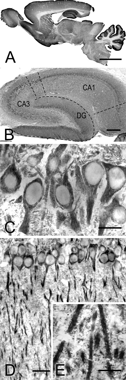
Immunoperoxidase staining for cortactin. A, Low-magnification view of a parasagittal section of adult rat brain. Staining is especially prominent in the cerebellar cortex and hippocampus and is also strong in other brain regions rich in dendritic spines. B, Micrograph showing the hippocampal formation. Staining is conspicuous in the molecular layer of the dentate gyrus (DG) and in the neuropil of CA1 (dashed lines mark borders of the main regions within the hippocampal formation). C, D, CA1 of hippocampus, showing stained somata and apical dendrites of pyramidal neurons. The detail in C reveals patchy staining in the somatodendritic compartment, surrounding immunonegative nuclei. E, High-magnification view of the stratum radiatum reveals intensely labeled dendrites; puncta in the neuropil may correspond to dendritic spines. Scale bars: A, 5 mm; B, 500 μm; C, E, 25 μm; D, 50 μm.
- Figure 3.
Subcellular distribution of cortactin in stratum radiatum of CA1. A, Confocal microscopy shows cortactin (red) and the presynaptic marker synaptophysin (green). The apposition of puncta immunopositive for the two markers (arrows in inset) suggests that cortactin may be postsynaptic. B, Double immunofluorescence with cortactin (red) and VGLUT1 (green). The pattern of punctate apposition suggests that cortactin is associated with glutamatergic synapses. C, Cortactin immunostaining was performed on material treated with the lipophilic dye DiO (green), which stains the plasma membrane of scattered pyramidal cells. Inset, Cortactin puncta (c1) can be seen within DiO-stained (c2) spines (merge; c3, arrowheads). D, Double fluorescence with cortactin (red) and phalloidin (green). Results suggest that cortactin localizes profiles also rich in actin. Inset, Arrowheads pointing to both cortactin-positive (d1) and phalloidin-positive (d2) sspines. Scale bars: A-C, 5 μm; D, 20 μm; insets, 5 μm.
- Figure 4.
Electron micrographs showing immunoperoxidase labeling of cortactin in CA1.A, Reaction product is visible in the cell bodies of two pyramidal neurons, associated with the endoplasmatic reticulum but sparing the Golgi complex. n, Nucleus. B-E, Reaction product accumulates in spines (asterisks) and dendrites (d) but not in presynaptic terminals. Arrowheads in D and E point to the region of immunostained spines, just beneath the PSD, that lacks immunostaining. Scale bars: A, 1 μm; B, 0.5 μm; C-E, 0.25 μm.
- Figure 5.
Preembedding immunogold labeling for cortactin in stratum radiatum of CA1. A, B, Electron micrographs show immunopositive dendrites (d). Most of the silver-enchanced gold particles visible in the apical dendrite in A are associated with microtubules. Gold particles in the dendrite of a possible interneuron (B) concentrate close to asymmetric synaptic appositions. C, Low-magnification view of synaptic neuropil. Arrows point to three immunolabeled spines; d marks an immunopositive dendritic shaft. D, E, High-magnification views showing immunopositive spines. Gold particles extend deep into the spine core (star in D indicates axon terminal with a gold particle). Scale bars: A-C, 0.5 μm; D, E, 0.2 μm.
- Figure 6.
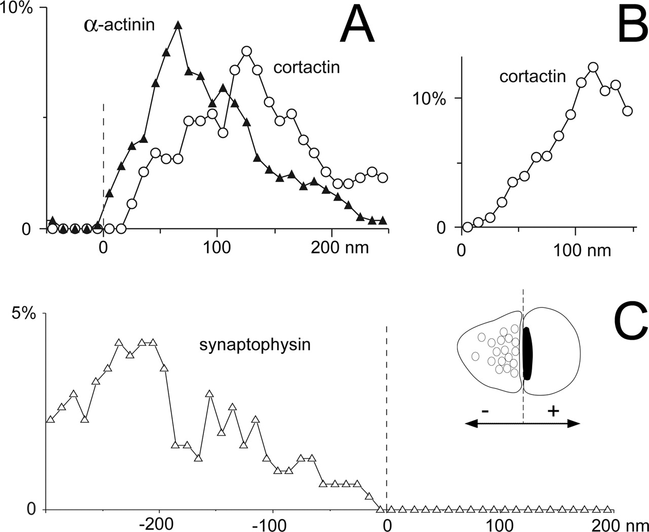
Axodendritic distribution of cortactin, α-actinin, and synaptophysin in axospinous synaptic profiles. A, Graph shows the axodendritic distribution of gold particles coding for cortactin (○) and α-actinin (▴) in the spine cytoplasm. The x-axis values represent the distance from the center of the gold particle to the synaptic membrane, along the axodendritic axis (negative values represent gold particles in the synaptic cleft or presynaptic terminal; see inset in C). The y-axis values represent the percentage of the total number of particles counted in each bin. Cortactin lies significantly farther from the PSD than α-actinin. B, Distances of gold particles coding for cortactin from the spine plasma membrane; cortactin concentrates in the spine core. Only particles lying within the spine were considered; the graph was truncated at 150 nm to permit inclusion of data from small spines. Labeling density was weighted to compensate for the reduction in perimeter, for particles lying deep in the cytoplasm. C, Graph shows that gold particles coding for synaptophysin (▵) are restricted to the presynaptic terminal. The dashed line represents the postsynaptic membrane, which corresponds to 0 in A and C (see inset for explanation). Random photos were taken of spines and terminals in the stratum radiatum of CA1 exhibiting a clear synapse and containing at least one gold particle. Data are from 65 profiles for α-actinin, 55 for cortactin, and 55 for synaptophysin. The bin width was 10 nm for A and B and 20 nm for C. Data were smoothed with a three-point weighted running average.


Tables
- Table 1.
Densities of gold particles coding for cortactin in different subcellular compartments
Density (particles/μm2) Nucleus 0.66 ± 0.05 (n = 11) Axon terminal 1.13 ± 0.45 (n = 30) Dendritic shaft 5.25 ± 0.50 (n = 14)* Spine 9.92 ± 1.25 (n = 44)* -
Randomly selected profiles identifiable as axon terminals, dendritic shafts, or spines were included in the analysis, regardless of whether they were immunopositive. All data were collected from CA1 stratum radiatum, 4.0 mm caudal to bregma. To assess noise, nuclear profiles from the stratum pyramidale were also examined. Only dendritic shafts and spines showed significant differences from the nuclear background. *p < 0.001; two-sided t test (mean ± SE; n = number of profiles).
-


