

Article Figures & Data
Figures
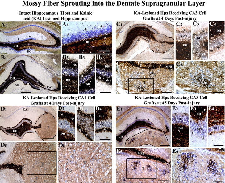
- Figure 1.
Extent of the mossy fiber sprouting in the dentate gyrus of control, lesion-only, and lesioned, grafted animals. A1, An example of the intact control hippocampus. A2 is an enlarged view of the upper blade of the dentate gyrus from A1 and demonstrates the absence of aberrant mossy fiber sprouting in the DSGL (black asterisks). B1, An example of the lesion-only hippocampus at 12 months after injury. B2–B4 are regions of the upper blade, the crest, and the lower blade of the dentate gyrus from B1 and demonstrate very robust aberrant mossy fiber sprouting into the DSGL (white asterisks). C1, An example of the lesioned hippocampus receiving CA3 cell grafts at 4 d after injury and analyzed at 1 year after grafting. The transplant (boundaries depicted by interrupted lines) is located in the CA3 region. C2–C4 are regions from the upper blade, the crest, and the lower blade of the dentate gyrus from C1 and show minimal aberrant mossy fiber sprouting into the DSGL (black asterisks). C5 illustrates the innervation of the transplant by host mossy fibers. C6 is an enlarged view of the boxed region of the transplant in C5 showing clusters of CA3 pyramidal neurons surrounded by mossy fiber terminals. D1, An example of the lesioned hippocampus receiving CA1 cell grafts at 4 d after injury and analyzed at 1 year after grafting. The graft (boundaries depicted by interrupted lines) is located in the CA3 region. D2–D4 are regions from the upper blade, the crest, and the lower blade of the dentate gyrus from D1 and show robust mossy fiber sprouting into the DSGL (white asterisks). D5 and D6 (enlarged view of the boxed region in D5) illustrate the absence of mossy fibers in regions of the CA1 transplant. E1, An example of the lesioned hippocampus receiving CA3 cell grafts at 45 d after injury and analyzed at 1 year after grafting. The graft (the boundaries depicted by interrupted lines) is located just below the CA3 region. E2–E4 are regions from the upper blade, the crest, and the lower blade of the dentate gyrus from E1 and demonstrate minimal aberrant mossy fiber sprouting into the DSGL (black asterisks). E5 shows the innervation of graft areas by host mossy fibers. E6 is an enlarged view of the boxed region in E5 showing clusters of CA3 pyramidal neurons surrounded by dense mossy fiber terminals. DH, Dentate hilus; GCL, granule cell layer; MFB, mossy fiber bundle; T, transplant. Scale bars: A1, B1, C1, D1, E1, 500 μm; A2, (in B4) B2–B4, (in C4) C2–C4, C6, (in D4) D2–D4, D6, (in E4) E2–E4, E6, 100 μm; C5, D5, E5, 200 μm.
- Figure 2.
Extent of the mossy fiber sprouting into the stratum oriens of CA3a subregion in lesion-only and lesioned, grafted animals. A1, An example from the intact control hippocampus. The mossy fiber terminals are sparsely distributed in the stratum oriens (SO) and stratum pyramidale (SP) under intact conditions. B1, An example from the lesion-only hippocampus. A robust sprouting of mossy fiber terminals is evident in both SO and SP. C1, An example from an injured hippocampus receiving fetal CA3 cell transplants. Note that the mossy fiber terminals are sparse in both SO and SP, akin to that in the intact hippocampus (A1). D1, An example from an injured hippocampus receiving fetal CA1 cell transplants. A robust sprouting of mossy fiber terminals is obvious in both SO and SP, similar to the condition observed in the lesion-only hippocampus (B1). MFB, Mossy fiber bundle; SR, stratum radiatum. Scale bar: (in D1) A1–D1, 100 μm.
- Figure 3.
Comparison of the width of sprouted area (A) and the density of sprouted terminals (B) in the DSGL between the intact hippocampus of control rats, the KA-lesioned hippocampus of lesion-only rats, and the KA-lesioned hippocampus of rats receiving CA3 or CA1 cell transplants. Note that the extent of the aberrant mossy fiber sprouting in animals receiving CA3 cell grafts is dramatically less than in both lesion-only animals and animals receiving CA1 cell grafts. The CA3 graft-mediated suppression of the aberrant sprouting is consistent in all regions of the dentate gyrus. LB, Lower blade; UB, upper blade. Error bars indicate SE. **p < 0.01; ***p < 0.001.
- Figure 4.
The pattern and extent of axon growth from a mouse CA3 cell graft into the host brain at 30 d after grafting, visualized with M6 immunostaining (A1). B1 shows the transplant indicated in A1 (by interrupted lines) with Nissl staining. Note the loss of CA3 pyramidal neurons (indicated by asterisks) in this hippocampus. B2 is a magnified view of the transplant region showing clusters of CA3 pyramidal neurons. The graft axon growth into the injured hippocampus is highly conspicuous in the strata oriens (SO) and radiatum (SR) of the CA1 subfield (A2), the inner molecular layer (IML) of the DG (A3), and the dentate hilus (DH) (A3). A sparse distribution of axons is also observed in the outer two-thirds of the molecular layer (A4). In addition, axons from CA3 cell grafts project into both ipsilateral and contralateral septal nuclei (A5, A6) and the CA3 region of the contralateral hippocampus (A7). GCL, Granule cell layer; T, transplant, SP, strata pyramidale. Scale bars: A1, B1, 500 μm; (in A6) A2–A7, B2, 100 μm.
- Figure 5.
The pattern and degree of axon growth from a mouse CA1 cell transplant placed into the injured CA3 region of the rat hippocampus (A1). B1 shows the transplant indicated in A1 (by interrupted lines) with Nissl staining. Note the loss of CA3 pyramidal neurons (indicated by asterisks) in the hippocampus. B2 is a magnified view of the transplant region showing the distribution of grafted CA1 cells. The graft axon growth occurs into the CA1 strata oriens (SO) and radiatum (SR) (A2); however, the extent of axon growth is considerably less than that observed with CA3 cell grafting (see Fig. 4). Note that the graft axon growth from CA1 cell graft into the inner molecular layer (IML) is conspicuously diminished (A3, A4) in comparison with the CA3 cell graft illustrated in Figure 4. A5 and A6 demonstrate axon growth from a CA1 cell graft into the subiculum (A5) and the ipsilateral septal nucleus (A6). DH, Dentate hilus; GCL, granule cell layer; T, transplant, SP, strata pyramidale. Scale bars: A1, B1, 500 μm; (in A6) A2–A6, B2, 100 μm.
- Figure 6.
Comparison of the overall graft axon growth from CA3 and CA1 cell grafts into the deafferented zones of the injured hippocampus and the septum. In comparison with the CA3 cell grafts, the overall axon growth from CA1 cell grafts is 43% less in the CA1 strata oriens and radiatum, 67% less in the dentate inner molecular layer, 48% less in the dentate hilus, and 44% less in the septum. DH, Dentate hilus; SO, strataoriens; SR, strata radiatum; IML, inner molecular layer. Error bars indicate SE. ***p < 0.001; ****p < 0.0001.


Tables
Transplant groups Location of transplants Transplant volume (mm3)a (mean ± SEM) CA3 transplants placed at 4 d after KA administration (n = 6) 50% of transplants in the CA3 region 0.57 ± 0.1 50% of transplants in the CA3 region and lateral ventricle CA3 transplants placed at 45 d after KA administration (n = 4) All transplants in the CA3 region and lateral ventricle 0.51 ± 0.05 CA1 transplants placed at 4 d after KA administration (n = 6) 50% of transplants in the CA3 region 0.48 ± 0.03 50% of transplants in the CA3 region and lateral ventricle -
↵ a The total volume of each graft was calculated using the Cavalieri method. Values represent means and SEs of four to six individual transplants in every group.
-


