

Article Figures & Data
Figures
- Figure 1.
Neuronal L-type channels activate over a wide voltage range and with fast kinetics. a, Normalized, averaged, current-voltage relationships for calcium currents recorded from tsA201 cells expressing CaV1.3 (▪), CaV1.2 (•), CaV2.2 (▴), and CaV3.1 (♦) subunit cDNAs. CaVα was coexpressed with CaVβ3 and CaVα2δ1 cDNAs. Average peak current amplitudes were the following: CaV1.3, -1.7 ± 0.4 nA (n = 12); CaV1.2, -0.5 ± 0.04 nA (n = 8); CaV2.2, -1.9 ± 0.3 nA (n = 6); CaV3.1, -1.3 ± 0.2 nA (n = 8). Activation midpoints (in millivolts) estimated from Boltzmann-GHK fits of data were the following: CaV1.3, -39.4 ± 0.6 mV (n = 8); CaV1.2, -17.6 ± 0.7 mV (n = 11); CaV2.2, -12.7 ± 0.8 mV (n = 8); and CaV3.1, -46.9 ± 1.2 mV (n = 8). b, Normalized representative current traces for CaV1.2, CaV1.3, CaV2.2 (gray trace), and CaV3.1 (gray trace) activated by step depolarization to activation midpoints: CaV1.3, -40 mV; CaV1.2, -15 mV; CaV2.2, -15 mV; and CaV3.1, -45 mV. c, Averaged macroscopic activation time constants (ln τact) at different test potentials estimated from exponential fits to currents recorded from cells expressing CaV1.2, CaV1.3, CaV2.2, and CaV3.1. Values are mean ± SE. The lines show regression fits to the data. Slopes and y intercepts are the following: CaV1.3, -0.02 ± 0.001 mV-1, 0.23 ± 0.02 (n = 9); CaV1.2, -0.02 ± 0.003 mV-1, 0.89 ± 0.09 (n = 11); CaV2.2, -0.02 ± 0.001 mV-1, -0.11 ± 0.01 (n = 7); and CaV3.1, -0.049 ± 5 × 10⁻⁴ mV-1, -0.63 ± 0.07 (n = 6). Student's t test on time constants at all test potentials: CaV1.3 to CaV2.2, p > 0.27; CaV1.3 to CaV1.2, p < 0.001.
- Figure 2.
CaV1.3 activates rapidly in response to action potential-like (AP) waveforms. Overlaid normalized (a) and non-normalized (b) representative current traces for CaV1.2, CaV1.3, and CaV2.2 channels in response to AP waveform are shown. The AP was recorded from a sympathetic neuron and was triggered by a brief current injection seen as a hump on the foot of the waveform. c, Average time delay between the peak of the action potential waveform (APPK) and peak inward current (IPK). Time delays were the following: CaV1.3, 0.65 ± 0.06 ms (n = 11); CaV1.2, 0.81 ± 0.05 ms (n = 8); and CaV2.2, 0.63 ± 0.05 ms (n = 8). CaV1.3 and CaV2.2 values were not significantly different; CaV1.2 and CaV1.3 values were significantly different (*p < 0.05). d, Averaged ratios of total charge moved during a single AP to peak inward current evoked from a 50 ms step depolarization. Average values were the following: CaV1.3, 0.84 ± 0.06 (n = 19); CaV1.2, 0.61 ± 0.05 (n = 8); and CaV2.2, 0.94 ± 0.11 (n = 6). Average AP peak currents for CaV1.2, CaV1.3, and CaV2.2 were 663 ± 137 pA (n = 8), 1282 ± 110 pA (n = 19), and 2898 ± 1273 pA (n = 10), respectively. IV, Current-voltage. Error bars represent SE.
- Figure 3.
Nifedipine inhibits CaV1.2 and CaV1.3 channels activated by step depolarization. The time courses of inhibition of CaV1.2 (a; •) and CaV1.3 (b; ▪) currents by 5 μm nifedipine (n = 7 and n = 8, respectively) are shown. Currents were evoked by 50 ms square pulse depolarizations to 0 mV (CaV1.2) and -20 mV (CaV1.3) from a holding potential of -80 mV. Depolarizations were applied once every 2 s. The bars show duration of nifedipine application. Insets, Representative CaV1.2 and CaV1.3 currents before (Con) and after exposure to nifedipine (Nif; 16 s time point). Calibration: 0.2 nA, 10 ms for CaV1.2 (a) and 0.5 nA, 10 ms for CaV1.3 (b). Error bars represent SE.
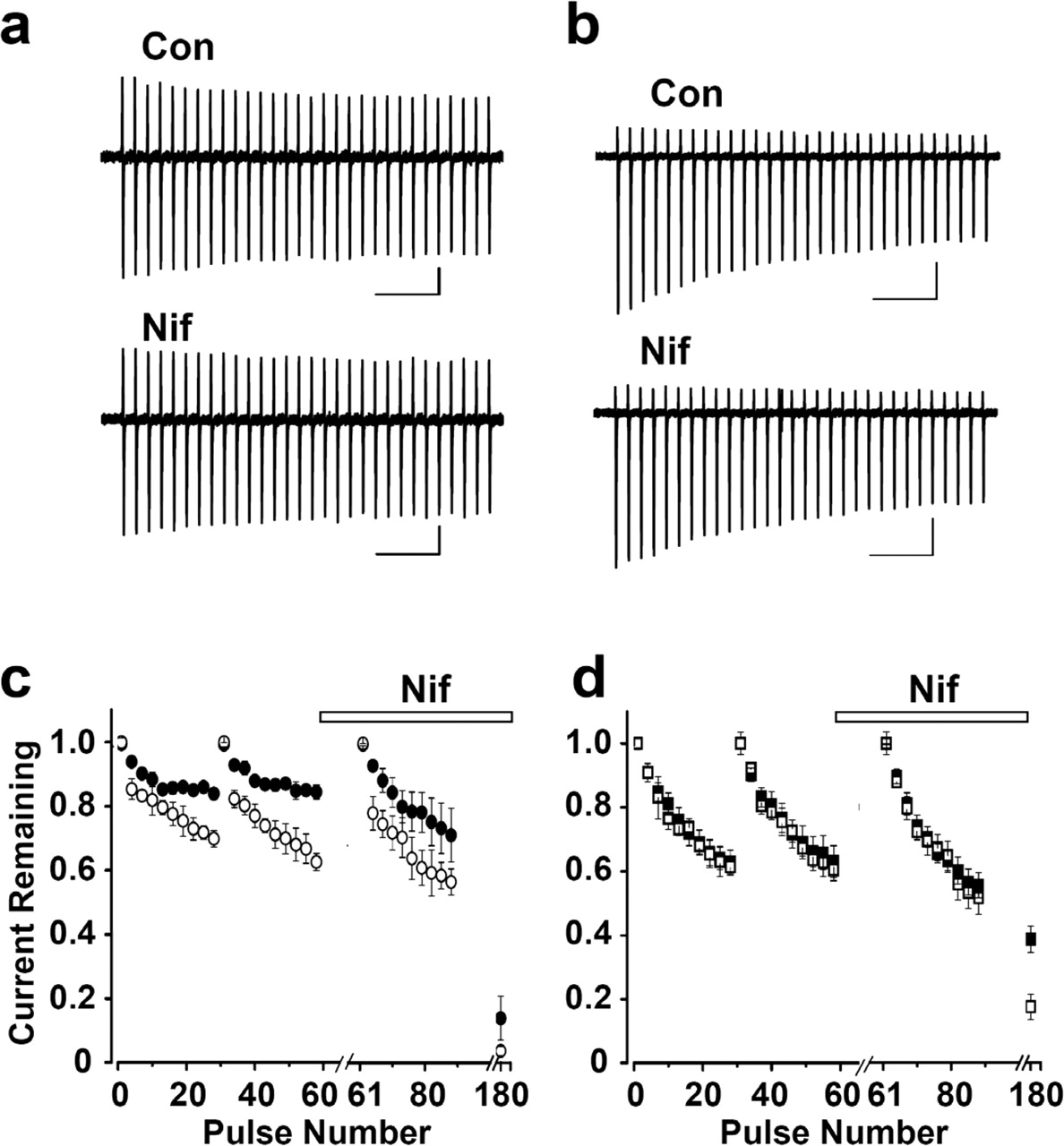
- Figure 4.
Nifedipine is weakly effective on CaV1.2 and CaV1.3 channels activated by action potential-like stimuli. Representative CaV1.2 (a) and CaV1.3 (b) currents evoked by a train of action potential waveforms, applied at 100 Hz from holding potentials of -80 mV, before (Con) and after a 10 s exposure to 5 μm nifedipine (Nif), are shown. Calibration: a, 0.2 nA, 50 ms; b, 0.5 nA, 50 ms. The action potential waveform used as command voltage was recorded from a sympathetic neuron and triggered by a brief current injection. Average peak current amplitudes for CaV1.2 (c; •, ○) and CaV1.3 (d; ▪, □) measured from currents induced by trains of 30 action potentials before and 10 s after exposure to 5 μm nifedipine (Nif) are shown. Currents were evoked from holding potentials of -80 mV (•, ▪) and -60 mV (○, □). Current amplitudes at the end of a series of four stimulus trains in the presence of nifedipine are shown for each series (180th pulse). Currents recovered completely after removal of nifedipine within three stimulus trains. Recovery was slowed approximately three fold when the membrane potential was depolarized to -60 mV. Error bars represent SE.


Additional Files
Supplemental data
Files in this Data Supplement:
- supplemental material - Supplemental Figure. CaV1.3, CaV3.1 and CaV2.2 channels can support calcium influx during action potential waveform stimulation. a, Normalized CaV1.3, CaV1.2 and CaV2.2 current traces evoked by action potential waveforms recorded from a cerebellar Purkinje neuron. b, Normalized CaV1.3, CaV3.1 and CaV2.2 current traces evoked by action potential waveform recorded from a sympathetic neuron. Vertical bar next to each waveform corresponds to 40 mV. The time course of CaV1.3 and CaV2.2 channel currents is similar. CaV3.1 channel currents activate with a slightly longer delay and continue to support calcium entry for several ms after the end of the stimulus. c, Average delay between the peak of the command voltage and the peak inward current for each current. Time intervals were for CaV3.1: 0.86 � 0.04 ms (n=8); CaV1.3: 0.62 � 0.01 ms (n=10); and CaV2.2: 0.63 � 0.05 ms (n=8). * denotes statistically significant difference between CaV3.1 and CaV1.3 (student�s paired t-test, p <_0.001.-- end="end" desc="desc" supplemental_fig_1.gif="supplemental_fig_1.gif" _--="_--">


