

Article Figures & Data
Figures
- Figure 1.
Activation of PKC accelerates recovery from action potential-induced increases in [Ca2+]i. [Ca2+]i was recorded from single rat DRG neurons using indo-1-based photometry as described in Materials and Methods. A, Brief trains of action potentials (3–5 s, 6–10 Hz) were delivered every 3 min as indicated by the filled triangles, and 0.5 μm PDBu was applied to the bath during the time indicated by the horizontal bar. To maintain comparable Ca2+ loads throughout the experiment, stimulation frequency in the presence of PDBu was increased by 20%. B, Representative [Ca2+]i responses in the absence and presence of PDBu, indicated by arrows in A, were superimposed on an expanded timescale. C, Rate constants describing the recovery kinetics for individual responses from experiments such as the one shown in A were normalized to the first response and plotted versus time. PDBu was applied after the fourth stimulus. Experiments were performed in the absence (filled circles; n = 11) and presence (open circles; n = 5) of 5μm GF109203x (GF) applied at time 0. Data points are means ± SEM.
- Figure 2.
PDBu stimulates SERCA-mediated recovery from action potential-induced increases in [Ca2+]i. [Ca2+]i was recorded from single rat DRG neurons using indo-1-based photometry. A–D, DRG neurons were cotransfected with plasmids containing an EGFP reporter construct and an antisense PMCA4 cDNA (AS4). Recordings are from EGFP-positive cells. A, Brief trains of action potentials (3–5s, 6–10 Hz) were delivered every 4 min to an AS4-expressing cell as indicated by the filled triangles. PDBu at 0.5 μm was applied to the bath during the time indicated by the horizontal bar. B, Representative [Ca2+]i responses in the absence and presence of PDBu, indicated by arrows in A, were superimposed on an expanded timescale. C, Rate constants describing the recovery kinetics for individual responses from experiments such as the one shown in A were normalized to the first response and plotted versus time. PDBu was applied after the fourth stimulus. Experiments were performed in the absence (open circles; n = 5) and presence (filled circles; n = 11) of 5 μm CPA applied 30 min before beginning the recording. Data points are means ± SEM. *p < 0.05 relative to the same time point when CPA was not added, one-way ANOVA with Bonferroni's post hoc test. Results in the presence of CPA are replotted from Usachev et al. (2002). D, Bar graph summarizes results of various combinations of SERCA and PMCA4 block on the PDBu effect. *p < 0.05, relative to untreated control (no AS4 and no CPA); #p < 0.05 relative to cells expressing AS4 (no CPA); Student's t test.
- Figure 3.
Direct recording of [Ca2+]ER to measure SERCA function. [Ca2+] in the lumen of the ER was recorded using Mag-indo-1, and [Ca2+]i was recorded using indo-1. A, Representative [Ca2+]i (top trace) and [Ca2+]ER (bottom trace) recordings obtained from two different cells are plotted on the same timescale. Depleting the Ca2+ stores by blocking SERCA with 5μm CPA in Ca2+-free buffer (indicated by horizontal bars) produced a leak of Ca2+ from the ER, resulting in a decrease in [Ca2+]ER and a corresponding increase in [Ca2+]i. Return of Ca2+ to the media allowed the ER to refill with Ca2+. B, Representative [Ca2+]ER recording shows that the refilling process could be evoked repeatedly. C, If CPA was not removed before the second application of Ca2+ to the bath, refilling was completely blocked. D, The two recovery phases from B were superimposed. The [Ca2+]ER recovery was well described by a single-exponential equation (heavy black line).
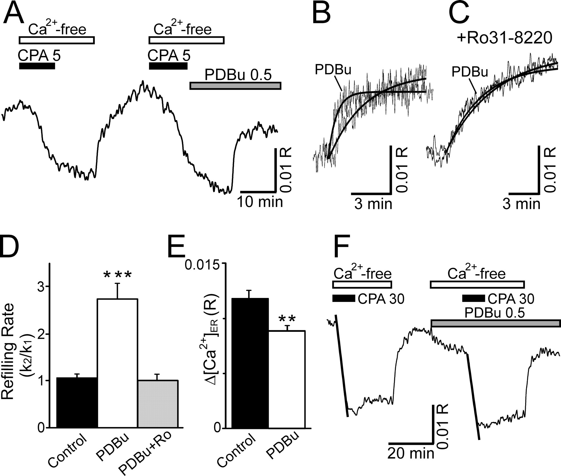
- Figure 4.
Activation of PKC accelerates Ca2+ uptake into the ER. [Ca2+] in the lumen of the ER was recorded using Mag-indo-1-based photometry. A, Representative recording shows the effects of PDBu on the rate of [Ca2+]ER recovery using the refilling protocol described in Figure 3B. CPA at 5μm, Ca2+-free buffer, and 0.5μm PDBu were applied to the bath by superfusion at the times indicated by the horizontal bars. B, [Ca2+]ER recordings from A were superimposed on an expanded timescale. Heavy lines show fitted single-exponential curves. C, [Ca2+]ER recordings from an experiment similar to that in A, except that the PKC antagonist Ro31–8220 (2μm) was applied 10 min before the application of PDBu. The initial control response and the response in the presence of Ro31-8220 plus PDBu were superimposed on an expanded time-scale. Heavy lines show fitted single-exponential curves. D, Bar graph summarizes the effects of PKC activation on refilling kinetics. ***p < 0.001 relative to untreated control, Student's t test. E, Bar graph illustrates difference in completeness of refilling in the absence (control) and presence (0.5 μm) of PDBu. Δ[Ca2+]ER was defined as the difference between the stable asymptote reached after refilling and the [Ca2+] in depleted stores. **p < 0.01 relative to untreated control in same cell, Student's paired t test. F, Representative recording shows the rapid depletion of Ca2+ stores by the application of 30μm CPA in Ca2+-free media. The rate of Ca2+ leaking from the store was quantified by measuring the slope of a linear regression fit to the first 5 min of the [Ca2+]ER trace in CPA (fits are indicated by heavy lines).
- Figure 5.
PKC activation reduces ER refilling by stimulating PMCA4. A–C, [Ca2+]i was recorded using indo-1-based photometry. CPA (5 μm) was applied in Ca2+-free media at the times indicated by the arrows. Because the refilling status of the Ca2+ stores varied at the start of the recording, the [Ca2+]i transients shown in A–C are the second and third CPA-evoked responses and correspond to the refilling status of the store after the first and second refilling under controlled conditions. The duration of the treatments are identical to those in Figures 3 (left column) and 4 (right column). Representative traces from nontransfected (Naive) cells (A), cells transfected with EGFP (B), and cells transfected with EGFP plus AS4 (C) are shown. The second and third CPA-evoked responses were superimposed for untreated (left column) and PDBu-treated (right column) cells. D, Bar graph summarizes the changes in the amplitude of the CPA-evoked response in the presence and absence of PDBu. *p < 0.05, PDBu compared with untreated, Student's t test.
- Figure 6.
Activation of PKC accelerates the rate of ER refilling with Ca2+ without reducing capacity in cells lacking the PKC-sensitive PMCA isoform. [Ca2+]ER was recorded using Magindo-1-based photometry. DRG neurons were cotransfected with plasmids containing an EGFP reporter construct and an antisense PMCA4 cDNA. Recordings are from EGFP-positive cells. A, Representative traces show [Ca2+]ER in AS4-treated DRG neuron during repeated application of the refilling protocol described in Figure 3. The rate and degree of refilling were reproducible. B, [Ca2+]ER traces recorded from AS4-treated DRG neurons before and after PDBu application were superimposed. PDBu accelerated the rate of refilling and increased the steady-state [Ca2+]ER.


Additional Files
Supplemental data
Files in this Data Supplement:
- supplemental material - Supplement 1. Combined [Ca2+]i and [Ca2+]ER recordings from DRG neurons. Cytosolic and ER luminal Ca2+ concentrations were simultaneously monitored using fura-2 (Kd ≈ 0.22 μM) and Mag-fluo-4 (Kd ≈ 22 μM), respectively. Cells were initially loaded with Mag-fluo-4/AM at a concentration of 7.5 μM in 0.02 % Pluronic F-127 at 37oC for 45 min and then washed 3 times in dye-free HEPES-buffered Hank�s solution (HHSS; see Materials and Methods); cells were further loaded with fura-2/AM (5 μM; 30 min at 22oC in 0.02 % Pluronic F-127), followed by a 30 min wash in dye-free HHSS buffer. Cells were imaged with a CCD-camera based microfluorimetry system (Till Photonics, Germany) mounted on an inverted microscope (Olympus IX-71) equipped with a 40x (oil immersion; NA=1.35) objective. Fura-2 and Mag-fluo-4 were alternately excited at 340/380 and 475 nm, respectively, and fluorescence measured at 525 (40) nm. Images were collected at each of the excitation wavelengths every 2 s (A) or every 5 s (B and C). The fluorescence ratio (R=F340/F380) of fura-2 was converted to [Ca2+]i according to the formula: [Ca2+]i = Kdβ(R-Rmin)/(Rmax-R) (Grynkiewicz et al., 1985). The dissociation constant (Kd) used for fura-2 was 224 nM. Ionomycin was used to determine the calibration constants, as described in Materials and Methods: Rmin=0.17, Rmax=2.71, β=5.6. Changes in [Ca2+]ER were expressed as ΔF/F=(F-Fo)/Fo, where F was the current Mag-fluo-4 fluorescence value and Fo was the fluorescence value before treatments. Fluorescence was always corrected for background. A, Caffeine (10 mM) induced Ca2+ release from ryanodine-sensitive stores. This led to a rapid rise in [Ca2+]i and a complementary decrease in [Ca2+]ER. After reaching its peak, [Ca2+]i began to recover despite the presence of caffeine and continuous Ca2+ flux from the stores. This is likely explained by the competition between Ca2+ release and Ca2+ clearance processes. As the stores deplete, the rate of Ca2+ release subsides; the Ca2+ clearance mechanisms turned on by the [Ca2+]i elevation begin to dominate leading to the recovery of [Ca2+]i. Indeed, the time course of [Ca2+]i changes correlated with the rate of Ca2+ mobilization from the stores, d[Ca2+]ER/dt (blue). A portion of the d[Ca2+]ER/dt trace during the refilling phase was truncated for clarity. B, A representative combined recording of [Ca2+]i (black) and [Ca2+]ER (green) during the standard refilling protocol used in the study. The stores were depleted by 5 μM CPA in Ca2+-free media. The maximal rate of CPA-induced Ca2+ mobilization from the stores was 15-20% of that induced by caffeine. The refilling process was initiated by switching to control HHSS buffer containing 1.3 mM Ca2+. The rate of refilling during the first and the second trial was highly reproducible: k1=0.29�0.03 min-1 and k2=0.28�0.03 min-1 (n=12). The d[Ca2+]ER/dt trace (blue) has been truncated during the refilling phase for clarity. C, CPA (5 μM) blocked the refilling phase, consistent with Ca2+ reuptake via SERCA. The same paired refilling protocol used in B was used here, except CPA remained in the solution during the second trial. The recording is representative of 5 experiments. Addition of extracellular Ca2+ during the second trial induced a rapid elevation of [Ca2+]i, whereas the stores remained depleted in the presence of CPA. [Ca2+]ER completely recovered after CPA was washed out. The recovery phase was slowed relative to the control refilling process because of the relatively slow (5-10 min) wash out of CPA. Note that during the control refilling protocol, CPA was washed from the bath for 10 min prior to initiating refilling. The [Ca2+]i overshoot above baseline likely resulted from capacitative Ca2+ influx (Usachev and Thayer, 1999) because the stores remained depleted in the presence of CPA. References Grynkiewicz G, Poenie M, Tsien RY (1985) A new generation of Ca2+ indicators with greatly improved fluorescence properties. J Biol Chem 260:3440-3450. Usachev YM, Thayer SA (1999) Ca2+ influx in resting rat sensory neurones that regulates and is regulated by ryanodine-sensitive Ca2+ stores. J Physiol 519:115-130.


