

In the article “β-Secretase-Cleaved Amyloid Precursor Protein Accumulates at Actin Inclusions Induced in Neurons by Stress or Amyloid: A Feedforward Mechanism for Alzheimer's Disease,” by Michael T. Maloney, Laurie S. Minamide, Andrew W. Kinley, Judith A. Boyle, and James R. Bamburg, which appeared on pages 11313–11321 of the December 7, 2005 issue, the most recent version of Figure 1 was not used. The correct version of this figure is printed here.
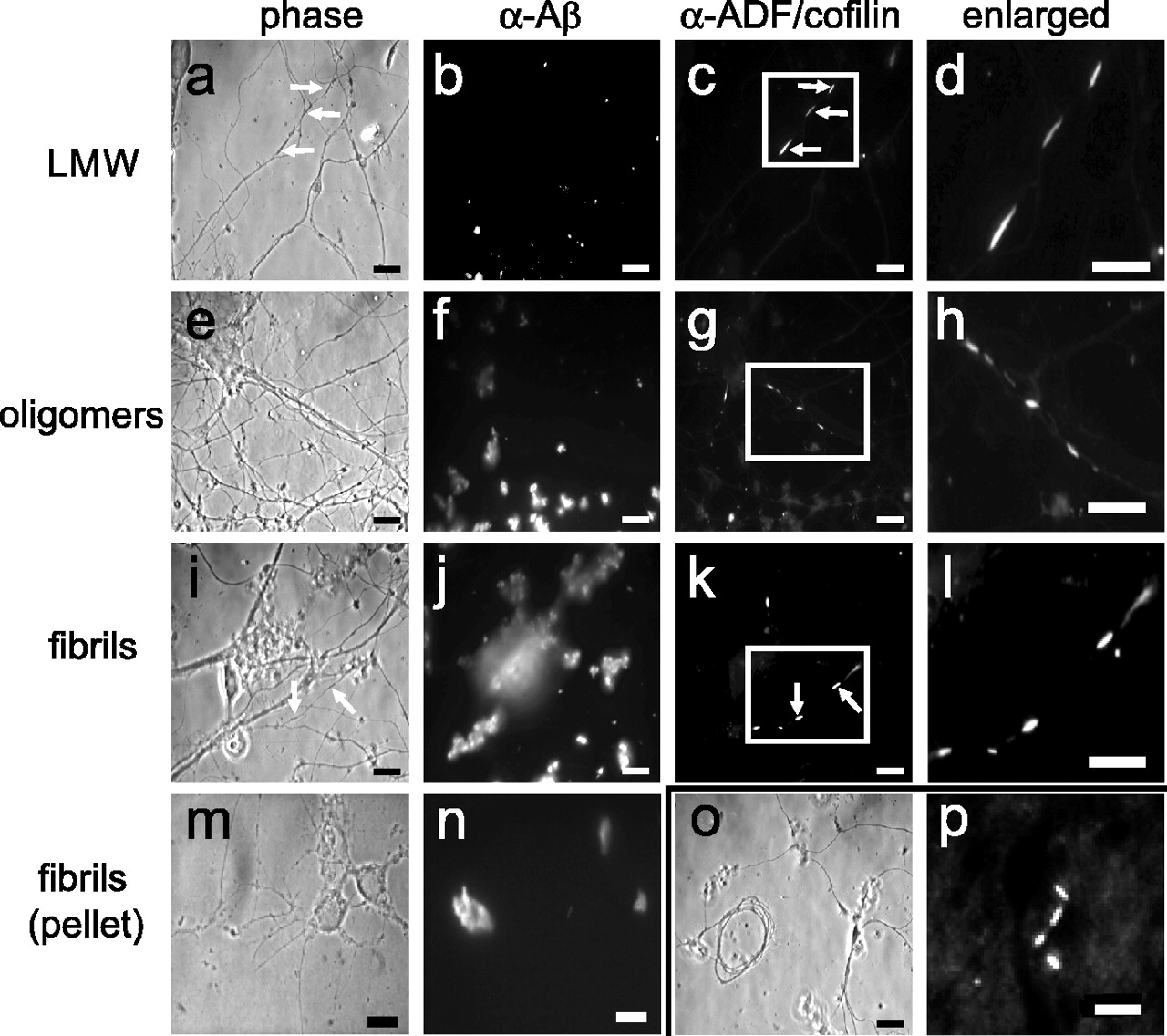
Aβ peptides induce rods in cultured neurons and in brains of transgenic mice. a–l, Rods in hippocampal neurons treated with 1μm Aβ1–42 LMW, large oligomer (oligomers) or fibril/insoluble aggregate (fibrils) preparations. The arrows in a, c, i, and k show the location of a rod in the phase and fluorescence image pairs. m, n, Neurons treated with the insoluble aggregates from the fibril preparations. Aβ immunofluorescence is more compact compared with whole fibril/insoluble aggregate preparations. o, Example of a dystrophic neurite commonly found in fibril/insoluble aggregate-treated cultures. p, Immunofluorescent rods stained for ADF/cofilin in the posterior cortex of a section of brain from a 4-month-old Tg2576 mouse. Scale bars:a–o, 10 μm; p, 5 μm.


