

Article Figures & Data
Figures
- Figure 1.
Structure of the human β4a A domain. A, Alignment of human and zebrafish β4a and human β3 N-terminal A domains. Identical residues and conservative substitutions are shaded blue and red, respectively. β4a-A predicted secondary structure shown as tubes (α helix) and arrows (β strand) (Protein Sequence Analysis server, http://bmerc-www.bu.edu/psa) (White et al., 1994). Numbers refer to β4a-A residues. B, Circular dichroism spectra of the purified β4a-A at pH 5 (⋄), 6 (□), and 7 (○). C, Solution structure of the β4a-A determined to a root mean square deviation of 0.73 Å. The structure contains two α helices (α1, α2) and two β strands (β1, β2) connected by three loops (L1–L3). The structure has been deposited in the Protein Data Bank (code 2D46). D, Combined β4a-A solution structure and β2a B–D crystal structure (Van Petegem et al., 2004). The figure is labeled as follows: A, A domain; SH3, Src homology 3, B domain; Linker, sequence connecting SH3 and GK domains, C domain; GK, guanylate kinase, D domain; AID, Ca2+ channel α1 subunit interaction domain; C-terminal Domain, beginning of the C-terminal domain (structure unknown). The software program “O” (version 8.0.11) was used to perform the merger of the two structures (Jones et al., 1991). PyMOL molecular graphics software version 0.96 was used to generate structure figures.
- Figure 2.
Electrophysiological comparison of full-length β4a and BCDE. A, Cav2.1 current size as a function of time after injection of no β (X), β4a (filled symbols), or BCDE (open symbols) cRNA. Inset, RNA gel showing α1, α2/δ mixtures with increasing amounts of β4 constructs (β4 to α1 ratios: squares, 1:1 β4 molar ratio relative to α1; diamonds, 4:1 ratio; circles, 12:1 ratio). B, Representative Cav2.1 current traces in the absence (dashed line on top of gray trace) and presence of β4a (black traces) or BCDE (gray traces). Effects of increasing injected amounts (in nanograms) of β4 constructs on rate of inactivation are indicated by arrows. C, Effects of increasing injected amounts (in nanograms) of β4a (top) and BCDE (bottom) on Cav2.1 activation. D, Effects of increasing amounts (in nanograms) of β4a (top) and BCDE (bottom) on Cav2.1 closed-state inactivation. Symbols represent mean values, and lines serve to connect data points.
- Figure 3.
Distribution of β subunit subtypes in mouse cerebellum. A, Splice variant-specific antibodies were created against unique β4a and β4b sequences (highlighted). B, Western blot showing that β4a-, β4b-, and β3-specific antibodies recognize 52–55 kDa proteins in sucrose step gradient-purified mouse cerebellar membranes. Numbers to the left indicate molecular mass in kilodaltons. C, Purkinje cells labeled with anti-calbindin antibody (scale bar, 20 μm) serve to distinguish the molecular (ML), Purkinje cell (PCL), and granule cell (GCL) layers (for orientation in D–G). D, E, Punctate labeling of the β4a subunit in the molecular layer of the cerebellum: D, confocal microscopy, 1-μm-thick image; E, light microscopy, 50-μm-thick section. F, The β4b subunit is expressed in Purkinje cell bodies and Bergman glia. G, The β3 subunit is expressed in basket cell structures surrounding Purkinje cell bodies and in granule cell layer puncta. Inset, Higher-power image (scale bar, 10 μm) of basket cell labeling (supplemental Fig. 2, available at www.jneurosci.org as supplemental material).
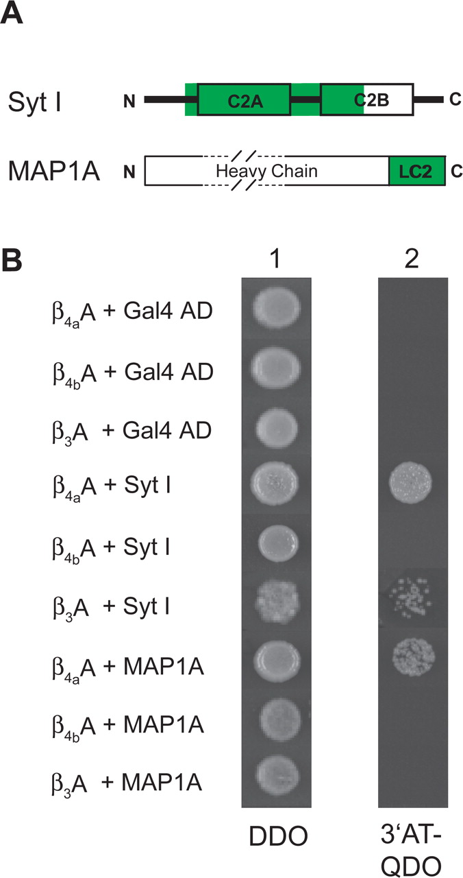
- Figure 4.
β4a-A protein–protein interactions. A, Yeast two-hybrid screening of a human cerebellar cDNA library identified Syt I and MAP1A as proteins that interact with the β4a-A. The green highlighted areas correspond to the protein regions encoded by the cDNAs isolated in the primary yeast two-hybrid screens. The Syt I cDNA codes for amino acids 95–337 of the full-length Syt I protein; the MAP1A cDNA codes for amino acids 2508–2775, corresponding to the complete LC2 domain of MAP1A. B, Characterization of β4a-A, β4b-A, and β3-A interactions with Syt I and MAP1A. Bait–target pairs were tested under two conditions: column 1, plates lacking leucine and tryptophan (DDO plates); column 2, plates lacking leucine, tryptophan, adenine, and histidine and supplemented with 30 mm 3′-AT. Growth of yeast colonies as shown in column 2 are indicative of interactions of β4a-A and β3-A with Syt I (rows 4 and 6, respectively) and interaction of β4a-A with MAP1A (row 7).
- Figure 5.
Synaptotagmin I labeling of cerebellar cortex and β4a-A interaction with 6His–C2AC2B in pull-down assays. A, Immunolabeling of mouse cerebellar cortex with anti-synaptotagmin antibody (scale bar, 20 μm). ML, Molecular layer; PCL, Purkinje cell layer; GCL, granule cell layer. B, CD spectrum of recombinant purified 6His–C2AC2B indicative of proper folding into a β-sheet structure. C, Pull-down assays analyzed by SDS-PAGE, using Coomassie blue-stained 5–15% polyacrylamide gels. Equal amounts (50 nmol each) of β4a-A and β4b-A (lanes 1, 2) were incubated with 6His–C2AC2B–Ni2+-NTA beads and washed until neither β4a-A or β4b-A appeared in the flow through (lanes 3, 4). Bound 6His–C2AC2B and any associated protein were eluted with 1 m imidazole. Lanes 5 and 6 show that β4a-A, but not β4b-A, associates with 6His–C2AC2B in the absence of added Ca2+. The interaction of β4a-A with 6His–C2AC2B does not occur when 10 mm Ca2+ is added before the wash step (PreW, lane 7). Addition of 10 mm Ca2+ to the assay after the wash step but before the imidazole elution competes off β4a-A (PostW, lane 8). Subsequent addition of 1 m imidazole elutes off 6His–C2AC2B (Imid, lane 9).
- Figure 6.
Relative surface potential modeling of the β4a A domain. Three surface views of the β4a-A with blue, red, and white surfaces corresponding to positive, negative, and neutral charge, respectively. PyMOL molecular graphics software, version 0.97, was used to generate the figures. A, Side view with the C terminus positioned to the right, oriented approximately as shown in Figure 1D. Examples of negatively and positively charged residues, E34 and R36, respectively, are identified for orientation. B, The β4a-A is rotated 180° in the horizontal plane relative to A. The N terminus is projecting outward, perpendicular to the plane of the page. Note that the N terminus contains glycine, serine, and histidine residues remaining from the pET-15b thrombin cleavage site. Two negatively charged residues, E34 and E37, are identified for orientation. C, The β4a-A is rotated 90° clockwise in the horizontal plane relative to B. This view could represent the protein interaction surface as seen from inside the cell. A single residue, R24, from loop 2, and another, R36, from loop 3, contribute positively charged surfaces. Three residues from loop 3, E34, E37 and E42, contribute negatively charged surfaces. Potential hydrophobic surfaces are formed by four loop 3 nonpolar residues, V30, L32, A38, and I39.
- Figure 7.
Alternative splicing of Ca2+ channel β subunit A domains. Splicing patterns of exons identified to date that code for the A domains of β subunits 1–4 (for review, see Takahashi et al., 2003; Foell et al., 2004). The diagram indicates that multiple A domain splicing variants have been identified for β subunits: β1, one variant; β2, five variants; β3, one variant; and β4, two variants. Exons are numbered relative to the positions of the seven exons on the β2 gene (for additional explanation of the relatedness of each exon, see Discussion). The first three amino acids (exons 1, 2, 4, 5, 6, and 7) or last two amino acids (exon 3) of individual exons are specified. The coding sequences for the SH3 (B) domain begin downstream of exon 7.


Additional Files
Supplemental data
Files in this Data Supplement:
- supplemental material - Figure S1. Testing for specificity of β subunit antibodies using purified recombinant A-domain proteins. A, Specificity of anti-β4a-A and anti-β4b-A affinity-purified polyclonal antibodies. Purified recombinant β4a-A and β4b-A were subjected to SDS-PAGE and either stained with Coomassie blue (Lanes 1 and 2 from left to right) or transferred to a nylon membrane for Western blot analysis (Lanes 3-6). Coomassie staining reveals that approximately equal amounts of β4a-A and β4b-A protein were loaded on to the gel. Western blot analysis demonstrates that the anti-β4a-A antibody (1:5000) labels only purified β4a-A protein (Lanes 3 and 4) and that the anti-β4b-A antibody (1:1000) labels only the β4b-A protein. Staining below each of the prominently labeled bands is indicative of some degradation of purified protein. B, The anti-β4a-A antibody does not cross-react with the 6His-β3 A-domain. Purified recombinant β4a-A and 6His-β3-A were subjected to SDS-PAGE and either stained with Coomassie blue (Lanes 1 and 2 from left to right) or transferred to a nylon membrane for Western blot analysis (Lanes 3-9). Coomassie staining reveals that approximately equal amounts of β4a-A and 6His-β3-A protein were present in the 1X stock solutions used to load the gel. Western blot analysis demonstrates that the anti-β4a-A antibody (1:1000) labels purified β4a-A protein (0.1X protein relative to stock solution, Lane 3), but does react with β3-A protein loaded on to the gel at 0.1X, 0.5X, and 1X concentrations relative to the stock solution (Lanes 4-6, respectively). Western blot analysis using an anti-6His antibody (1:1000) demonstrates that the 6His-β3-A protein was successfully transferred to the nylon membrane, with staining evident at 0.5X and 1X concentrations of 6His-β3-A (Lanes 7-9). Coomassie staining of the polyacrylamide gel after protein transfer verified that both β4a-A and 6His-β3-A proteins transferred with equal efficacy (data not shown). Numbers to the left of each panel indicate molecular mass in kDa.
- supplemental material - Figure S2. Specificity of β subunit antibody labeling in cerebellar cortex. As a partial test to determine whether β subunit antibody labeling in cerebellar cortex is due to specific binding to β subunit A-domains, anti-β4a-A and anti-β4b-A affinity-purified polyclonal antibodies were pre-incubated with purified recombinant β4a-A and β4b-A protein (1:1 mass ratio) prior to standard immunolabeling procedures. Anti-β3 antibody was pre-incubated with the β3 subunit C-terminal peptide antigen that was included with the purchase of the antibody (Chemicon). In each case (β4a, top two panels; β4b, middle two panels; and β3, lower two panels), pre-absorption with protein antigen eliminated specific labeling; however, as can be seen in the right-hand panels, light shadows of Purkinje cell bodies were still visible.


