

Article Figures & Data
Figures
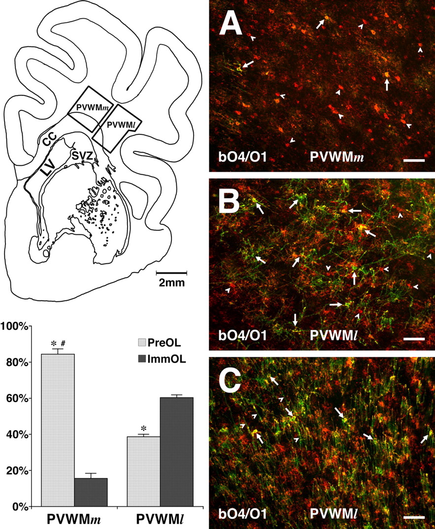
- Figure 1.
Spatial heterogeneity of OL maturation in 0.65 gestation ovine brain. Top left, A representative frontal section from the ovine brain shows schematically the approximate boundaries of the medial (PVWMm) and lateral (PVWMl) regions of the frontal PVWM. See Materials and Methods (Measurement of regional cerebral blood flow) for the definition of the boundaries of the PVWM analyzed for cell counts and blood flow measurements. A–C show the differences in OL maturation between the PVWMm and the PVWMl by immunohistochemical double labeling with bO4 (red) and O1 (green) antibodies. A, The PVWMm contains few immature OLs (arrows) (O4+, O1+) and numerous single-labeled preOLs (arrowheads) (O4+, O1−). B, The PVWMl contains more differentiated cells with larger, more developed processes. The majority are immature OLs (arrows), and the minority are preOLs (red; arrowheads). C, Consistent with the more mature state of the white matter in the PVWMl, numerous O1-labeled myelinated axons (arrowheads) are also seen in association with the immature OLs (arrows) and the smaller population of preOLs (red). The graph (bottom left) shows the relative percentages of preOLs and immature OLs in the PVWMm and the PVWMl. PreOLs predominate in the PVWMm, composed of >84% of the total OLs, and immature OLs composed the remaining 16%. In the PVWMl, immature OLs are markedly increased and comprise ∼62% of total OLs (*p < 0.001 preOLs vs immature OLs for PVWMm and PVWMl; #p < 0.001 preOLs in PVWMm vs PVWMl; unpaired two-tailed t tests). CC, Corpus callosum; LV, lateral ventricle. Scale bars: A–C, 200 μm.
- Figure 2.
Quantification of fetal cerebral blood flow in situ under conditions of basal cerebral blood flow and global ischemia. A, Nissl-stained coronal frontal section that corresponds to the ICM image (middle) and convolved image of blood flow (right). B–E, The center image represents a 3D surface reconstruction (Amira; TGS) of magnetic resonance images of a 0.65 gestation ovine control brain that indicates the frontal and parietal levels at which the blood flow analysis was done in B–E. B, C, Representative pseudocolor basal flow images show higher blood flow (light blue) in cortical gray matter areas (arrowheads) and lower flow (dark blue) in the PVWM (arrows). The pons (C, double arrowheads) had higher basal flow rates than any region of the cerebrum. D, E, During global ischemia, blood flow was dramatically reduced in all regions and approached zero (dark blue/black) in the PVWM (arrows). F, G, Blood flow measured by digital dissection in the frontal (F) and parietal (G) cortex (CTX) and PVWM during basal blow and ischemia. *p < 0.001, basal flow versus ischemia; #p < 0.001, basal flow CTX versus basal flow PVWM; † p < 0.001, ischemia CTX versus ischemia PVWM; paired two-tailed t tests.
- Figure 3.
Histopathological features of PVWM lesions generated after global ischemia of 37 or 45 min duration, as visualized with isolectin B4. A, After 37 min of ischemia, activated microglial were localized to a lesion at the apex of the lateral ventricle (LV; arrows). The typical morphology of the activated microglia is shown in the inset at the top right. The inset at the bottom left shows the typical morphology of ramified microglia and resting macrophages in control PVWM. A focal collection of reactive macrophages/microglia localized to the lateral body of the corpus callosum (arrowheads) is shown. B, After 45 min of ischemia, activated microglia were diffusely localized to a lesion at the external angle of the lateral ventricle (arrows). Focal collections of reactive phagocytic macrophages/microglia localized to medial and lateral regions in the PVWM (arrowheads). The typical morphology of the reactive phagocytic macrophages is shown in the inset at the bottom right. Note the presence of a focal lesion in the caudate nucleus (CN; arrowheads). C, D, After 45 min of ischemia, large lesions were also generated in the SWM (C, arrowheads) and in the cerebral cortex (CTX) (D, arrowheads). A detail of the cortical lesion is shown in the inset. Scale bars: A, B, 500 μm; C, D, 300 μm; A, B, insets, 30 μm.
- Figure 4.
Regional distribution of TUNEL-labeled nuclei in cerebral lesions at 24 h after 30, 37, or 45 min of global ischemia. A and E show major anatomical structures at frontal and parietal levels of the PVWM. B–D and F–H show composite pseudocolor TUNEL maps of the distribution of TUNEL-labeled nuclei plotted in two sections from each of four animals. The pseudocolor probability scale (1–7) in H indicates the number of sections in which overlapping lesions were found for any given region. Note that at 30 and 37 min, cerebral injury was mostly restricted to cerebral white matter. However, at 45 min, injury to the cerebral cortex and basal ganglia greatly increased, but the SVZ and hippocampus remained relatively spared. I–L show representative examples of the magnitude and distribution of TUNEL-labeled nuclei in cerebral lesions after either 37 min (I–K) or 45 min (L) of ischemia. I, Large numbers of TUNEL-labeled nuclei were typically localized to the PVWM even after 37 min of ischemia. J, Within the SVZ, TUNEL-labeled nuclei typically localized to the dorsal extent of the SVZ (arrows) and to small extensions of the SVZ (arrowheads) into the adjacent caudate nucleus. K, Focal cortical lesions were uncommon after 37 min of ischemia. L, Large cortical lesions were common after 45 min of ischemia and contained a high density of TUNEL-labeled nuclei. CC, Corpus callosum; CN, caudate nucleus; CL, claustrum; EC, external capsule; HIP, hippocampus; IC, internal capsule; LV, lateral ventricle; PU, putamen. Scale bars: A–H, 2 mm; I–L, 200 μm.
- Figure 5.
Regional heterogeneity of cerebral injury at 24 h after cerebral ischemia of 30, 37, or 45 min duration relative to control. Cell degeneration in the PVWM was compared with that in the cortex (CTX), caudate nucleus (CN), and SVZ by quantification of TUNEL-labeled cells as a percentage of total cells in each region. *p < 0.05 (PVWM vs CTX; LSD test); # indicates not significant.
- Figure 6.
Differential vulnerability of glia and axons in PVWM lesions at 24 h after ischemia. Although astrocytes and microglia appeared mostly resistant to injury from prolonged ischemia (45 min; A–C), numerous degenerating OL precursors were detected after more mild ischemia (37 min; D–F). Axons were also mostly resistant to prolonged ischemia (G–L). A, A lesion with numerous astrocytes visualized by staining for GFAP (red; arrows) that did not overlap with TUNEL-labeled nuclei (green). B, A higher-power image shows intact-appearing vimentin-labeled astrocytes (red) that did not colocalize with TUNEL-labeled nuclei (green). C, Most isolectin B4-labeled microglia/macrophages (red) were TUNEL negative (arrowheads), but an occasional shrunken TUNEL-labeled cell appeared to be degenerating (arrow). D, A low-power image in which many degenerating O4+ cells (red) double labeled for TUNEL (green; arrows). D, Inset, High-power image shows several degenerating O4+ cells (arrows) with pyknotic Hoechst 33324-labeled nuclei (blue) that also labeled with TUNEL. Note the intact cells with normal-appearing nuclei (arrowheads). E, Low-power image in which many degenerating O4+ cells (green) labeled for activated caspase-3 (red; arrows). E, Inset, A triple-labeled high-power image shows a degenerating O4+ cell (arrow) that labeled for activated caspase-3 (red) and had a pyknotic Hoechst-labeled nucleus (blue). An intact cell (arrowhead) did not label for activated caspase-3. F, A low-power image of O4+ cells (green) in which degenerating cells labeled with the fractin antibody (red). F, Inset, A degenerating cell that labeled with fractin (arrow). Intact cells had normal-appearing nuclei (arrowheads). G–I, Low-power images of SMI 312-labeled axons (green) and Ham 56-labeled microglia/macrophages (red) in the PVWM from control (G) and animals subjected to 37 min (H) or 45 min (I) of ischemia. Note the increased labeling for Ham 56 in the ischemic animals relative to control (insets) but the similar axonal staining pattern for all conditions. J, Apparent degenerating axons were rarely detected in the PVWM after prolonged ischemia (45 min). K, L, Higher-power images of the axons in the boxes in J. Note that the degenerating axons had a fragmented staining pattern and focal swellings (arrows). Scale bars: A, 200 μm; B, 100 μm; C, 20 μm; D–F, 100 μm; G–J, 100 μm; K, L, 25 μm.
- Figure 7.
Differential injury in two adjacent regions of the PVWM, the PVWMm and PVWMl, coincides with the extent of preOL degeneration. A, Many degenerating O4-labeled cells (arrowheads) localized to the PVWMm. Note the lack of early myelination in this region. B, High-power image that corresponds to the inset in A shows typical degenerating cells with pyknotic Hoechst-labeled nuclei (arrowheads). Intact O4-labeled cells are indicated (arrows). C, The low-power distribution of O4-labeled cells and early myelinated axons in the PVWMl. Note that most premyelinating cells localized to the border of the myelinated tract, where occasional pyknotic cells (arrowheads) were observed. D, High-power image that corresponds to the inset in C shows a degenerating cell (arrowhead) and adjacent intact cells (arrows). Several myelinated axons are indicated (red arrows). E, The percentage of total OLs degenerating at 24 h after ischemia. The PVWMm had a markedly higher (26 ± 1%) percentage of cells that degenerated than the PVWMl (8 ± 2%). F, The total density of 04-labeled cells at 24 h after ischemia relative to control in the PVWMm and PVWMl. Note that the total density of OL lineage cells, defined by labeling with the O4 antibody, was very similar in the control PVWMm and PVWMl. A significant loss of O4-labeled cells of ∼40% occurred in the PVWMm, whereas the number of cells in the PVWMl decreased by only ∼20%. *p < 0.001 for control versus 37 min ischemia; #p < 0.01 for ischemia PVWMm versus PVWMl; unpaired two-tailed t tests. Scale bars: A, C, 200 μm; B, D, 25 μm. Error bars represent 1 SEM.
- Figure 8.
In situ quantification of cerebral blood flow by digital dissection of microsphere density in the PVWMm and PVWMl. No significant differences in blood flow between the PVWMm and PVWMl were detected under conditions of basal flow (n = 8), ischemia (n = 7), or at 15 min (n = 5) or 60 min (n = 7) of reperfusion. During ischemia, blood flow decreased to similar levels of 14 ± 9% of basal in the PVWMm and 17 ± 11% in the PVWMl. After 15 min of reperfusion, there was a modest hyperemia in both the PVWMm and PVWMl. By 60 min, blood flow returned to near basal levels again in the PVWMm and PVWMl. Error bars represent 1 SEM.


Tables
- Table 1.
Fetal MABP, HR, paH, blood gases, arterial oxygen content, and Hct 10 min before ischemia (basal), at 30 min of ischemia, and 15 min after ischemia
Basal Ischemia 15 min after ischemia HR 197 ± 16 196 ± 23 206 ± 24 MABP 36 ± 4 37 ± 4 35 ± 4 paH 7.39 ± 0.03 7.39 ± 0.02 7.38 ± 0.03 paCO2 (mmHg) 46 ± 3 45 ± 4 47 ± 4 paO2 (mmHg) 24 ± 2 28 ± 3* 25 ± 3 HCO3− (mmol/L) 28 ± 2 27 ± 2 27 ± 2 THb (mmol/L) 10 ± 1 10 ± 1 9 ± 1 SatO2 % 73 ± 7 81 ± 5* 73 ± 9 CaO2 (Vol%O2) 9 ± 2 10 ± 1* 9 ± 1 Hct % 29 ± 4 30 ± 3 28 ± 4 -
Data are mean ± SD. Basal, n = 33; ischemia, n = 28; 15 min after ischemia, n = 33
-
↵(*p < 0.001 vs control; one-way ANOVA with post hoc Bonferroni for multiple comparisons). Hct, Hematocrit; HR, heart rate; MABP, mean arterial blood pressure; THb, hemoglobin content.
-
Supplemental data
Files in this Data Supplement:
- supplemental material - Supplemental material
- supplemental material - Supplemental Figure 1. Focal PVWM lesions contain activated microglia and phagocytic macrophages identified with isolectin B4 or Ham 56. Panels A-C show higher power details of immunofluorescent staining for isolectin B4 in the control PVWM (A) relative to PVWM lesions (arrowheads) generated after 37- (B) or 45-min (C) of ischemia. Microglia/macrophage-rich lesions were also confirmed using a Ham 56 antibody (D-F). Asterisk denotes apex of lateral ventricle. Scale Bars: A-F, 200 �m.
- supplemental material - Supplemental Figure 2. Time course for appearance of TUNEL-labeled nuclei in frontal PVWM (A) and cerebral cortex (B) after 30 or 45 min of global ischemia (n=4 animals per group at 24 h or 72 h survival). The density of TUNEL-labeled nuclei was consistently higher 24 h after global ischemia than after 72 h. In A, B, # p <_0.001 xmlns:recovery="urn:x-prefix:recovery" _30="_30" min="min" h="h" i="i" _24="_24" vs.="vs." _72="_72" recovery="recovery" _10013="_10013" p0.05="p0.05" _45="_45" recovery:_="recovery:_" unpaired="unpaired" two-tailed="two-tailed" t="t" tests.--="tests.--" end="end" desc="desc" supp_figure_2resized.gif="supp_figure_2resized.gif" _--="_--">


