

Article Figures & Data
Figures
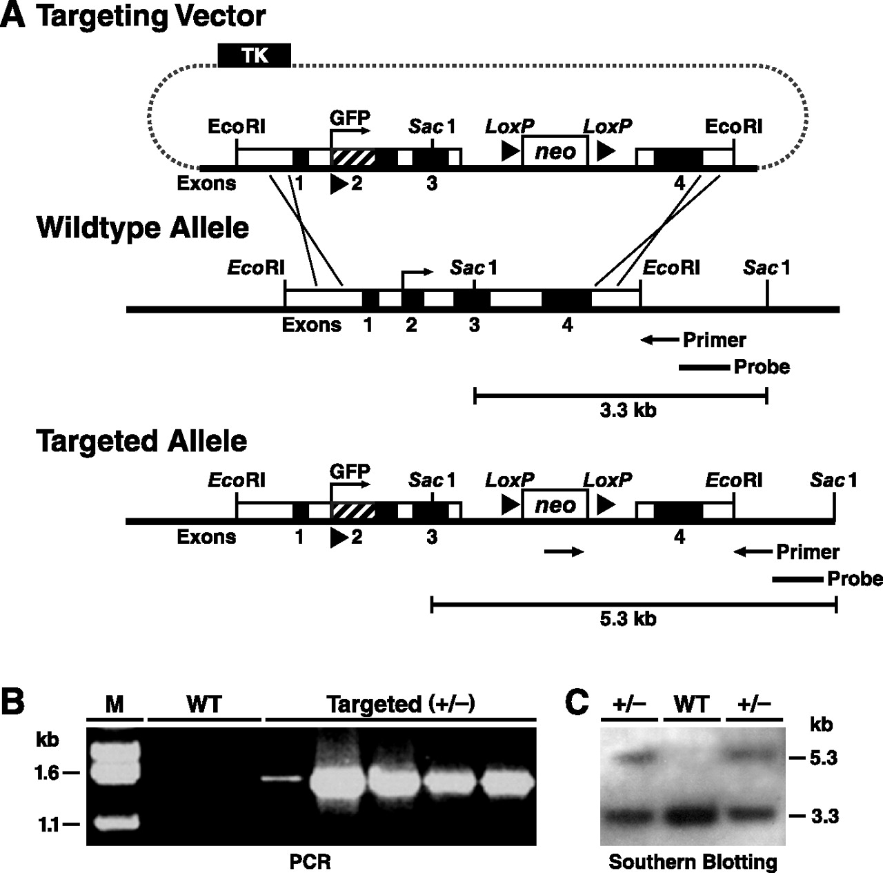
- Figure 1.
Generation and characterization of EGFP-targeted mouse ES cells. A, An EGFP cDNA with start and stop codons and a poly-A sequence (∼1 kb) was inserted into the basic gene-targeting vector used to generate apoE-Arg-61 knock-in mice at the Gladstone Institutes (Raffaï et al., 2001). Homologous recombination between the gene-targeting vector and the Apoe locus in ES cells introduces the EGFP cDNA. A Neo cassette was placed in intron 3. Targeted ES cell clones and mice were identified by PCR with primers and by digestion of genomic DNA with SacI and subsequent Southern blotting with an Apoe 3′ flanking sequence probe, which reveals an expanded 5.3 kb fragment; the wild-type fragment is 3.3 kb. B, PCR screening for ES cell clones with homologous recombination of EGFP revealed a 1.5 kb band in targeted (+/−) ES cells but no band in wild-type (WT) ES cells. C, Southern blotting revealed both 5.3 kb and 3.3 kb DNA fragments in targeted (+/−) ES cells but only a 3.3 kb fragment in wild-type ES cells.
- Figure 2.
Heterozygous EGFPapoE mice express apoE at ∼50% of the level in wild-type mice. A, Northern blotting analysis of apoE and actin mRNA in brains of wild-type and heterozygous EGFPapoE mice at 5 months of age. B, Western blotting analysis of apoE and actin in brains of wild-type and heterozygous EGFPapoE mice at 5 months of age.
- Figure 3.
Expression of EGFP in hepatocytes and peritoneal macrophages in a heterozygous EGFPapoE mouse at 2 months of age. A, Expression of EGFP in hepatocytes as determined by confocal microscopy. B, Peritoneal macrophages were cultured in vitro for 1 or 4 d and analyzed by confocal microscopy. Alternatively, after 4 d of culture, macrophages were incubated with acetylated LDL (AcLDL; 100 μg/ml) for 16 h and analyzed by confocal microscopy.
- Figure 4.
Expression of EGFP in hippocampal astrocytes in heterozygous EGFPapoE mice at 4–6 months of age before (A–C) and 6 d after (D–F) kainic acid (KA) treatment (25 mg/kg body weight). Astrocytes were identified by anti-GFAP immunostaining and confocal microscopy. In the merged image, yellow indicates colocalization of EGFP (green) and GFAP (red).
- Figure 5.
Expression of EGFP in hippocampal microglia in heterozygous EGFPapoE mice at 4–6 months of age before (A–C) and 6 d after (D–F) kainic acid (KA) treatment (25 mg/kg body weight). Microglia were identified by anti-CD11b immunostaining and confocal microscopy. In the merged image, yellow (arrow) indicates colocalization of EGFP (green) and CD11b (red).
- Figure 6.
Hippocampal CA3 neurons express EGFP, representing apoE, in response to excitotoxic injury. Heterozygous 5-month-old EGFPapoE mice received peritoneal injections of kainic acid (KA; 25 mg/kg) (C–G), and the brains were collected 1 d (E–G) or 6 d (C, D) later. Untreated age-matched heterozygous EGFPapoE mice served as controls (A, B). Confocal images of immunostained brain sections were collected for EGFP (green) and anti-NeuN (red), a neuronal marker, or anti-GFAP (red), an astrocytic marker. Images in A–D and G are merged, and yellow indicates colocalization.
- Figure 7.
EGFP and apoE protein and mRNA are present only in injured hippocampal neurons in kainic acid-treated mice. Heterozygous 5-month-old EGFPapoE mice received peritoneal injections of kainic acid (KA; 25 mg/kg) (D–F), and the brains were collected 6 d later. Untreated age-matched heterozygous EGFPapoE mice served as controls (A–C). A, D, Gallyas silver staining of the hippocampal CA1 region. B, E, Merged confocal images of EGFP (green) and anti-apoE (red) in the CA1 region. C, F, In situ hybridization of mouse apoE mRNA in the CA1 region.
- Figure 8.
Expression of EGFP in smooth muscle cells in large blood vessels and cells surrounding small blood vessels in brains of heterozygous EGFPapoE mice. A–C, Merged confocal images of EGFP (green) and anti-α-actin (red), a smooth muscle cell marker, were collected from a 5-month-old heterozygous EGFPapoE mouse. A, Small blood vessels in the hippocampus. B, A small blood vessel in the cortex. C, A large blood vessel in the cortex. D, In situ hybridization shows mouse apoE mRNA along the wall of a large blood vessel in the cortex. E, F, Merged confocal images of EGFP (green) and anti-GFAP (red), an astrocyte marker, were collected from a 5-month-old heterozygous EGFPapoE mouse.
- Figure 9.
Expression of EGFP in choroid plexus cells of heterozygous EGFPapoE mice. A, Merged confocal image of EGFP (green) and anti-GFAP (red) from a 5-month-old EGFPapoE mouse. B, In situ hybridization revealed mouse apoE mRNA in the choroid plexus.


