

Article Figures & Data
Figures
- Figure 1.
Injection site placements. a, Dorsolateral view of the brain showing the DLPFC injection sites in yellow; b, medial view showing the dACC and vmPFC injection sites in orange and red, respectively; c, ventral view showing the OFC injection sites in dark orange. Note that the injection sites cover a relatively small, restricted part of each functional region. cgs, Cingulate sulcus; cs, central sulcus; iar, inferior arcuate sulcus; lorb, lateral orbital sulcus; morb, medial orbital sulcus; ps, principal sulcus; ros, rostral sulcus; sar, superior arcuate sulcus.
- Figure 2.
Schematic chartings and microphotographs of labeled fibers after injections into different prefrontal regions. a, vmPFC injection site (area 25); b, dACC injection site (area 24b); c, OFC injection site (area 11); d, DLPFC injection site (area 9/46). The focal projection fields (black area) and the diffuse projections are shown in charts at three anteroposterior levels, rostral to the anterior commissure. e illustrates microphotographs at low and high magnification (1.6×, scale bar, 5 mm; 10×, scale bar, 100 μm) of the dense terminal field charted in d (the most caudal section) with the border of the focal projection. Cd, Caudate nucleus; ic, internal capsule; Pu, putamen nucleus.
- Figure 3.
3-D renderings of the combined projection fields from the PFC regions. a–c, Frontal view of the striatum showing inputs from the vmPFC (a), from both dACC and OFC (b), and from all PFC areas, including DLPFC (c); d, e, medial (d) and lateral (e) views showing all PFC inputs. White lines in e indicate the level of sections illustrated in f–j. f–j, Coronal slices through the 3-D model with their corresponding Nissl section: red/purple, Inputs from vmPFC; dark orange, inputs from OFC; light orange, inputs from dACC; yellow, inputs from DLPFC. ac, Anterior commissure; Cd, caudate nucleus; ic, internal capsule; Pu, putamen nucleus.
- Figure 4.
Photomicrographs of adjacent coronal sections of the striatum showing the labeled terminals after dual tracer injections into different PFC areas. a, b, Labeled striatal fibers after injections into dACC and OFC (area 24b, a; area 13, b). Scale bar, 500 μm. c, d, Labeled striatal fibers after injections into vmPFC and OFC (area 14, c; area 13, d). Scale bar, 500 μm. e, f, Labeled striatal fibers after injections into dACC and OFC (area 32, e; area 11, f). The scale bar is shown in c. White outline on a, c, and e indicate the focal projection from areas 24b, 14, and 32, respectively. Note the converging and interdigitation of the labeled clusters. The white arrowheads indicate the correspondent blood vessels in adjacent sections.
- Figure 5.
Charts of labeled terminals at different anteroposterior levels of the striatum. a, b, Collective diffuse projections from the ACC and OFC. c, The focal projection fields are represented as colored areas in the small schematics; d–f, interface between ACC/OFC diffuse projections and those from the DLPFC; g–j, diffuse projections are superimposed onto the dense projection showing the interface of the diffuse and focal projections. Cd, Caudate nucleus; ic, internal capsule; Pu, putamen nucleus.
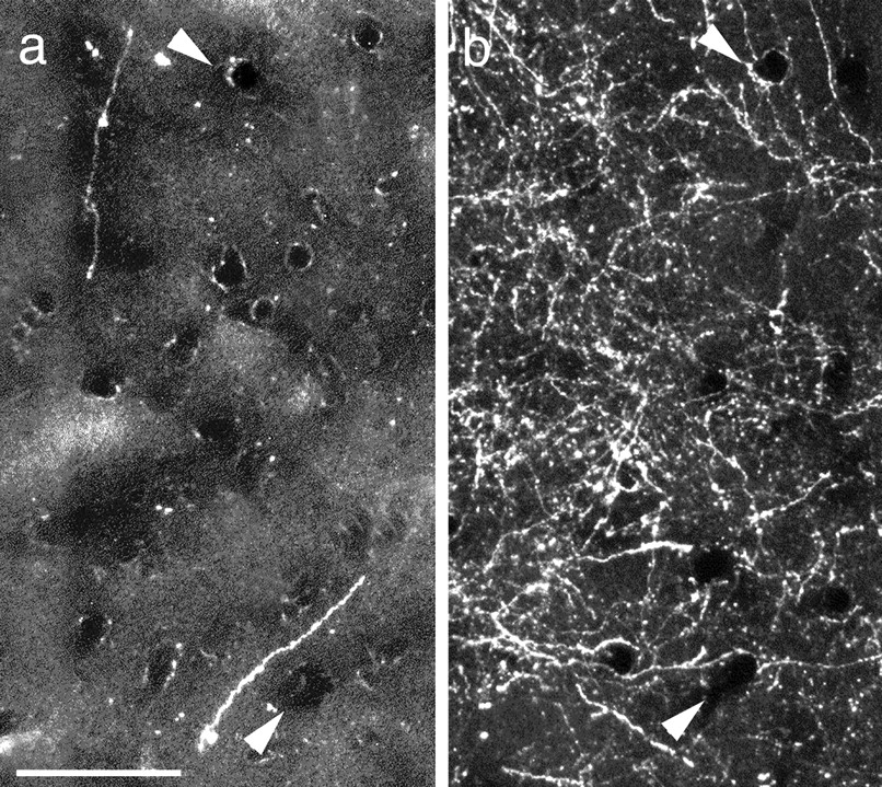
- Figure 6.
After a small PHA-L injection into area 24a, single fibers are found in the dorsal striatum (a), in the area that receives its focal input from area 9m (b). Scale bar, 100 μm.


