

Article Figures & Data
Figures
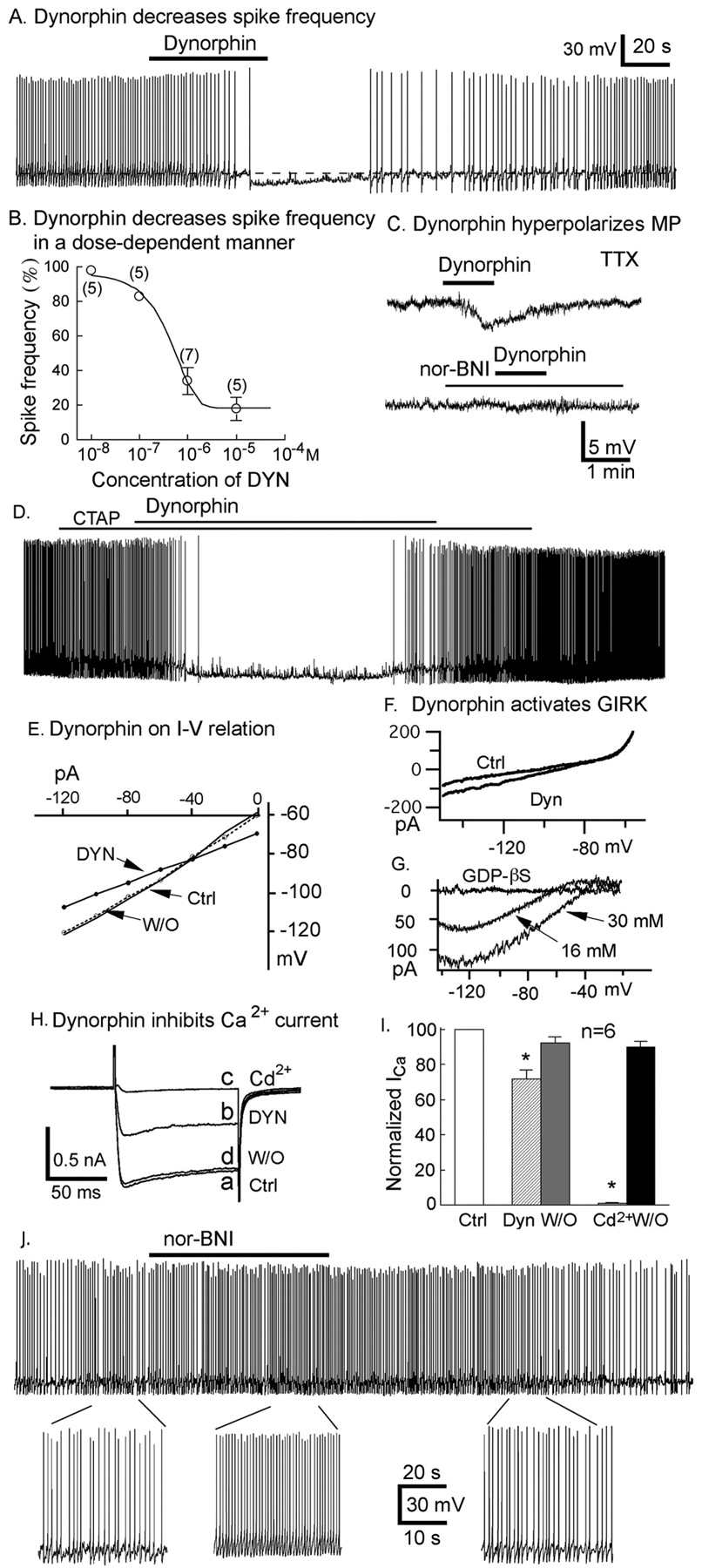
- Figure 1.
Dynorphin inhibits hypocretin neurons: mechanisms. A, A typical hypocretin neuron showing that flow-pipe application of dynorphin (10 μm) decreased spike frequency. B, Dynorphin decreased spike frequency in a dose-dependent manner. Dynorphin (0.01, 0.1, 1, and 10 μm) was applied by flow pipe. C, nor-BNI (1 μm) blocked dynorphin-induced hyperpolarization. Top, Dynorphin hyperpolarized membrane potential. Bottom, Pretreatment with κ-receptor antagonist nor-BNI blocked the dynorphin-mediated hyperpolarization. MP, Membrane potential. D, Dynorphin (10 μm) inhibited spikes of hypocretin neurons in the presence of μ receptor antagonist CTAP (1 μm), suggesting that μ receptors are not responsible for actions of dynorphin on hypocretin cells. E, Dynorphin decreases input resistance. F, Dynorphin-activated GIRK. Representative current induced by ramp pulse before and during dynorphin application. External potassium concentration was 16 mm. G, The net dynorphin-induced current was potassium and GDP-β-S dependent. The approximate reversal potential was −90, −60, and −40 mV when external potassium concentration was 3, 16, and 30 mm, respectively. H, A typical cell showing that dynorphin (10 μm) depressed and Cd2+ (200 μm) blocked the Ca2+ current. The letters a–d refer to the order of application and proceed in alphabetical order. I, A bar graph showing that dynorphin (10 μm) decreased Cd2+-blockable Ca2+ current. DYN, Dynorphin; W/O, washout; Ctrl, control. J, κ receptor antagonist nor-BNI (1 μm) increased spike frequency.
- Figure 2.
Dynorphin inhibits glutamate release presynaptically. A, A representative cell showing that dynorphin (flow pipe, 10 μm) decreased sEPSC frequency. B, Bar graph of dynorphin effect on sEPSC frequency. C, Representative cell showing cumulative probability of dynorphin action on the amplitude of sEPSCs. D, Typical cell showing dynorphin decreases mEPSC frequency. E, Bar graph showing that dynorphin decreased mEPSC frequency (n = 5; p < 0.05). F, A typical cell where dynorphin (10 μm) did not change the amplitude of mEPSCs. G, A representative cell in which dynorphin (10 μm) depressed activity and thus could block a hypocretin-induced increase in spike frequency. H, A bar graph summarizing group data demonstrating that the hypocretin-induced increase in spike frequency was not found in the presence of dynorphin. DYN, Dynorphin; W/O, washout; Ctrl, control.
- Figure 3.
Dynorphin release from dorsal lateral hypothalamus. A, Photomicrograph of dynorphin immunostaining using Texas Red. Three arrows indicate immunoreactive cells. B, Same cells also express GFP driven by hypocretin promoter. Three arrows indicate GFP-positive hypocretin neurons. Scale bar, 20 μm. C, ELISA results showing that electrical field stimulation (Elect. Stim.) increased dynorphin release from microslices of dorsal lateral hypothalamus. Ctrl, Control; ES, electrical stimulation; W/O, recovery washout. *p < 0.05.
- Figure 4.
Dynorphin desensitization. A, Under current clamp, hypocretin (1 μm) increased spike frequency continuously during 2.5 min of flow pipette application without desensitization. B, The mean response to hypocretin of seven hypocretin neurons over time. C, In contrast, before the end of a similar 2.5 min application of dynorphin (1 μm), substantial desensitization is shown. D, The mean response to dynorphin of six hypocretin neurons. E, A typical trace showing dynorphin desensitizes during 6 min bath application. F, A mean response (n = 8 cells) to dynorphin application. The graph shows the group data. G, Hypocretin (200 nm) increased spike frequency until the end of application. H, The mean (n = 7) response to hypocretin application. The graph shows the group data. Little desensitization is shown. I, Hypocretin (HCRT) and dynorphin (DYN) were coapplied to hypocretin neurons in current clamp. The bar graph shows the spike responses to three coapplications (gray bars). The other bars show control and recovery. The first coapplication reduced spike frequency, whereas later coapplications increased spike frequency.
- Figure 5.
Lateral hypothalamic MCH neurons: dynorphin responses desensitize faster than hypocretin responses. A, Flow-pipe application of dynorphin (10 μm) hyperpolarizes the membrane potential of an MCH neuron. B, The outward current evoked by dynorphin application desensitizes with repeated application (30 s followed by a 2 min recovery). C, The inward current in response to hypocretin (1 μm) application shows little or no desensitization in this example neuron. See F for group data. D, Coapplication of dynorphin and hypocretin shifts from an outward current to an inward current with repeated coapplication from a single flow pipe. E, Bar graph shows the attenuation of outward current with repeated application of dynorphin (n = 6 neurons). F, Bar graph shows the minimal desensitization with repeated application of hypocretin (n = 8 neurons). G, Multiple coapplications of dynorphin (DYN) and hypocretin (HCRT) reverse from inducing an outward current to inducing an inward current (n = 11). Data are means ± SEM.
- Figure 6.
Arcuate nucleus NPY neurons: synergistic responses to hypocretin and dynorphin. A, A representative trace showing that hypocretin (1 μm) increases spike frequency of an NPY neuron under current clamp. B, An example cell showing 1 μm hypocretin-induced inward current in the presence of TTX (0.5 μm), AP-5 (50 μm), CNQX (10 μm), and bicuculline (30 μm) under voltage clamp. C, A typical cell showing reduction of sIPSC frequency induced by dynorphin (2 μm). D, A bar graph showing that dynorphin strongly decreased sIPSC frequency. E, A typical cell showing that dynorphin (2 μm) decreased the mIPSC frequency. F, A bar graph presenting the mean reduction of mIPSC frequency induced by dynorphin. G, The cumulative distribution of mIPSC amplitude was not altered by dynorphin (2 μm). DYN, Dynorphin; W/O, washout; Ctrl, control; HCRT, hypocetin.


