

Article Figures & Data
Figures
- Figure 1.
Odor stimulation evokes an LFP response specific to a single glomerulus. A, The field potential recording pipette (filled with Oregon Green), was precisely placed in the center of the labeled glomerulus under visual control using TPLSM. In the GL, odor- evoked LFP responses that consisted of rapid and repetitive negativities locked to respiration. Note that LFP responses evoked by four successive applications of benzaldehyde (0.5%) were very similar. Occasionally β oscillations were recorded (inset, star). B, Odor-evoked fast negativities were exclusive to the glomerular volume. Unlike the field potential negativity evoked by nerve stimulation (C), odor-evoked fast negativities (same as in A) disappeared in the EPL a few tens of micrometers below the GL, leaving a slow and long-lasting negativity. Inset shows another recording in which brief application of two different odors [almond, 0.5% and propionic acid (PA), 0.3%] evoked rapid negativities followed by slower ones of various shapes. Rapid negativities also disappeared, and the slower ones diminished in the EPL. C, Depth profile of LFP responses to olfactory nerve stimulation. D, LFP responses to 2-butanol (0.5%) were glomerular-specific and showed little volume conduction. E, Glutamate receptor antagonist NBQX (250 μm dissolved in fluorescent saline) blocked LFP responses to limonene (0.4%). TPLSM was used to image drug diffusion within glomerulus boundaries. Partial (first application) and full block (second application) occurred within seconds after application. ONL, Olfactory nerve layer; MCL, mitral cell layer; GCL, granule cell layer.
- Figure 2.
LFP responses are correlated to principal cell activation and associated with a local increase in blood flow. A, A mitral cell that was impaled in the soma, labeled with Oregon Green BAPTA-1, and imaged with TPLSM. This cell fired a burst of action potentials on odor inhalation [isoamyl acetate (AA) at 0.6%]. After the micropipette was withdrawn, an extracellular pipette was placed in the labeled glomerulus and used to record the LFP response. LFP negativities were locked to respiration as action potential bursts. B1, In another cell, odor (hexanal 1.2%) evoked a large EPSP crowned with action potential firing. Both the EPSP and the action potentials generated calcium ([Ca2+]i) increases in the dendritic tuft. B2, Left, Subsequent and simultaneous recordings of tuft [Ca2+]i, LFP, and RBC velocity revealed that mitral cell activity underlies LFP responses and is associated with an increase in RBC velocity. Right, Raw data of blood flow recordings at the time indicated by red triangles in the graphs. Note that [Ca2+]i measurements were performed using movie acquisition (B1) or with line-scan acquisition, allowing simultaneous measurement of RBC velocity (B2).
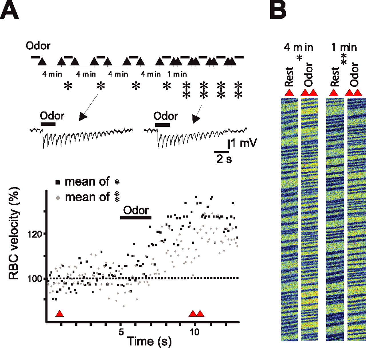
- Figure 3.
LFP and vascular responses uncouple on repetitive odor stimulation. A, Top, Experimental protocol: benzaldehyde (0.5%) was repetitively applied every 4 min (single star) and every 1 min (double stars). LFP responses remained stable with both interstimulus intervals (single traces are illustrated). In contrast, vascular responses decreased with 1 min interstimulus intervals. Averages of four vascular responses recorded with 4 (in black) and 1 (in gray) min interstimulus intervals are illustrated. Bottom, Single and double triangles indicate the times at which raw data in B are extracted. B, Raw data of RBC flow recordings obtained before odor (single triangle) and during odor (double triangle).
- Figure 4.
A, Blockade of local postsynaptic activity does not affect the vascular responses. Top, Experimental protocol: Hexanal (1.2%) was repetitively applied every 4 min in the control condition and 30–60 s after intraglomerular applications of glutamate ionotropic receptor antagonists (250 μm NBQX, 500 μm d-APV diluted in fluorescent saline). TPLSM was used to image drug diffusion within glomerulus boundaries. Glutamate antagonists reversibly and reproducibly blocked LFP negativities but left vascular responses to hexanal unchanged. Single trial LFPs and average vascular responses are illustrated. Single and double triangles indicate the times at which raw data in B are extracted. B, Raw data of RBC flow recordings obtained before odor (single triangle) and during odor (double triangle) in control condition (single star) and in the presence of glutamate antagonists (double star). C, Blockade of local glutamate release does not affect vascular responses. Top, Local application of 1 μm TTX (diluted in fluorescent saline) fully blocked LFP responses but did not affect vascular responses to ethyl propionate (0.005%). Bottom, Application of a higher concentration of TTX (5 μm) outside glomerular boundaries decreased vascular responses to hexanal (1.2%). In each case, single trial LFPs and average vascular responses are illustrated. Single and double triangles indicate the times at which raw data in D are extracted. D, Raw data of RBC flow recordings obtained before odor (single triangle) and during odor (double triangle) in control condition (single triangle) and in the presence of 1 μm TTX (top) and 5 μm TTX (bottom).
- Figure 5.
Odor induces Ca2+ signals in glomeruli from G-CaMP2 mice. A, Two-photon imaging of three fields of view obtained in the glomerular layer (glomerular contours are outlined). G-CaMP2 fluorescence is expressed in mitral cells and in some periglomerular cells: arrows point to two longitudinal mitral cell apical dendrites and double arrowheads to two apical dendrite sections (AI); arrowheads point at several periglomerular cells (AII, AIII). Scale bars, 100 μm. B, Odorant-dependent increases in Ca2+ fluorescence. Movies were acquired over the field of view illustrated in AII with a lower spatial and higher time resolution. Responses to ethylpropionate (EP), benzaldehyde (BA), and isoamylacetate (AA) varied according to each glomerulus. For each glomerulus, responses to two odor applications are overlaid. As odor-evoked LFP responses, Ca2+ responses were occasionally locked to respiration, in particular at low odor concentration. C, Ca2+ responses are concentration dependent. Movies were acquired over the field of view illustrated in AIII. At a high concentration, isoamylacetate (AA) activated all glomeruli in the field of view. Only two of them responded when the odor was diluted. For each glomerulus, responses to two odor applications are overlaid. Note the increasing delay (in gray) of Ca2+ responses with odor dilution.
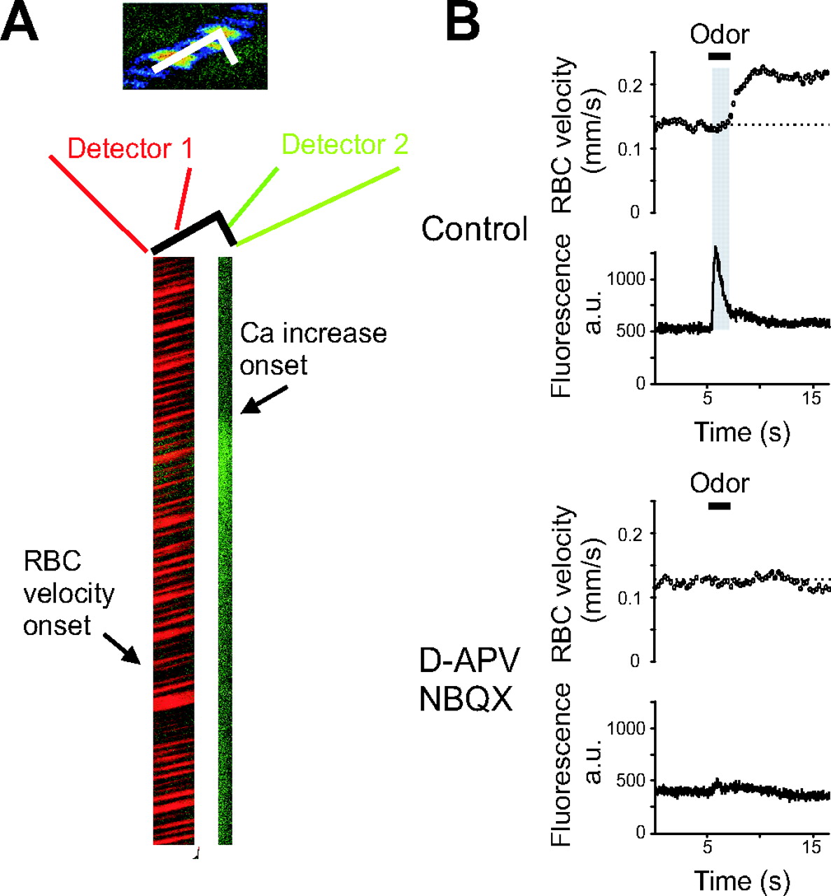
- Figure 6.
Postsynaptic activation of glomerular dendrites is required to trigger blood flow responses in G-CaMP2 expressing mice. A, Left, Odor (ethyl butyrate at 0.004%) triggers a Ca2+ signal followed by an increase in CBF. Line scans were acquired on a segment going along the longitudinal axis of the capillary for its first part and through the neighboring neuropil for its second part. RBC velocity measurements (the first part of the line scan) were acquired on one detector and Ca2+ fluorescence (the last part of the line scan) on the second one. Right, Both neuronal and vascular responses were blocked in the presence of 250 μm NBQX and 500 μm d-APV. Note the delay (in gray) between neuronal and vascular responses.


