

Article Figures & Data
Figures
- Figure 1.
Neuroprotective effect of AIF knockdown against oxygen–glucose deprivation injury in primary hippocampal/cortical neurons. A, Western blots show that AIF expression was decreased time dependently in neurons infected with AAV expressing the AIF targeting sequence (AAV–AIFt) (Table 1). The expression levels of COX IV, VDAC, and HSP60 were unchanged. B, Semiquantitative data of A, based on four independent experiments. *p < 0.05, ***p < 0.001 versus empty AAV-infected cells. C, Confocal images showing AIF expression (red) in neurons coinfected with AAV–AIFs (scramble control sequence) and AAV–GFP (a–c) or AAV–AIFt and AAV–GFP (d–f). The arrows in e and f point to infected neurons showing decreased AIF expression. D, Hoechst 33258 (top) and dual TUNEL/Cyto11 green fluorescent nuclear staining (bottom) in normal unstressed neurons (a, d) and neurons at 24 h after 60 min of OGD (b, c, e, f). After OGD, many neurons showed condensed nuclei (b) and TUNEL-positive nuclei (yellow–red, e); condensed nuclei and TUNEL-positive nuclei were present less frequently in cells infected with AAV–AIFt (c, f). E, Percentages of condensed nuclei were quantified at 24 h after OGD of different duration in neurons either nontransfected or infected with AAV–AIFs or AAV–AIFt. F, Cell viability was measured based on Alamar blue fluorescence at 24 h after OGD of different duration. G, Cell viability was measured at 48 and 72 h after OGD (60 min). *p < 0.05, **p < 0.01 versus nontransfected neurons. Data are mean ± SE; n = 12 per experimental condition from three independent transfection experiments.
- Figure 2.
Translocation of a truncated AIF in neurons after OGD. A, Western blots show that endogenous AIF in brain or cultures of primary neurons or astrocytes has a molecular size of ∼62 kDa. Recombinant proteins AIF67 (proform AIF), AIF62 (lacking the first 50 amino acids), and AIF57 (lacking the first 100 amino acids) served as size controls. B, Western blots after subcellular fractionation show that a truncated AIF at the size of 57 kDa was translocated to both nucleus and cytosol after OGD (60 min), whereas the levels of mitochondrial AIF (62 kDa) were decreased after OGD. Recombinant proteins AIF62 and AIF57 served as size controls. The graph at the bottom illustrates the time-dependent changes of AIF in three fractions after OGD. Data are based on four independent experiments. *p < 0.05 versus non-OGD neurons. C, Western blots based on total-cell extracts show that AIF was truncated to yield the 57 kDa fragment in neurons at 2, 6, and 12 h after OGD (60 min). D, Western blots show that both antibodies raised against amino acids 105–118 (AIF105–118) and amino acids 65–80 (AIF65–80) of pro-AIF were able to detect endogenous AIF in brain extracts and cultured neurons. Recombinant proteins AIF67, AIF62, and AIF57 served as positive controls. E, AIF that translocated into nucleus at 2, 6, and 16 h after OGD (60 min) was detectable using the AIF105–118 antibody but not the AIF65–80 antibody. All blots are representative of at least three sets of independent data with similar results.
- Figure 3.
Calpain I induces truncation and release of AIF from mitochondria in cell-free assays. A, Western blot shows that active calpain I (Calp-I) (Ca2+ at 5 μm) was capable of truncating AIF and inducing its release from isolated brain mitochondria. Immunoblotting was performed using both pellets (mitochondria, 35 μg) and supernatant from the cell-free assays. The recombinant proteins (Rp) AIF62 and AIF57 served as positive control. B, The addition of recombinant calpastatin (micrograms per milliliter) in the assay inhibited calpain I (0.3 U/5 μm Ca2+)-induced AIF release from isolated brain mitochondria. Blot for VDAC from total mitochondria served as loading control. C, The addition of the MPT inhibitor cyclosporin A (CSA, 1 μm) or bongkrekic acid (BA, 1 μm) in the cell-free assay failed to inhibit calpain I (0.3 U/5 μm Ca2+)-induced AIF release from isolated brain mitochondria. D, Left, Mitochondrial swelling assays show that calpain I at the concentration (1 U/5 μm calcium) that induces AIF release did not induce mitochondrial swelling. Right, CSA or BA at the concentration (1 μm) that did not inhibit calpain I-induced AIF release was sufficient to block calcium (500 μm)-induced mitochondrial swelling. E, CSA or BA at the concentration (1 μm) that did not inhibit calpain I-induced AIF release was sufficient to block calcium (500 μm)-induced cytochrome c release. Blot for VDAC from total mitochondria served as loading control. F, Cell-free assays were performed using isolated brain mitochondria (35 μg) from Bid-deficient (−/−), Bid/Bax-double deficient, or wild-type (Wt) mice. Blot for VDAC from total mitochondria served as loading control. G, The synergism between calpain I and Bid to induce AIF release. Mitochondria (35 μg) were incubated with recombinant tBid (3 or 30 ng) or Bax (30 or 300 ng) in the absence or presence of calpain I (0.1 or 1 U) for 60 min, and AIF and cytochrome c release was detected by Western blot. The relative density of AIF or cytochrome c band under each experimental condition is indicated at the bottom. Note that tBid enhanced AIF release induced by calpain I. All data are representative of at least three independent experiments with similar results.
- Figure 4.
Calpain inhibition prevents OGD-induced AIF release in neurons. A, Primary neurons were infected for 3 d with empty AAV or AAV carrying the calpastatin cDNA (AAV–Cps), and the expression of Cps (tagged with triple HA) was confirmed by Western blot using antibodies against Cps and HA, respectively. B, Neurons infected with empty AAV or AAV–Cps were subjected to OGD (60 min), and Western blot was performed to detect α-fodrin cleavage, a marker for calpain activation, at 2 and 6 h after OGD. C, Neurons infected with empty AAV or AAV–Cps were subjected to OGD (60 min), and calpain activity was measured using substrate-based assays at 2, 6, and 16 h after OGD, and the data are expressed as fold increase over non-OGD controls (Con). *p < 0.05, **p < 0.01 versus empty AAV-infected neurons, from three independent experiments. D, Representative confocal images show the inhibitory effect of Cps overexpression on OGD-induced AIF release in neurons. Triple-label immunofluorescent staining for AIF (green), COX IV (red), and 4′-6′-diamidino-2-phenylindole (DAPI; blue) was performed at 6 h after OGD (60 min) in neurons infected with empty AAV or AAV–Cps. The arrows in the middle point to neurons that showed nuclear translocation of AIF after OGD. E, Percentages of neurons showing nuclear translocation of AIF at 2, 6, and 16 h after OGD (60 min). **p < 0.01, ***p < 0.001 versus empty AAV-infected neurons, from four independent experiments. F, Western blots for AIF using total cell extracts or nuclear (Nuc) and mitochondrial (Mito) fractions from non-OGD control neurons (C) or 2 and 6 h after OGD in neurons infected with empty AAV or AAV–Cps. The recombinant proteins AIF62 and/or AIF57 served as positive controls. The blots are representatives of three independent experiments. G–I, Cps overexpression attenuated OGD-induced neuronal cell death. Neurons were infected for 3 d with empty AAV or AAV–Cps and then subjected to OGD for 60 min. Cell death was measured at 24 h after OGD using Alamar blue fluorescence (G), LDH release (H), and phosphatidylinositol (PI) uptake (I). Data are mean ± SE; n = 12 per experimental condition from three independent experiments. **p < 0.01, ***p < 0.001 versus empty AAV-infected neurons.
- Figure 5.
Calpain I activation contributes to OGD-induced AIF release in neurons. A, Left, Neurons were transfected with empty plasmid (lane 1), calpain I scramble shRNA sequence (Calp-Is; lanes 2, 3), or calpain I targeting shRNA sequence (Calp-It) for 12 d and then immunoblotted against calpain I. **p < 0.01 versus plasmid controls; n = 3. Right, Neurons were transfected with GFP expression plasmid alone, both GFP and Calp-Is, or both GFP and Calp-It for 12 d. Transfected neurons were collected by GFP cell sorting and then immunoblotted against calpain I and GFP. ***p < 0.001 versus GFP alone; n = 3. B, Transfected neurons were subjected to OGD for 60 min, and, at 6 h after OGD, nuclear (Nuc), mitochondrial (Mito), or total-cell extracts were immunoblotted against AIF. The graph at the bottom illustrates the semiquantitative results of nuclear AIF from three experiments. *p < 0.05 versus non-OGD controls; #p < 0.05 versus empty plasmid-transfected OGD neurons. C, D, Knockdown of calpain I attenuated OGD-induced neuronal cell death. Cell viability and cell death were measured at 24 h after OGD (60 min) using Alamar blue fluorescence (C) and LDH release (D), respectively. Data are mean ± SE; n = 12 per experimental condition from three independent experiments. *p < 0.05 versus empty plasmid-transfected neurons. E, Western blot detects endogenous calpain I in brain mitochondria. Purified brain mitochondria were subfractionated in the presence of digitonin at either 0.4% (outer membrane broken but no membrane protein dissolving) or 1.0% (resulting in membrane protein dissolving) and then immunoblotted against calpain I and AIF and fraction marker proteins COX IV (IM), VDAC (OM), and AK2 (IMS). Note that, under 0.4% digitonin, calpain I is present exclusively in the IMS, whereas AIF is associated with the IM; under 1.0% digitonin, both AIF and VDAC are dissolved from membranes and appear in all fractions, whereas calpain I remains in the IMS. For calpain I and AIF, the recombinant proteins (Rp) served as markers. The blots are representatives of three experiments. F, Translocation of calpain I and AIF within mitochondria after OGD. Neurons were subjected to OGD (60 min); 2 h later, mitochondria were purified and subfractionated under 0.4% digitonin. Note that calpain I is present in IM, IMS, and, to a lesser extent, in OM fractions after OGD; AIF appears in the IMS fraction at the size of 57 kDa after OGD. G, Semiquantitative analysis of calpain I and AIF levels in total or subfractions of mitochondria after OGD, based on three independent experiments described in F. Levels are expressed as fold increase over non-OGD neurons. *p < 0.05, **p < 0.01 versus non-OGD controls. H, Calpain activity measured in isolated whole mitochondria (30 μg protein/reaction) before or 2 h after OGD from neurons transfected with empty plasmid, Calp-Is, or Calp-It. #p < 0.05 versus non-OGD controls (con); *p < 0.05 versus empty plasmid-transfected neurons, from three experiments. I, Calpain activity measured in subfractions (IMS, 30 μg protein/reaction; OM or IM, 20 μg protein/reaction) of isolated mitochondria before or 2 h after OGD. *p < 0.05 versus non-OGD controls, from three experiments. J, Active calpain I is capable of entering mitochondria. Isolated brain mitochondria were incubated for 20 min with either inactive calpain I (1 U) or active calpain I (1 U) [preincubated with calcium (10 μm)], and subfractions (0.4% digitonin) were immunoblotted against calpain I and AIF. Note that calcium-activated calpain I translocates to IM and OM fractions, whereas the IM-bound AIF is truncated (57 kDa) and released to the IMS. The blots are representatives of three experiments.
- Figure 6.
Calpain-dependent truncation of AIF is required for its release after OGD. A, Neurons were infected for 3 d with AAV carrying either the AIF-GFP fusion construct (a–l) or the calpain-resistant mutant (L101/103G) AIFm-GFP construct (m–t) and then subjected to OGD for 60 min. Confocal images were taken on triple-label immunofluorescent neurons (GFP, green; COX IV, red; Hoechst 33258, blue) in non-OGD cultures (a–d, m–p) or at 2 h (e–h, q–t) and 6 h (i–l) after OGD. In non-OGD neurons, AIF–GFP and AIFm–GFP are localized in mitochondria; after OGD, AIF–GFP is translocated into the nucleus (e–l), whereas AIFm–GFP remains in the mitochondria (q–t). B, Percentages of transfected neurons showing nuclear localization of AIF–GFP or AIFm–GFP at 2, 6, and 16 h after 60 min OGD. *p < 0.05, **p < 0.01 versus non-OGD neurons, from four independent experiments. C, Western blots of the mitochondrial and nuclear fractions using anti-GFP antibody in non-OGD and OGD neurons (lanes 1, 4, empty AAV; lanes 2, 5, AIF–GFP transfected; lanes 3, 6, AIFm–GFP transfected). Note that AIF–GFP, but not AIFm–GFP, is cleaved and translocated into the nucleus after OGD. The whole-cell extracts were immunoblotted with the anti-AIF antibody, showing the truncation of endogenous AIF after OGD. All blots are representative of three independent experiments. D, Neurons were infected with AAV carrying either C-terminal HA-tagged AIF construct (a–h) or the calpain-resistant mutant (L101/103G) AIFm–HA (i–l) and then subjected to OGD for 60 min. Confocal images were taken on triple-label immunofluorescent neurons (HA, red; COX IV, green; Hoechst 33258, blue) in non-OGD cultures (a–d) or at 2 h (e–h) after OGD. AIF–HA is translocated into the nucleus after OGD (e–h, arrows), whereas AIFm–HA remains in the mitochondria (i–l). E, Western blots using anti-HA antibody on whole-cell extracts or nuclear and mitochondrial fractions of transfected neurons at 2, 6, and 16 h after OGD. AIF–HA, but not AIFm–HA, is subjected to truncation and nuclear translocation after OGD. Blots are representative of three independent experiments.
- Figure 7.
Truncation of AIF enables its disassociation with HSP10 in the mitochondria. A, HSP10 is an endogenous binding protein for AIF in neurons. Left, Proteins from cultured neurons were first immunoprecipitated with anti-HSP60 or anti-HSP10 antibody and then immunoblotted with anti-AIF, anti-HSP60, and anti-HSP10 antibodies. Right, Proteins were first immunoprecipitated with anti-AIF or anti-HSP60 antibody and then immunoblotted with anti-AIF, anti-HSP60, and anti-HSP10 antibodies. B, Autoradiograph shows direct protein–protein interaction between AIF and HSP10. The protein-binding assay was performed using in vitro translated proteins (the Flag–HSP10 and [35S]AIF). The reaction products were immunoprecipitated using the anti-Flag antibody, electrophoresed, and then autoradiographed. C, Autoradiograph for [35S]AIF shows that Flag–HSP10 can directly bind to AIF62 but not to AIF57 and that calpain I, which cleaves AIF, inhibited AIF62/HSP10 binding but not the binding between HSP10 and the calpain-resistant AIFm. The protein-binding assay was performed as described in B. D, Western blots show decreased AIF/HSP10 binding after OGD in empty AAV-infected neurons. Cell extracts were immunoprecipitated using the anti-HSP10 antibody and then immunoblotted with the anti-AIF antibody. The AIF/HSP10 binding was restored in neurons transfected with AAV–Cps. The graph at the bottom illustrates the relative changes of three independent experiments. *p < 0.05 versus non-OGD controls. E, Neurons were infected with AAV–AIF–HA or AAV–AIFm–HA (calpain-resistant mutant) and then subjected to OGD (60 min). Immunoprecipitation (IP) was performed using the anti-HA antibody and then immunoblotted with HSP10 and HA antibodies. Note that the binding of HSP10/AIF, but not HSP10/AIFm, was decreased after OGD. Blots are representative of three experiments.
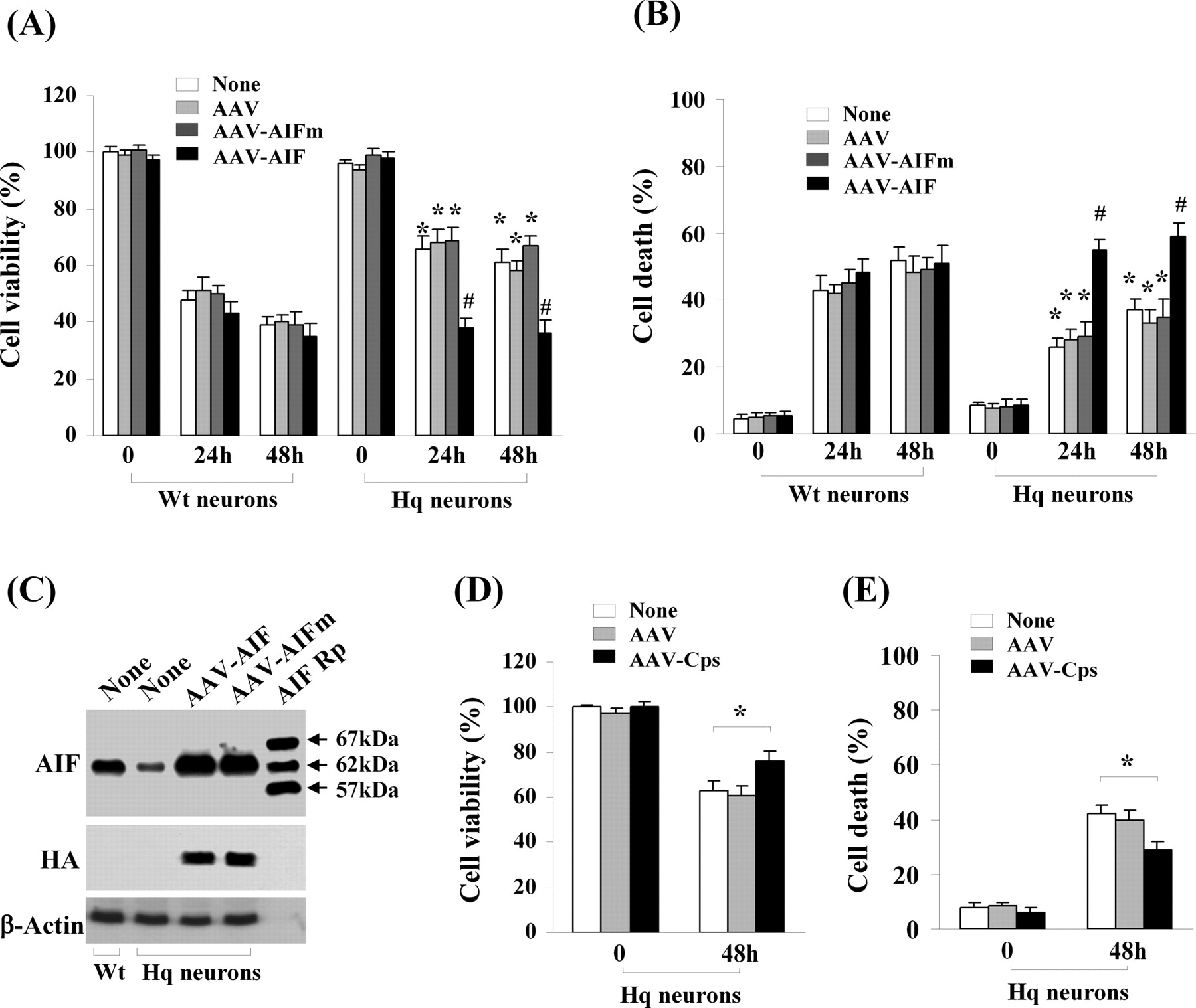
- Figure 8.
The wild-type AIF, but not the calpain-resistant mutant AIF, restores OGD-induced cell death of AIF-deficient neurons. A, B, Neurons from Hq mice or wild-type (Wt) mice were infected with AAV–AIF, AAV–AIFm, or empty AAV for 3 d and then subjected to OGD for 60 min. Cell viability (A) and cell death (B) were measured at 24 and 48 h after OGD using Alamar blue fluorescence and phosphatidylinositol/Hoechst 33258 staining, respectively. *p < 0.05 versus wild-type neurons after OGD; #p < 0.05 versus Hq neurons infected with AAV–AIFm. n = 12, from three independent experiments. C, Western blots (anti-AIF and anti-HA) show the expression levels of AIF or AIFm in Hq neurons 3 d after AAV infection. AIF recombinant proteins (AIF Rp) served as size markers. D, E, AAV-mediated overexpression of calpastatin (Cps) in Hq neurons resulted in additive prosurvival effect against OGD. Neurons from Hq mice were infected with AAV–Cps or empty AAV for 3 d and then subjected to OGD for 60 min. Cell viability (D) and cell death (E) were measured at 48 h after OGD using Alamar blue and phosphatidylinositol/Hoechst 33258 staining, respectively. *p < 0.05 versus noninfected Hq neurons, n = 14 per condition, from three independent experiments.
- Figure 9.
Calpastatin overexpression inhibits AIF translocation in CA1 neurons after global ischemia. A, Overexpression of calpastatin (Cps) after AAV infection in the rat brain. AAV was infused into the hippocampus using convection methodology to achieve widespread expression in the CA1 sector. Images show HA immunoreactivity (HA–Cps) at 14 d after AAV–Cps infection (b); preabsorption of the antibody with Cps protein abolished the signals (c). No Cps immunoreactivity is seen in CA1 after empty vector infection (a). Western blots (anti-HA or anti-Cps) confirm the expression of Cps protein in the CA1 sector at 7–14 d after AAV–Cps infection; n = 5 per group. B, C, AAV–Cps infection inhibited global ischemia-induced calpain activation in CA1. Western blot (B) was performed at 36 h after ischemia to detect α-fodrin cleavage, which was inhibited by AAV–Cps (lanes 6, 7) but not by empty AAV (lanes 4, 5). Substrate-based activity assays (C) show that ischemia-induced increases in calpain activity were inhibited by AAV–Cps. *p < 0.05 versus non-ischemic controls; n = 6 per group. D, Representative immunofluorescence of AIF (red) from non-ischemic CA1 (a) or 72 h after global ischemia (b–d). AAV–Cps (c, d) or the empty vector (b) was infused 14 d before ischemia, and brain sections were double-label immunostained for AIF (red) and HA (green, d). Note that majority of CA1 neurons lost normal localization of AIF after ischemia (b, arrows), but AIF translocation was rare in Cps-overexpressed CA1 (c, d, arrows). Scale bars, 50 μm. E, Western blots of CA1 whole-cell extracts or subcellular fractions after sham operation (S) or at 36 and 72 h after ischemia. AAV–Cps infection inhibited ischemia-induced truncation and nuclear translocation of AIF. F, Quantitative analysis of AIF nuclear translocation 72 h after ischemia, based on the immunostaining experiments described in D. **p < 0.01 versus empty vector group or non-infection group; n = 5 per group. G, Semiquantitative analysis of AIF nuclear translocation at 36 and 72 h after ischemia, based on Western blots. *p < 0.05, **p < 0.01 versus empty AAV-infected brains; n = 6 per group. H, I, Cps expression promoted CA1 cell survival after global ischemia. Viable cells (H) and DNA-damaged cells (I) in CA1 of the dorsal hippocampus were quantified stereologically at 7 d after ischemia or sham operation after AAV–Cps or empty vector infection. **p < 0.01 versus non-infected or empty vector-infected brains; n = 9 per group.
- Figure 10.
AIF knockdown is neuroprotective in CA1 against transient global ischemia. A, CA1 was coinfected for 14 d with AAV–GFP and the AIF-targeting sequence AAV–AIFt (a, c) or the AIF scramble sequence AAV–AIFs (b). d–f show double-label immunofluorescence of AIF (red) and GFP (green) in AAV–AIFs/AAV–GFP-infected CA1 neurons; g–i show reduced AIF expression (red) in AAV–AIFt/AAV–GFP-infected CA1 neurons. j and k show double-label immunofluorescence of AIF (red) and Hoechst 33258 (blue) in AAV–AIFs- and AAV–AIFt-infected CA1, respectively. B, Western blots show the expression levels of AIF in CA1, either non-infected (lanes 1–3) or at 14 d after infection with AAV–AIFs (lanes 4–6) or AAV–AIFt (lanes 7–9). The graph illustrates the relative changes of AIF expression in CA1 after AAV–AIFt infection. **p < 0.01 versus AAV–AIFs-infected brains; n = 6 per group. C, D, Knockdown of AIF expression promoted CA1 cell survival after transient global ischemia. Viable cells (C) and DNA-damaged cells (D) in CA1 of the dorsal hippocampus were quantified stereologically, respectively, at 7 d after ischemia or sham operation after AAV–AIFs or AAV–AIFt infection. *p < 0.05, **p < 0.01 versus non-infected or AAV–AIFs-infected brains; n = 9 per group. E, Knockdown of AIF expression attenuated ischemia-induced large-scale DNA fragmentation in the CA1 sector, determined using DNA pulse-field gel electrophoresis at 3 d after global ischemia. Lanes 1, 2, Size markers; lane 3, non-ischemic control; lanes 4, 5, non-infected ischemic brains; lanes 6, 7, AAV–AIFs-infected ischemic brains; lanes 8, 9, AAV–AIFt-infected ischemic brains. Note that ischemia-induced formation of both 50 and 10 kbp fragments was reduced in AAV–AIFt-infected brains. F, Western blots show the expression levels of AIF in CA1, either non-infected (lanes 1, 2) or at 14 d after infection with AAV–AIFt (lanes 3, 4), coinfection with AAV–AIFt/hAIF (lanes 5, 6), or coinfection with AIFt/hAIFm (lanes 7, 8). G, H, Overexpression of human AIF, but not the calpain-resistant hAIFm, ameliorated the prosurvival effect of AIF knockdown after transient global ischemia. Viable cells (G) and DNA-damaged cells (H) in CA1 of the dorsal hippocampus were quantified stereologically, respectively, at 7 d after ischemia (n = 10 per group) or sham operation (n = 6 per group). *p < 0.05 versus con-infection of AAV–AIFt/AAV–hAIF.


Tables
Construct Sequences AIFt Targeting sequence 1, 5′-GAGAAACAGAGAAGAGCCA-3′ Targeting sequence 2, 5′-GTCACGTCCCTTTCCTGCT-3′ AIFs Scramble sequence, 5′-AACAGGAACAGACGGAGAA-3′ GFPt Targeting sequence, 5′-AACAGCTGCTAGGATTACACA-3′ calp-It Targeting sequence 1, 5′-CAGTTTCTCATAGCTTGGGCTTCAA-3′ Targeting sequence 2, 5′-ATCAAGTGGAAGCGTCCTACGGAA-3′ calp-Is Scramble sequence, 5′-TTAAGCTTACCTTGGATAGTGCCT-3′ calp-IIt Targeting sequence 1, 5′-AAAGCTCAGGAAGCCTGCAGAAATT-3′ Targeting sequence 2, 5′-CAAATACCTTCTGGATGAACCCTCA-3′ calp-IIs Scramble sequence, 5′-TTTGAAGAACAAGCAGACAAGGCCT-3′ -
calp-IIt, Calpain II-targeting sequence; calp-IIs, calpain II scramble sequence.
-
Supplemental Data
Files in this Data Supplement:
- supplemental material - Supplemental Legend
- supplemental material - Supplemental Data 1
- supplemental material - Supplemental Data 2
- supplemental material - Supplemental Data 3
- supplemental material - Supplemental Data 4
- supplemental material - Supplemental Data 5


