

Article Figures & Data
Figures
- Figure 1.
Domain structure of human parkin and Myc-tagged wild-type and FLAG-tagged mutant constructs used to generate transgenic flies.
- Figure 2.
Mutant parkin produces age-dependent deficits in motor performance. Three independent assays were used to analyze motor behavior. A , Using the ddc-GAL4 driver, parkinQ311X and parkinT240R produce age-dependent impairments in climbing performance compared with control and parkinwt. For each genotype at each time point, at least five cohorts consisting of 16–18 flies each were tested. B , At 5 weeks, the righting reflex test demonstrates postural instability of parkinQ311X and parkinT240R flies compared with parkinwt or control. Twenty flies of each genotype were analyzed. C , Rotarod performance demonstrates normal postural stability for all genotypes at 2 weeks. D , By 4 weeks, postural instability is apparent in parkinQ311X and parkinT240R compared with parkinwt and control flies; the <1 score for the arbitrary index at higher angles indicates flies that fall more frequently. For each genotype at each time point, ≥12 flies were tested. Values shown represent mean ± SEM. **p < 0.01, ***p < 0.001 relative to control ddc [two-way ANOVA with Bonferroni's multiple comparison test ( A , C , D ) or unpaired t test ( B )].
- Figure 3.
Analysis of activity-dependent G-CaMP signal in living brains at 1 week after eclosion. GFP signal intensity was converted into thermally coded color images. A , In the control ddc::G-CaMP brain, intense GFP signal was detected. B , parkinT240R expressed in ddc::G-CaMP flies resulted in markedly reduced GFP signal. C , Expression with parkinwt resembled the control. D , Analysis of GFP pixel density of at least five brains for each genotypes showed decreased G-CaMP activity in the parkinT240R brain (one-way ANOVA with Bonferroni's multiple comparison test; n ≥ 5; **p < 0.01). The images are represented using a pseudocolor scale as indicated in A , where blue is low intensity and yellow–red is high intensity. Scale bar, 40 μm.
- Figure 4.
Expression of mutant human parkin in Drosophila DA neurons causes age-dependent neurodegeneration. A , Schematic representation of DA neurons in the central brain of Drosophila. a , The projected confocal image shows anti-TH and phalloidin staining spanning optical sections 40 μm in depth from posterior to anterior. Two major DA clusters, DM (circles) and DL (rectangles), are indicated. b , A projected image shows anti-TH (red) and anti-GFP (green) colocalized in all DA neurons in both DM and DL (enlarged images to the right) clusters. Arrows indicate 5-HT neurons expressing GFP but not TH. B , Reductions in TH immunoreactivity at 5 weeks after eclosion are apparent in ddc::parkinQ311X ( b ) and ddc::parkinT240R ( c ) but not ddc control ( a ) or ddc::parkinwt ( d ) brains. Circles, DM; rectangles, DL. C , Quantification of DA neurons at 0, 3, and 5 weeks after eclosion in DM and DL clusters. Progressive loss of TH-immunoreactive neurons is induced in both clusters by ddc::parkinQ311X (blue bars) and ddc::parkinT240R (red bars) compared with the ddc control (black bars) and ddc::parkinwt (green bars). Values represent the mean ± SEM; n = 12. *p < 0.05, **p < 0.01, ***p < 0.001 relative to the ddc control. D , At 4 weeks, mCD8-GFP signal (green) is reduced in parkinT240R ( b ) compared with parkinwt ( a ) brain, which is similar to changes observed in corresponding images showing reduced TH immunoreactivity. Measurement of GFP pixel density (corresponding to the left y-axis) and GFP cell counts in the DM cluster (corresponding to the right y-axis) from 4-week-old brains of both genotypes showed a significant decrease (unpaired t test; n = 5; *p < 0.05) of GFP signal in parkinT240R brains compared with parkinwt ( c ). The GFP signal at eclosion was indistinguishable between parkinT240R and parkinwt (data not shown). d , e , Confocal images of ddc::parkinT240R ( e ) and ddc::parkinwt ( d ) brains stained with an anti-parkin antibody (green) reveals reduced parkin signals in ddc::parkinT240R compared with ddc::parkinwt brains. f , Analysis of brains at 4 weeks indicates that cell counts stained with anti-parkin for parkinT240R are decreased compared with parkinwt (unpaired t test; n = 6; *p < 0.05 for DM; ***p < 0.001 for DL). Parkin staining at eclosion was indistinguishable between parkinT240R and parkinwt (data not shown). a , b , d , and e are confocal images of adjacent brains imaged simultaneously in the same slide but reoriented for presentation. E , Anti-5-HT staining at 5 weeks after eclosion shows reduced neuritic immunoreactivity in 5-HT neurons expressing mutant parkin ( b , c ) compared with those expressing wild-type parkin ( d ) or controls ( a ). Scale bar, 40 μm.
- Figure 5.
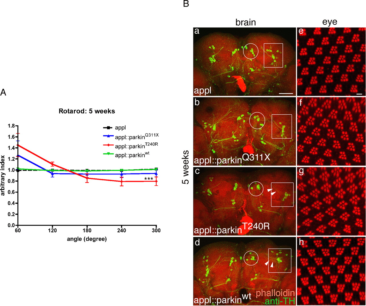
Pan-neuronal expression of parkinT240R induces motor dysfunction and degeneration of DA neurons, whereas photoreceptor neurons are resistant. A , Rotarod assays at 5 weeks demonstrate postural instability of appl:: parkinT240R flies, which fall more frequently at higher angle positions compared with the appl-GAL4 control and appl::parkinwt. appl:: parkinQ311X flies show a modest trend toward postural instability. For each genotype at each time point, >12 flies were scored. The values shown represent mean ± SD. ***p < 0.001 relative to control appl (two-way ANOVA with Bonferroni's multiple-comparison test). Ba–Bd , Confocal images of brains aged 5 weeks stained with anti-TH (green) and phalloidin (red). Circles, DM; rectangles, DL. Reductions in TH immunoreactivity are apparent in the DL cluster in appl::parkinT240R brain ( c ) compared with appl ( a ), appl::parkinQ311X ( b ), and appl::parkinwt ( d ). Arrowheads indicate reduced TH-immunoreactive processes ( c ) in brains expressing mutant parkin compared with wild type ( d ). Be–Bh , No differences in morphology of rhabdomeres within individual ommatidia morphology occur in ddc control ( e ) or flies expressing parkinQ311X ( f ), parkinT240R ( g ), or parkinwt ( h ). Scale bar, 40 μm.
- Figure 6.
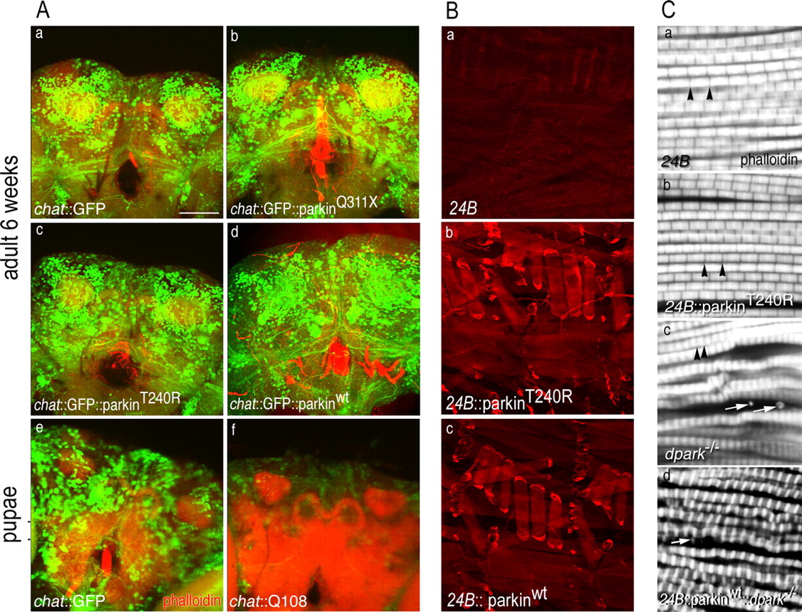
Cholinergic neurons and indirect flight muscle are resistant to mutant parkin. A , Images show signals for TRITC (tetramethylrhodamine isothiocyanate)-phalloidin (red) and GFP (green). a–d , Confocal images of chat::GFP brains coexpressing mutant or wild-type parkin transgenes at 6 weeks. Expression of either parkinQ311X ( b ) or parkinT240R ( c ) has no effect on GFP signal that is distinguishable from the expression of parkinwt ( d ) or control ( a ). e , f , In contrast to the robust fluorescence of mutant parkin brains ( a–d ), expression of a toxic polyglutamine construct, Q108 ( f ), markedly reduces GFP signal in midpupal brains compared with controls ( e ). (Images show pupal brains for Q108 as expression of this transgene is late pupal lethal.) B , Staining of larval muscle verified expression of human parkin under control of 24B-GAL4. a–c , The driver-alone control ( a ) shows only background staining with PRK8 monoclonal antibody, whereas a robust signal in larval muscle is observed for both mutant ( b ) and wild-type human parkin transgenes ( c ). C , Confocal image of sarcomeres from indirect flight muscle stained with phalloidin. a , b , 24B-GAL4 control ( a ) and 24B::parkinT240R ( b ) show normal organization of sarcomeres. Black arrowheads show normal Z-bands. c , The control dpark null mutant shows abnormal deposition of actin-containing debris in indirect fly muscle (white arrows) with irregularly organized sarcomeres. d , 24B-GAL4::parkinwt fails to rescue the muscle phenotype of the dpark null mutant. The white arrow shows abnormal debris. Scale bar, 40 μm.
- Figure 7.
DVMAT modulates mutant parkin-induced degeneration and motor phenotypes. A , Rotarod performance reveals early onset of postural instability in ddc::parkinT240R::DVMATi flies at 2 weeks (gray lines) compared with ddc::parkinT240R (red lines) and ddc::parkinT240R::DVMAT (green lines). At least six flies were scored for each genotype. B , The righting reflex demonstrates earlier onset of postural instability in ddc::parkinT240R::DVMATi flies (gray bars at 1 week) compared with ddc::parkinT240R (red bar at 1 week). At 2 weeks, the progressively impaired righting ability of ddc::parkinT240R (red bar) was ameliorated in ddc::parkinT240R::DVMAT flies (green bar), whereas ddc::parkinT240R::DVMATi flies continued to show a more pronounced motor deficit compared with ddc::parkinT240R (red bar). The values represent the mean ± SEM; n = 20. *p < 0.05, **p < 0.01 relative to control ddc::parkinT240R by one-way ANOVA with Bonferroni's multiple comparison correction. All genotypes at individual stages were compared with ddc::parkinT240R. C , Modulation of DVMAT affects the incomplete pupal lethality phenotype produced using two copies of ddc-GAL4 driver to express mutant parkin. Overexpression of parkinT240R (solid red bar) or parkinQ311X (solid blue bar) causes incomplete pupal lethality, whereas parkinwt (solid green bar) produces an ∼100% eclosion rate. Knockdown of DVMAT significantly worsens the parkinT240R lethality phenotype (red checked bar; *p < 0.05), whereas overexpressing DVMAT significantly suppresses both parkinT240R-induced (red striped bar; **p < 0.01) and parkinQ311X-induced (blue striped bar; *p < 0.05) pupal lethality. Modulation of DVMAT expression has no effect on wild-type parkin (green bars; one-way ANOVA with Bonferroni multiple comparison test; n ≥ 4). D , Confocal images of ddc::parkinT240R ( a , b ) or ddc::parkinwt ( d , e ) coexpressed with either DVMAT ( a , d ) or DVMATi ( b , e ) brains at 2 weeks. A marked reduction of TH immunoreactivity is observed in ddc::parkinT240R::DVMATi ( b ) compared with ddc::parkinT240R::DVMAT ( a ). At this stage, ddc::parkinT240R::DVMAT and ddc::parkinT240R brains are indistinguishable (data not shown). Quantitation of TH-positive neurons shows significant decreases in both DM and DL clusters in mutant parkin brains coexpressing DVMATi ( c ) (mean ± SEM; unpaired t test; n = 4; *p < 0.05; **p < 0.01). Modulation of DVMAT expression had no evident effect on wild-type parkin brains ( d – f ). Confocal images were reoriented from adjacent brains imaged simultaneously for paired comparison ( a , b , d , e ). E , Confocal images of ddc::parkinT240R ( a ) coexpressed with DVMAT ( b ) brains at 4 weeks. Significant increases in TH immunoreactivity are observed in ddc::parkinT240R::DVMAT ( b ) compared with ddc::parkinT240R ( a ). Quantitation of TH-positive neurons showed significant increases in both DM and DL clusters in mutant parkin brains coexpressing DVMAT ( c ) (mean ± SEM; unpaired t test; n = 8; **p < 0.01; ***p < 0.001). Confocal images were reoriented from adjacent brains imaged simultaneously for paired comparison.


Additional Files
HTML Page - index.htslp
Files in this Data Supplement:
- supplemental material - Figure legends
- supplemental material - Figure 1
- supplemental material - Figure 2
- supplemental material - Figure 3
- supplemental material - Figure 4
- supplemental material - Figure 5
- supplemental material - Figure 6
- supplemental material - Figure 7
- supplemental material - Figure 8


