

Article Figures & Data
Figures
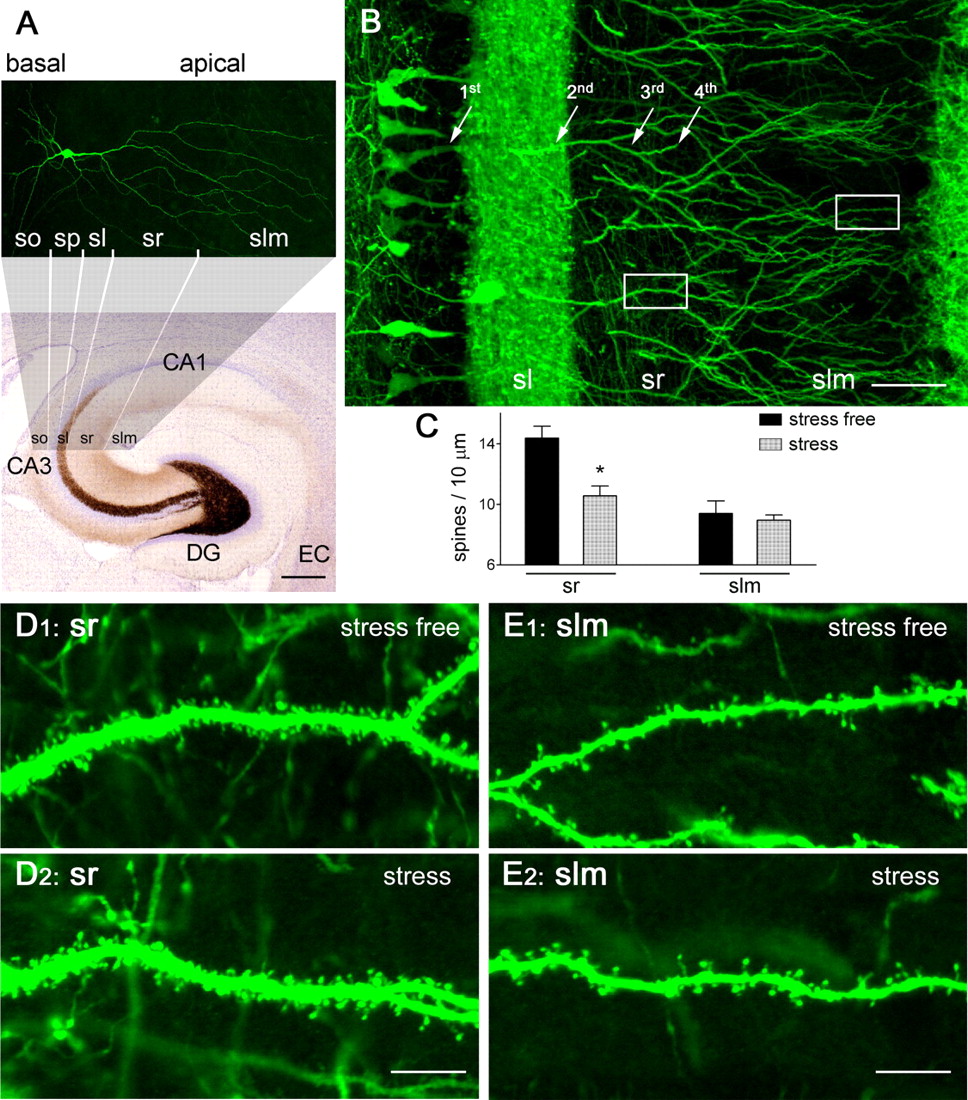
- Figure 1.
Reduced hippocampal dendritic spine density after a 5 h stress. A, Structure and organization of the hippocampus, clarifying the location and subcellular domains of a YFP-expressing CA3 pyramidal neuron. The location of the apical dendrites, emerging from stratum pyramidale (sp) and spanning stratum lucidum (sl), radiatum (sr), and lacunosum-moleculare (slm), is shown. Basal dendrites are in stratum oriens (so). EC, Enthorinal cortex. Scale bar, 260 μm. B–D2, Stress leads to spine loss in vulnerable domains of apical dendrites (third- and fourth-order branches in sr). In a 3-month-old, Thy1-YFP-expressing mouse, density of spines on the third/fourth dendritic branches was high under stress-free conditions (D1), and was significantly reduced immediately after a 5 h combined restraint/noise stress (D2), as quantified in C (*p < 0.05). Density of spines on the distal dendritic branches located in slm was not influenced by the acute stress (C, E1, E2). The frames in B denote areas magnified in D1 and E1. Error bars indicate SEM. Scale bars: B, 50 μm; D1–E2, 7 μm.
- Figure 2.
Deletion of CRH receptor CRFR1 enhances spine density. A, Apical dendrites of a CA3 pyramidal neuron expressing YFP (see Materials and Methods) from a 14-d-old mouse with a normal complement of the CRH receptor CRFR1 (wild type). B, The genetic absence of CRFR1 (CRFR1−/−) promoted both complex dendritic branching and increased spine density (F(1,68) = 19.89; p < 0.0001). C, The overall increased spine density in CRFR1−/− dendrites resulted from significant differences of spine densities in the third/fourth-order dendritic branches (*p < 0.05; n = 5 mice per group; Bonferroni's post hoc test). The framed areas in the top images are magnified in the bottom images, to show dendritic spines (filled arrowheads), including those with abnormal shapes (arrows and open arrowhead presented filopodia and big-head spine, respectively). Error bars indicate SEM. Scale bars: 50 μm (low magnification); 5.5 μm (high magnification).
- Figure 3.
CRH application reduces spine density in hippocampal organotypic slice cultures, whereas neurons grown in the presence of a CRH receptor antagonist have increased spine density. A, Apical dendrites of a CA3 pyramidal neuron in control condition. B, Treatment of hippocampal slices with CRH (100 nm; 13 d) decreased spine density (F(1,60) = 10.74; p = 0.001), an effect resulting primarily from lower spine density on the third- and fourth-order dendritic branches (*p < 0.05; n = 10 neurons per group; Bonferroni's post hoc test) (D). C, Growing hippocampal slices in the presence of the CRH receptor antagonist NBI 30775 (1 μm; 13 d) led to higher spine density (F(1,58) = 4.17; p = 0.046), also stemming from increased density of spines on the third- and fourth-order dendritic branches (*p < 0.05; n = 10 neurons per group; post hoc test) (D). The framed areas are magnified to show individual spines (arrowheads) and filopodia (C, arrow) on fourth-order branches. Hippocampi were cultured on P1 and grown for 14 d. Error bars indicate SEM. Scale bars: 55 μm (low magnification); 6 μm (high magnification).
- Figure 4.
Dendritic spine dynamics in hippocampal pyramidal neurons, visualized using live, time-lapse two-photon microscopy. A, A series of images shows that the structure of the dendrites remained constant over the 60 min of imaging. B, During this period, new dendritic spines appeared (red arrowheads) and existing ones disappeared (retracted; yellow arrowheads) within minutes. C, Under the imaging conditions, spine formation and disappearance occurred at similar rates (F(1,48) = 0.02; p = 0.90). D, Spine density (left panel), expressed as numbers of total spines per unit dendritic length, was stable (p = 1), with little percentage change of spine density over the imaging period (right panel). A total of 2006 μm of dendritic branches from 12 neurons were imaged and analyzed blindly. Hippocampi were cultured on P1 and imaged on in vitro days 5–6. Error bars indicate SEM. Scale bars: A, 21.5 μm; B, 6.6 μm.
- Figure 5.
CRH alters spine dynamics rapidly and reversibly. A, Infusion of CRH (100 nm) for 30 min did not affect dendritic structure appreciably. B, High-magnification imaging revealed a rapid effect of CRH on the rate of spine retraction (yellow arrowheads): accelerated spine disappearance was apparent already by 5 min after CRH exposure, with little change in the rate of spine formation (red arrowheads; 0 min vs CRH 5 min). CRH-induced spine elimination was partially reversed by a 30 min washout. C, The effect of CRH (F(1,60) = 15.79; p = 0.0002, compared with vehicle infusion) was attributable primarily to increased retraction rates at the 5 min and 10 min time points (*p < 0.05; +p = 0.07). D, Accelerated spine disappearance with stable spine formation resulted in a significant net reduction of spine density (*p < 0.05, compared with the 0 min time point); this is presented as actual spine density (left panel) and percentage change of spine density (right panel). A total 2650 μm of dendrites was imaged and analyzed in six experiments, two to four neurons each. Hippocampi were cultured on P1 and imaged on in vitro days 5–6. Error bars indicate SEM. Scale bars: A, 21.5 μm; B, 6.6 μm.
- Figure 6.
Spine disintegration induced by CRH requires activation of the receptor CRFR1. A, Application of the CRFR1 antagonist NBI 30775 (1 μm) for 30 min did not influence spine formation or elimination (F(1,36) = 0.1298; p = 0.7208, compared with untreated cultures). The red arrowheads denote newly formed spines, and the yellow ones show eliminated spines. B, Infusion of CRH (100 nm) accelerated spine disappearance (F(1,36) = 20.81; p < 0.0001). *p < 0.05, +p = 0.07, compared with formation at the same time point. C, This effect was blocked by infusion of the CRFR1 antagonist commencing 5–10 min before (and continuing during) CRH application (F(1,36) = 0.2529; p = 0.618, CRH+ antagonist compared with antagonist alone). Data are derived from five experiments, and 1367–2650 μm of dendrites were studied in each group. Organotypic cultures were initiated on P1 and grown for 5–6 d, followed by two-photon microscopy live imaging. Error bars indicate SEM. Scale bar, 11 μm.
- Figure 7.
CRH leads to rapid dephosphorylation (activation) of the F-actin-regulating protein, cofilin, within dendritic spines. A, Confocal images (1 μm) of YFP-expressing dendrites labeled also for phosphorylated (inactive) cofilin (pCofilin) demonstrate the presence of pCofilin immunoreactive puncta (red) within spine heads (arrows). B–D, CRH exposure (100 nm) for 5–30 min resulted in a significant reduction of pCofilin puncta (*p < 0.05). The CRH-induced reduction of pCofilin immunoreactive puncta per spine was still significant when the reduction of spine density by the peptide was taken into consideration (D, right panel). Note the similar time course of CRH-induced dephosphorylation of cofilin and of CRH-induced spine loss. Hippocampi were cultured on P1 and grown for 6 d. Data are derived from 20–40 sections per group, obtained in five experiments (n = 5). Error bars indicate SEM. Scale bar, 5 μm.


