

Article Figures & Data
Figures
- Figure 1.
Visual stimulus setup and analysis methods. A, Motion stimuli were presented to the bee using a computer monitor (black rectangle), whereas color stimuli were presented using an LED array with 20 blue, green, and violet LEDs each arranged in a semirandom pattern. Both stimuli were presented to the bee's right eye, but are presented on either side here for visualization purposes. B, The motion stimuli used are represented in i–v, which included black or white large oriented bars (i, ii), small moving targets (iii), a large moving edge (iv), and a high-contrast grating (v). Each stimulus was moved in eight different directions, which are indicated in vi. C, We measured the precision and reliability of the spike times across five trials of repeated light flashes. i, ii, We calculated a PSTH (bin width, 0.1) from the raster plot (i) and convolved the PSTH with a Gaussian curve (half-width, 8 ms). iii, iv, We detected events three SDs above the mean (black peak, iii) and measured the spike-time precision and response reliability of the spikes across trials during the event (black spikes under the asterisk; iv).
- Figure 2.
Bee brain structure. Ai, The bumblebee brain. Aii, A horizontal section of the bee brain stained with osmium taken from the level of the dashed lines in Ai. Specific areas of the optic lobe subdivisions are indicated: the retina (ret), the lamina (la), the medulla (me), and the lobula (lo), as well as the central protocerebrum (prot). B, Bombus impatiens lobula structure. Reduced silver Bodian staining, six layers can be seen within the lobula, which progress from the outer portion of the lobula (i.e., layers 1–3) to the inner, or proximal, regions of the lobula (i.e., layers 4–6). The distal layers (1–4) contain a spatially ordered arrangement interspersed with horizontal branching. In addition, larger processes dominate layer 4. The arrangement breaks down in layers 5 and 6, where the large processes form a complex network. C, Input to the lobula, which follow the internal structure of the layers (site of dye injection at the asterisk). D, In this mass fill of medullar neurons, the input to the lobula is clearly columnar in the outer, or distal, regions of the lobula, which disappears in the inner layers 5 and 6. E, Multiple sections of the brain can be used to reconstruct a three-dimensional representation of the brain, with the optic lobes (lo and me), the antennal lobes (al), the central complex (cc), and the mushroom bodies (mb). F–H, The lobula and the component layers were three-dimensionally reconstructed from individual sections (F) using a custom Matlab program. G is the same view as in A–D, whereas H is a rotated view. Note how the outer layers (1 and 4) form rinds around the inner layers (5 and 6). We also reconstructed the tracts leaving different regions of the lobula (gray, orange, green, yellow, red, and purple tracts). aott, Anterior optic tubercle tract.
- Figure 3.
A–F, Localization of GABA-like and serotonin-like immunoreactivity within the lobula in frontal (A, C, E) and horizontal (B, D, F) views of the lobula layers. A, B, Frontal (A) and horizontal (B) three-dimensional views of the bee brain. C, D, GABA is distributed differentially among the layers in the lobula, with higher concentrations in layer 5 and in subregions of layers 1–3. E, F, Serotonergic neurons are also distributed differentially among the layers, with a high concentration of processes in layers 2 and 4, with diffuse processes in layers 5 and 6. Abbreviations are as defined in Figure 2 and Results.
- Figure 4.
Lobula neuron morphology. A, B, Reconstructed layer 1–4 neurons filled with fluorescent dye. Note that the neurons project to the rim of the lobula and have highly regular projections. C, D, Reconstructed layer 5–6 neurons. These neurons do not project to the rim of the lobula and have less regular branching patterns. E, An example of the six recorded and filled columnar neurons. F, Multiple neurons filled in the same brain. The blue- and green-labeled neurons project to layers 5 and 6, whereas the red-labeled neuron projects to layers 3 and 4. G, H, Some of these neurons were reconstructed in three dimensions to examine their branching pattern relationships to the layers. Note how the layer 1–4 neuron (red) forms a rim around the layer 5–6 neuron (blue). A–F, The lobula is outlined with white dotted line. Scale bars, 100 μm. Abbreviations are as defined in Figure 2 and Results.
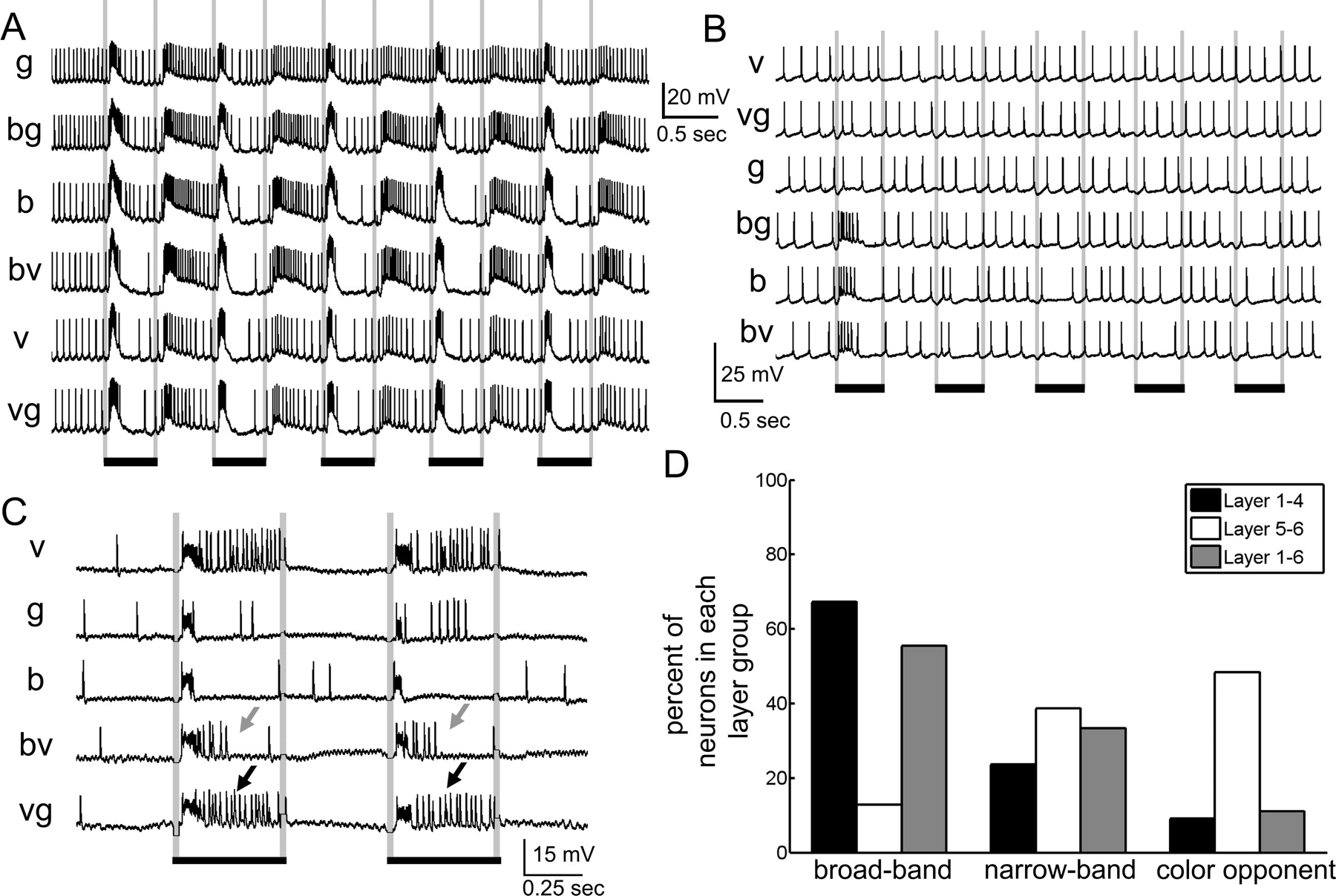
- Figure 5.
Responses to chromatic cues. We found three chromatic response types. A, Broad-band chromatic selectivity, where the neuron responded similarly to all colors and color combinations regardless of color. B, Narrow-band chromatic selectivity, where the neuron responded to one or two of the colors, but not all three. C, Chromatic opponency, where the responses to one color alter the response to another color. In this case, the neuron responds with a phasic burst to all colors, but with tonic activity to violet and somewhat to green. When blue and violet are presented together, the tonic activity is inhibited (gray arrows), but not when green and violet are presented together (black arrows). In all cases, black bars signify duration of the light flash. D, The percentage of chromatic response types among the responsive neurons in the layers. The layer 5–6 neurons have the highest percentage of color-sensitive neurons (narrow-band and color opponent), whereas most of the layer 1–4 and layer 1–6 neurons are mostly broad-band chromatically selective.
- Figure 6.
Motion response types. A–D, In these examples, the neurons were presented a large moving bar (A, B) or a wide-field moving grating (C, D). The spike rates significantly changed in response to the motion cues presented. To examine the directionality, we plotted the spike rates for each direction on a polar plot for each stimulus. B, D, We found clear directionality sensitivities for some neurons. A, C, Other neurons did not exhibit clear directionality preferences. In all cases, black bars signify the duration of the motion cue. Numbers signify the direction of movement in degress relative to the eye. E, When we compared the response properties of neurons where color and motion cues were tested among the layers, we found different percentages of neurons which were sensitive to color, to motion, or to both. Notably, the layer 5–6 neurons were color and motion sensitive, whereas most of the layer 1–4 neurons were motion sensitive but color insensitive. The remaining tested neurons had no motion responses and were broadly sensitive to the different colors.
- Figure 7.
The four main response types. A, Phasic–tonic responses included a burst of excitation followed by tonic activity for the duration of the light flash. B, Phasic responses only involved a short response during the light flash. C, Tonic responses involved a tonic increase or decrease in the membrane potential during the light flash, which did not correlate with a significant change in spike rate. D, The on–off response type was characterized by a burst of excitation at the onset of the light flash and when the light was turned off. Calibration: 10 mV, 0.5 s (duration of the stimulus). E, The percentages of each lobula layer group exhibiting each of the response types. The majority of the tested layer 1–4 neurons (72%) exhibited a clear on–off response, whereas the majority of layer 5–6 neurons had tonic or phasic responses (72%).
- Figure 8.
Precision and reliability. A, B, Rasters of spike times across multiple trials of light flashes (indicated by the black bars) for five light flashes of each color (indicated by the b, g, v, and color combinations). A, The cell fires sparsely, but has highly precise spikes occurring at the onset and in particular the offset of the light flash. B, Another neuron only spikes in response to the onset and offset of the light flash, but with a barrage of activity, not necessarily with precise spikes. C, D, The spike-time precision and response reliability are calculated and compared across the layers for each color and color combination. C, Spike-time precision. The layer 5–6 neurons (white bars) have significantly higher spike-time precision values compared with layer 1–4 (black bars) neurons for all colors (b, g, v, b-v, and v-g) except the blue–green (b-g) color combination (for the significantly different values, p < 0.05). D, Response reliability. The layer 1–4 neurons (black bars) have significantly higher response reliability values for all colors (b, g, and v) and color combinations (b-v, b-g, and v-g) than the layer 5–6 neurons (white bars) (p < 0.05). Error bars represent SE. Asterisks indicate whether the layer 5–6 neurons are significantly different from the layer 1–4 neurons for each comparison.
- Figure 9.
Summary of the anatomical segregation of function in the bee lobula. A–D, In these schematics in a frontal (A, C) and horizontal (B, D) view, the different layout of GABA (white bands) are combined with the layout of the wide-field layer 1–4 (red), layer 5–6 neurons (blue), and columnar layer 1–6 neurons (green). These neurons predominantly exit the lobula via different tracts, which include the aott, psot, piot, and lot. E–G, The results are summarized in these diagrams. E, The layer 1–4 neuron pathway would likely be activated during the presentation of achromatic motion cues (black and white moving grating). Their responses would be reliable, less precise, and would have an on–off response to flicker. F, The layer 5–6 neuron pathway would probably be stimulated during color (blue, green, or violet light) and chromatic motion cues, and would produce highly precise responses and have phasic responses to flicker. G, The layer 1–6 neurons had variable responses, but were sensitive to color and/or motion stimuli and could have precise and/or reliable responses overall. Abbreviations are as defined in Figure 2 and Results.


Additional Files
Supplemental Data
Files in this Data Supplement:
- supplemental material - Supplemental Material


