

Article Figures & Data
Figures
- Figure 1.
Central termination of the peptidergic and nonpeptidergic afferents in the mouse and rat and their relationship to the PKCγ interneurons of inner lamina II. A–C , CGRP-immunoreactive afferents ( A ) terminate dorsal to the PKCγ-positive neurons ( B , C ). D , Nonpeptidergic afferents, labeled with the IB4 lectin, also terminate mainly dorsal to the PKCγ interneurons. E , F , In the rat, there is some overlap with the PKCγ neurons. G–I , In contrast, in the mouse, there is no overlap between the IB4 terminals ( G ) and PKCγ-immunoreactive neurons ( H , I ). Scale bar, 50 μm.
- Figure 2.
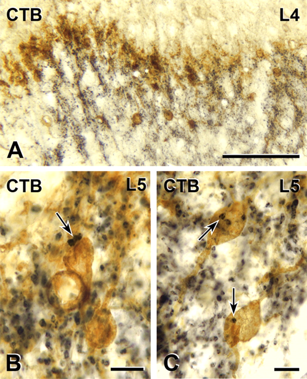
Myelinated primary afferent terminations in the rat and their relationship to the distribution of PKCγ interneurons of inner lamina II. A–C , Transganglionic transport of CTB (black) reveals dense termination in the neck of the dorsal horn, laminae III and IV as well as IIi, which includes the dense band of PKCγ-immunoreactive neurons (brown; A ). Some of the CTB-labeled terminals form close appositions (arrows) on PKCγ-immunoreactive cell bodies (brown) in lamina IIi ( B , C ). Scale bars: A , 100 μm; B , C , 10 μm.
- Figure 3.
Myelinated primary afferents that express VGLUT1 (in the rat) terminate among PKCγ interneurons. A–C , VGLUT1-immunoreactive axons occur in the layer containing PKCγ-immunoreactive interneurons (yellow; C ). D , Two VGLUT1-immunoreactive terminals (arrow) form close appositions on the cell body of a PKCγ-immunoreactive neuron (brown) that lies within the dense band of PKCγ neurons in lamina IIi (taken from the L3 segment). E , In segment L3 as in other spinal segments, the dense band of PKCγ neurons in lamina IIi is heavily innervated by VGLUT1-immunoreactive axons. Scale bars: A–C (in C ), D , 10 μm; D, 10 μm; E , 50 μm.
- Figure 4.
VGLUT1 terminals in the rat are presynaptic to PKCγ interneurons. A , B , VGLUT1-immunoreactive axon terminals form synapses (arrowheads) onto the dendrites of some PKCγ neurons in the dorsal horn of spinal cord segments C6 ( A ) and L3 ( B ). VGLUT1 immunoreactivity is denoted by the amorphous electron-dense peroxidase reaction product and PKCγ immunoreactivity by the presence of TMB-tungstate crystals (stars). Unlabeled dendrites (d), as well as a PKCγ-immunoreactive dendrite, receive synapses (arrowheads) from each VGLUT1 terminal. Scale bars, 500 nm.
- Figure 5.
Nerve-injury-triggered expression of the transneuronal tracer WGA in myelinated sensory neurons of the mouse results in transneuronal labeling of spinal PKCγ interneurons. A , Before injury, WGA (red) is expressed at very low levels in DRG neurons of the ZW2 transgenic mouse. B , Axotomy induces expression of WGA in large number of sensory neurons, ipsilateral to the injury. C , Most of the WGA-immunoreactive neurons are myelinated (i.e., are N52 positive; yellow). D , Consistent with the central projection of myelinated DRG neurons, the WGA-positive terminals (red) are concentrated in the medial regions of the deep laminae of the dorsal horn. However, the WGA immunostaining also overlapped extensively with the band of PKCγ interneurons (green) in lamina IIi. E , F , Higher magnification of the white boxes in D shows that some of these PKCγ interneurons contain the WGA tracer (arrows), indicating that they are directly postsynaptic to myelinated primary afferent fibers. Note that some of the scattered PKCγ interneurons in both laminae I (arrow in D ) and IIi also receive inputs from myelinated sensory neurons. Scale bar: (in F ) A–D , 100 μm; E , F , 6.25 μm.
- Figure 6.
Walking on a rotarod induces Fos expression in PKCγ interneurons of the rat. Double-label immunocytochemistry for Fos (black) and PKCγ (brown) is shown. A , B , The number of Fos-positive neurons in the superficial and deep dorsal horn in mice that walked on the rotating rod ( B ) was significantly greater than the number recorded in mice that rested in their cages ( A ). C , Higher-magnification images demonstrate that there is no overlap between Fos (arrow) and PKCγ-immunoreactive interneurons in control mice (arrowhead). D , There is, however, a significant increase in the number of neurons that double label for Fos and PKCγ in animals that walked on the rotarod (arrows). Note that not all PKCγ-immunoreactive neurons express Fos in the rotarod animals ( D , arrowheads). Scale bars: (in B ) A , B , 50 μm; (in D ) C , D , 10 μm.
- Figure 7.
In mouse and rat, noxious stimulation does not induce Fos expression in PKCγ interneurons. Ninety minutes after unilateral injection of formalin to the hindpaw, Fos expression is increased in the spinal cord in both superficial and deep laminae of the spinal cord. A , B , Although we observed a few Fos-immunoreactive neurons in the layer of interneurons that contain PKCγ cells in the rat, we never found double-labeled cells. C , D , In the mouse, all noxious stimulus-evoked Fos immunoreactivity was outside of the PKCγ layer of interneurons. Scale bars, 100 μm.
- Figure 8.
Schematic diagram illustrating the discrete laminar organization of the central projections of unmyelinated and myelinated afferents in the superficial dorsal and their relationship to the band of PKCγ-expressing interneurons. Peptidergic primary afferents denoted by the CGRP (green) population target projection neurons and interneurons in laminae I and outer II. Nonpeptidergic primary afferents, denoted by their binding of IB4 (blue), target neurons just ventral to the CGRP terminal zone, in inner lamina II. The IB4 terminals do not target the PKCγ band of interneurons. Instead, myelinated afferents that express the vesicular glutamate transporter (VGLUT1; red) target and synapse onto the PKCγ interneurons, which are located in the most ventral part of inner lamina II. The current results provide further evidence for the remarkable stratification of the superficial dorsal horn and indicate that non-noxious stimuli conveyed by myelinated fibers provide input to the PKCγ interneurons.


Additional Files
Supplemental Data
Files in this Data Supplement:
- supplemental material - Supplemental Figure


