

Article Figures & Data
Figures
- Figure 1.
Atoh1, Neurog1, and CMV enhancers direct GFP expression to commissural neurons/axons in the embryonic chick spinal cord. A, B, Unilateral electroporation of Atoh1tauGFP (A) or Neurog1tauGFP (B) reporter constructs into E2.5 (stage 18–19) chick embryos labels dorsal domains of the spinal cord that contain commissural neurons, which extend axons toward and across the FP in transverse vibratome sections derived from these embryos at E5 (stage 25–26). C, In contrast, unilateral electroporation of a CMV–GFP construct labeled a large number of cells broadly distributed along the D-V axis of the spinal cord. VF, Ventral funiculus; LF, lateral funiculus. D, E, Transverse vibratome sections containing dorsolateral spinal cord regions of chick embryos electroporated as in A and B were labeled with either anti-Lhx2/9 (D) or anti-Lhx1/5 (E). In each of these rows, the first panel represents GFP expression, the second panel represents either Lhx2/9 or Lhx1/5 expression, and the third and fourth panels represent the merge of the first two panels. As shown at high magnification (of white boxed regions in third panels) in the fourth panels of each row, GFP labeling of dI1 (Atoh1tauGFP) and dI2 (Neurog1tauGFP) cell bodies surrounds Cy3 nuclear Lhx2/9 or Lhx1/5 expression, respectively. Scale bars: (in C; D, first panel) A–C; D, E, first three panels, 100 μm; (in D, fourth panel) D, E, fourth panels, 25 μm.
- Figure 2.
dI1 and dI2 commissural axons elaborate ILC and MLC projections on the contralateral side of the FP. A, Schematic representing an open-book view of the spinal cord after the electroporation of GFP reporter constructs. In wild-type embryos, postcrossing MLC axons extend alongside the contralateral margin of the FP and ILC axons project a significant distance away from the FP. a, Anterior; p, posterior; fp, floor plate; rp, roof plate. B–D, Open-book preparations derived from E5 chick embryos unilaterally electroporated with Atoh1tauGFP (B), Neurog1tauGFP (C), or CMV–GFP (D) reporter constructs. In each case, the labeled postcrossing axons elaborate both MLC (arrowheads) and ILC (arrows) projections. Scale bar, 100 μm.
- Figure 3.
Robo1 and Robo2 are preferentially expressed on postcrossing segments of commissural axons that arise from dorsal interneurons in the chick spinal cord. A, B, Fluorescent in situ hybridization performed on transverse cryosections derived from E4 chick spinal cords with chick robo1 (A) and robo2 (B) riboprobes reveals domains of mRNA expression that coincide with the positions of dorsal interneurons (arrows). C, E, Both anti-cRobo1 and anti-cRobo2 strongly label the decussated commissural axon-rich marginal zone (arrows), and weakly label precrossing commissural axons (arrowheads) in transverse cryosections derived from E5 chick embryos. D, F, Preincubating anti-cRobo1 and anti-cRobo2 with their peptide immunogens for 1 h significantly diminished the binding of these antibodies to E5 chick spinal cord sections. Conversely, anti-cRobo1 and anti-cRobo2 staining is not attenuated by preabsorption with the Robo2 and Robo1 peptide immunogens, respectively (data not shown). Scale bars, 100 μm.
- Figure 4.
Subsets of dI1 and dI2 commissural neurons express robo1 and robo2 mRNA. A–D, Transverse cryosections derived from E4 chick embryos electroporated with either Atoh1tauGFP or Neurog1tauGFP were subjected to fluorescent in situ hybridization to detect robo1 and robo2 mRNA followed by anti-GFP labeling to identify dI1 and dI2 commissural neurons. In each row, the first panel represents GFP expression within the dorsal quadrant of the left side of the spinal cord (Fig. 1A,B), the second panel represents Cy3-labeled robo1 or robo2 mRNA expression, and the third panel is the merge of the first two panels. dI1 (A, B) and dI2 (C, D) dorsal commissural neurons express robo1 (A, C) and robo2 (B, D) mRNA (see arrows in third panel). Scale bar: (in D) A–D, 50 μm.
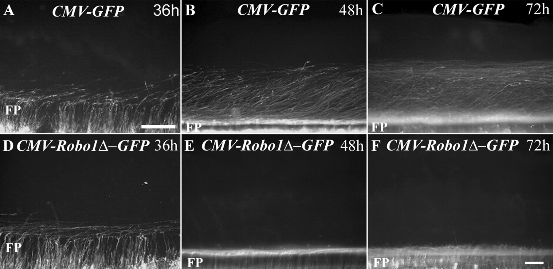
- Figure 5.
Postcrossing commissural axons expressing a truncated, cytoplasmic domain-lacking form of Robo1 fail to project away from the FP along ILC trajectories in the embryonic chick spinal cord. CMV–GFP (control) and CMV-Robo1Δ–GFP constructs were separately and unilaterally electroporated into stage E2.5 chick embryos, and open-book preparations were derived from these embryos 36, 48, or 72 h after electroporation. A–C, In embryos electroporated with the CMV–GFP reporter construct, a significant number of commissural axons have crossed the floor plate and begun to extend away from the VM at E4, 36 h after electroporation (A). Between E5 (48 h) (B) and E6 (72 h) (C), the postcrossing segments of these axons elaborate ILC and MLC projections. D–F, In CMV-Robo1Δ–GFP-treated embryos, GFP-labeled axons successfully cross and turn into the longitudinal plane alongside the FP at E4, 36 h after electroporation (D) (observed in 10 of 10 embryos). Although these axons continue to extend adjacent to the contralateral margin of the FP along MLC trajectories, they do not project away from the VM between E5 (48 h) (E) (observed in 11 of 11 embryos) and E6 (72 h) (F) (observed in 6 of 6 embryos). Scale bars, 100 μm.
- Figure 6.
Postcrossing dI1 and dI2 commissural axons fail to project away from the FP in chick embryos unilaterally electroporated with a truncated form of Robo1. A, D, Open-book preparations derived from E5 chick embryos that had been coelectroporated with CMV–GFP and Atoh1taumCherry (A) or Neurog1taumCherry (D) at E2.5 contain labeled, postcrossing dI1 and dI2 axons, which have projected away from the FP along ILC and MLC trajectories. In an E5 chick embryo that had been coelectroporated with CMV-Robo1Δ–GFP and Atoh1taumCherry (B, C) (8 of 8 embryos) or Neurog1taumCherry (E, F) (5 of 5 embryos) at E2.5, both GFP- (B, E) and mCherry-labeled (C, F), postcrossing axons exclusively project adjacent to the contralateral margin of the FP along MLC trajectories. Scale bars, 100 μm.
- Figure 7.
Postcrossing commissural axons on either side of the spinal cord fail to project away from the FP after unilateral electroporation of a truncated form of Robo1. A, Schematic depicting an open-book view of the spinal cord after the coelectroporation strategy described below. The dashed lines represent wild-type ILC projections, and the solid lines represent the trajectories that these axons adopt after unilateral electroporation of truncated Robo1. a, Anterior; p, posterior; fp, floor plate; rp, roof plate. A, B, Neurog1taumCherry (red axons in schematic) was unilaterally electroporated into E2.5 chick embryos, and ∼2 h later the opposite side of the spinal cord was electroporated with CMV-Robo1Δ–GFP (green axons in schematic). At E5, postcrossing GFP-labeled axons misexpressing a truncated form of Robo1 do not project away from the FP (B) and postcrossing dI2 mCherry-labeled axons emanating from cell bodies on the opposite side of the spinal cord also fail to project away from the FP (observed in 11 of 11 embryos). C, Open-book preparations derived from embryos coelectroporated with CMV–GFP and Neurog1taumCherry. Postcrossing axons on either side of the spinal cord elaborate wild-type MLC and ILC projections. Scale bar, 100 μm.
- Figure 8.
Commissural axons fail to cross the VM after unilateral electroporation of FL Robo2. A, C, Open-book preparations derived from E4–E4.5 chick embryos electroporated with a control CMV-Del-Npn1-myc plasmid (A) (8 of 8 embryos) or the Neurog1tauGFP construct (C) (9 of 9 embryos) at E2.5 display wild-type ILC and MLC axons. B, In embryos electroporated with CMV-Robo2FL-myc, essentially all myc-labeled axons fail to cross the FP (6 of 6 embryos). The two axons that do cross the FP follow aberrant trajectories on the contralateral side of the midline and this is indicative of the pathfinding behavior displayed by these rare axons. D, After the coelectroporation of Neurog1tauGFP and CMV-Robo2FL-myc, fewer labeled axons crossed the FP (D, inset) (9 of 9 embryos) compared with embryos electroporated with only Neurog1tauGFP (C, inset). E, Bar graph quantifying the number of axons that cross the FP in embryos electroporated with Neurog1tauGFP or Atoh1tauGFP or coelectroporated with Neurog1tauGFP or Atoh1tauGFP and CMV-Robo2FL-myc. Approximately twofold fewer axons cross the FP in the embryos coelectroporated with Neurog1tauGFP or Atoh1tauGFP and CMV-Robo2FL-myc. Error bars indicate SEM. *p < 0.0001; **p < 0.0001. Scale bar, 100 μm.
- Figure 9.
In embryos coelectroporated with FL Robo2 and Neurog1tauGFP, an increased number of longitudinally oriented, GFP-expressing axons are present on the ipsilateral side of the spinal cord. A, In wild-type Neurog1tauGFP-electroporated embryos, a small number of longitudinally projecting axons are present on the ipsilateral side of the VM (A, arrow). B, In embryos coelectroporated with Neurog1tauGFP and CMV-Robo2FL-myc, there is an increase in the number of longitudinally oriented axons on the ipsilateral side of the spinal cord (B, arrow) (9 of 9 embryos). A′, B′, Vertical line scans, represented by the white dashed lines in each open book in A and B, were performed to obtain YZ planes that captured, in cross section, axons projecting in the longitudinal plane (note: the dashed lines in A and B are superficial, and the line scans shown in A′ and B′ are not all derived from the images in A and B). Three examples of vertical line scans are shown for Neurog1tauGFP- and Neurog1tauGFP/Robo2FL-myc-electroporated embryos together with their respective “object” overlay images (i′–vi′) (see Materials and Methods). The green outlines represent the objects isolated for each line scan. The object analysis was performed to obtain a measure of the relative number of longitudinally projecting axons, to omit random noise or debris, and to avoid counting the rare transversely projecting axons that happen to overlap with an individual YZ line scan (A′i′, B′vi′, asterisks). The orientation of each line scan is demarcated by the position of the FP. We observed approximately twofold more longitudinally projecting axons in embryos electroporated with Neurog1tauGFP/Robo2FL-myc compared with those electroporated with Neurog1tauGFP (Table 1). Scale bars: (in A) A, B, 100 μm; (in B′) A′, B′, 20 μm. ipsi, Ipsilateral.


Tables
- Table 1.
In embryos coelectroporated with Neurog1tauGFP and CMV-Robo2FL-myc (Rb2), compared with embryos electroporated with Neurog1tauGFP (WT), more axons turn into the longitudinal plan on the ipsilateral side of the FP
Groups WT mean Rb2 mean WT median Rb2 median Sample size Vertical objects 8.25 19.45 8a* 16a* WT: 24 Rb2: 31 Horizontal objects 34.83 38.25 33.5a** 32a** WT: 6 Rb2: 8 Ratio: vertical/horizontal 0.24 0.51 0.24b*** 0.50b*** WT: 24 Rb2: 31 Rb2 ratio/WT ratio: 2.13 Rb2 ratio/WT ratio: 2.08 -
We compared the number of GFP-positive axons projecting into the longitudinal plane on the ipsilateral side of the FP in open-book preparations obtained from embryos electroporated with Neurog1tauGFP (WT) or coelectroporated with Neurog1tauGFP/CMV-Robo2FL-myc (Rb2). This was done by first counting the number of objects captured in the vertical line scans (which represent longitudinally projecting axons) and the number of objects captured in the horizontal line scans (which represent electroporated cell bodies). The ratios of vertical-to-horizontal objects were then calculated using the means and medians of these values for the two different groups. In all cases, object analysis was performed as described in Materials and Methods.
-
↵a,bStatistical significance of the medians and the median value ratios: atwo-tailed and bone-tailed.
-
Highly significant: *p = 0.009, ***p < 0.0001; not significant: **p = 0.9497.
-
Supplemental Data
Files in this Data Supplement:
- supplemental material - Supplemental Legend
- supplemental material - Supplemental Figure 1
- supplemental material - Supplemental Figure 2
- supplemental material - Supplemental Figure 3
- supplemental material - Supplemental Figure 4
- supplemental material - Supplemental Figure 5
- supplemental material - Supplemental Figure 6
- supplemental material - Supplemental Figure 7
- supplemental material - Supplemental Figure 8


