

Article Figures & Data
Figures
- Figure 1.
The C-terminal 7 aa of GluR1 are required for its association with SAP97; both proteins are found in the neonatal spinal cord. A , HEK 293 cells were cotransfected with expression vectors for SAP97 + GFP-tagged GluR1 or GFP-tagged GluR1Δ7, and lysates were immunoprecipitated with anti-SAP97 and probed with anti-GFP, anti-GluR1, or anti-SAP97. Full-length GluR1 (detected with either anti-GFP or anti-GluR1), but not GluR1Δ7, could be pulled down with anti-SAP97. B , Spinal cord lysates from WT or GluR1Δ7 P7 mouse pups were immunoprecipitated with anti-SAP97 and blotted with anti-GluR1 or anti-SAP97. Endogenous full-length GluR1, but not GluR1Δ7, is pulled down with anti-SAP97. Levels of expression of GluR1, GluR1Δ7, and SAP97 are equivalent in the mice of either genotype. C , Spinal cord lysates from WT P7 pups were immunoprecipitated with anti-SAP97 or no primary antibody and probed with anti-SAP97, anti-GluR1, or anti-CASK. Protein complexes containing GluR1, SAP97, and CASK were pulled down only when immunoprecipitating with anti-GluR1. The level of GluR1 in the starting material (input) was equivalent. D , Lysates from HEK 293 cells heterologously expressing the α-isoform or β-isoform of SAP97 were run along with P7 spinal cord lysates and blotted for SAP97. Spinal cord SAP97 migrates at the same molecular weight as the β-isoform, indicating that this is the predominantly expressed endogenous species. E , SAP97 is present in lysates from WT mouse spinal cord at P7, P14, and P21 and mouse brain at P14 and P21. GluR1 is expressed at a lower level in the spinal cord at P7–P14 in comparison with the brain. F , Immunostaining of the P7 spinal cord with anti-SAP97 reveals staining of motor neurons in the ventral horn. (Photomicrograph printed with permission from Dr. Maria Rubio, University of Connecticut, Storrs, CT.) IP, Immunoprecipitation; GM, gray matter; WM, white matter.
- Figure 2.
GluR1Δ7 traffics normally to the plasma membrane, where it properly oligomerizes with other AMPA receptor subunits, whereas SAP97 is not detectable in synaptic membranes of GluR1Δ7 mice. A , An EndoH-sensitive pool of GluR1 and other GluR subunits is found in the P3 fraction but not SPM fraction of P7 spinal cord lysates from WT animals and animals bearing the GluR1Δ7 allele (1 or 2 copies; +/Δ7 or Δ7/Δ7, respectively). B , Whereas GluR1, GluR1Δ7, and GluR2/3 are found in the P3 and, to a greater extent, SPM fractions regardless of genotype, SAP97 is not detected in the SPM fraction from the GluR1Δ7 mice. C , Two other components of the postsynaptic density, PSD-95 and stargazin, are enriched in the SPM fraction regardless of genotype. The cytosolic protein HSP90 was excluded from the SPM fraction and is a minor contaminant of the P3 fraction. HSP90 is equivalently abundant in the total lysate and the cytosolic fraction. D , Using SPM as the material source, immunoprecipitation with anti-GluR2/3 reveals that GluR1 is in association with AMPA receptor subunits GluR2 and/or GluR3 but not the kainate receptor subunit KA2. E , Lysates of P7 spinal cord from WT and GluR1 null mice were fractionated and subjected to Western blot analysis. No GluR1 is detected in any fraction from the null mice, whereas in the WT mouse, GluR1 is found in the initial lysate, SPM, and, to a lesser extent, the P3 fraction. GluR2/3 immunoreactivity is present in all three fractions of WT and null mice. SAP97 is found in all fractions of WT mice but not in the SPM fraction of the null mice. cyto, Cytosolic; IP, immunoprecipitation.
- Figure 3.
Immunocytological localization of GluR1 or GluR1Δ7 with SAP97. Spinal cord cultures were transfected with expression vectors for N-terminally myc-tagged GluR1 or GluR1Δ7 and SAP97. Five days later, live labeling with anti-myc was performed to identify cell surface GluR1 or GluR1Δ7, and after fixation, this was combined with anti-SAP97 staining. Left, Representative laser confocal images (at low and high power) of the localization of WT GluR1 and SAP97. At low power (top triptych), puncta of cell surface GluR1 (red) decorate neurons, and SAP97 has a similar punctate distribution (green). Scale bar, 40 μm. At higher magnification (bottom triptych), accumulation of cell surface GluR1 into discrete puncta (arrows) along a dendrite can be seen. The SAP97 (green) images reveal numerous puncta of immunoreactivity, and some coregister with the GluR1 puncta (arrows). The bottom image (merge of GluR1 and SAP97 immunocytology) shows that many GluR1 puncta have close association with SAP97 (circles). Scale bar, 3 μm. Right, Representative laser confocal images (at low and high power) of the localization of GluR1Δ7 and SAP97. At low power (top triptych), puncta of cell surface GluR1 (red) decorate neurons, and SAP97 similarly has a punctate distribution (green). Scale bar, 40 μm. At higher magnification (lower triptych), accumulation of cell surface GluR1 into discrete puncta (arrows) along a dendrite can be seen. The SAP97 (green) images reveal numerous puncta of immunoreactivity, but in contrast with WT GluR1, there is no coregistration of the GluR1 puncta with the SAP97 puncta (arrows). The bottom image (merge of GluR1 and SAP97 immunocytology) shows that most GluR1 puncta are independent of SAP97 puncta. A single potential colocalization of GluR1Δ7 with SAP97 (merge; arrow) is different in appearance from colocalization of WT GluR1 and SAP97 and is likely to have occurred by chance. Scale bar, 3 μm.
- Figure 4.
Overexpression of SAP97 with WT GluR1 (but not GluR1Δ7) leads to synergistic dendrite growth-promoting effects. Top, Representative camera lucida images of neurons expressing GFP alone, SAP97 alone, WT GluR1 + SAP97, and GluR1Δ7 + SAP97. Scale bar, 35 μm. Bottom, The chart provides quantitative analysis of dendrites as well as statistical analysis using ANOVA. The number of neurons drawn is noted in parentheses next to the column title. There is a statistically significant increase in branching when SAP97 is expressed in neurons compared with GFP-expressing neurons. Coexpression of WT GluR1 + SAP97 leads to a marked increase in dendrite branching as well as increase in overall arbor size and average length of dendrites. This growth-promoting effect was not seen when GluR1Δ7 was coexpressed with SAP97.
- Figure 5.
Selective knockdown of SAP97 causes dendritic tree atrophy and prevents GluR1-dependent dendrite growth. Top, Western blots from HEK 293 cells transfected with expression vectors for PSD-93, PSD-95, SAP97, or SAP102 and either the active shRNA to SAP97 or a scrambled sequence shRNA. The active shRNA led to the selective knockdown of SAP97 and did not affect the expression of other members of the postsynaptic density family of proteins. Middle, Representative camera lucida images of neurons expressing GFP + scrambled shRNA, GFP + active shRNA, WT GluR1 + scrambled shRNA, or WT GluR1 + active shRNA. Scale bar, 40 μm. Bottom, The chart provides quantitative analysis of dendrites as well as statistical analysis using ANOVA. The number of neurons drawn is noted in parentheses next to the column title. There is a statistically significant decrease in branching, overall arbor, and average dendrite size in the GFP + active shRNA-expressing neurons in comparison with neurons expressing GFP + scrambled shRNA. There was a statistically significant increase in dendrite branching in neurons expressing WT GluR1 + scrambled shRNA in comparison with neurons expressing GFP + scrambled shRNA as seen previously. This growth-promoting effect was eliminated when WT GluR1 was coexpressed with the active shRNA. There were no statistically significant differences in the dendritic tree of neurons expressing GFP + active shRNA versus WT GluR1 + active shRNA.
- Figure 6.
Synergistic dendrite growth when P-SAP97 is coexpressed with WT GluR1 or GluR1Δ7. Top, Representative camera lucida images of neurons expressing GFP alone, P-SAP97 alone, WT GluR1 + P-SAP97, or GluR1Δ7 + P-SAP97. Scale bar, 22 μm. Bottom, The chart provides quantitative analysis of dendrites as well as statistical analysis using ANOVA. The number of neurons drawn is noted in parentheses next to the column title. There is a statistically significant increase in branching when P-SAP97 is expressed in neurons compared with GFP-expressing neurons. Coexpression of P-SAP97 + WT GluR1 leads to a synergistic increase in branching as well as overall arbor and average dendrite growth. In contrast with observations in Figure 4, coexpression of P-SAP97 + GluR1Δ7 also leads to a synergistic increase in branching as well as overall arbor and average dendrite growth. There are no statistically significant differences between the dendritic arbors of P-SAP97 + WT GluR1- and P-SAP97 + GluR1Δ7-expressing neurons.
- Figure 7.
Mutations in PDZ2 of SAP97 disrupt its interaction with GluR1 but do not prevent the synergistic effects on growth if mutant SAP97 is targeted to the plasma membrane. A , Western blots of HEK 293 cells cotransfected with expression vectors for wild-type GluR1 + one of the following constructs: WT SAP97, mutant SAP97 (mutSAP97), palmitoylated (pal) wild-type SAP97, or palmitoylated mutant SAP97. Immunoprecipitation (IP) using anti-SAP97 and immunoblotting (IB) for GluR1 reveal that only WT SAP97 and palmitoylated SAP97 physically associate with GluR1. IP for SAP97 and IB for SAP97 demonstrate that all constructs immunoprecipitate equivalently and the input controls of all constructs express equivalently. B , Representative camera lucida images of neurons expressing GFP alone, GluR1 alone, GluR1 + palmitoylated WT SAP97, or GluR1 + palmitoylated mutant SAP97. Scale bar, 25 μm. C , The chart provides quantitative analysis of dendrites as well as statistical analysis using ANOVA. The number of neurons drawn is noted in parentheses next to the column title. GluR1 leads to enhancement of dendrite branching and overall tree growth, in comparison with neurons expressing GFP alone. Both palmitoylated WT SAP97 and palmitoylated mutant SAP97, when coexpressed with GluR1, cause synergistic dendrite growth. There are no statistically significant differences between the GluR1 + palmitoylated WT SAP97 and GluR1 + palmitoylated mutant SAP97.
- Figure 8.
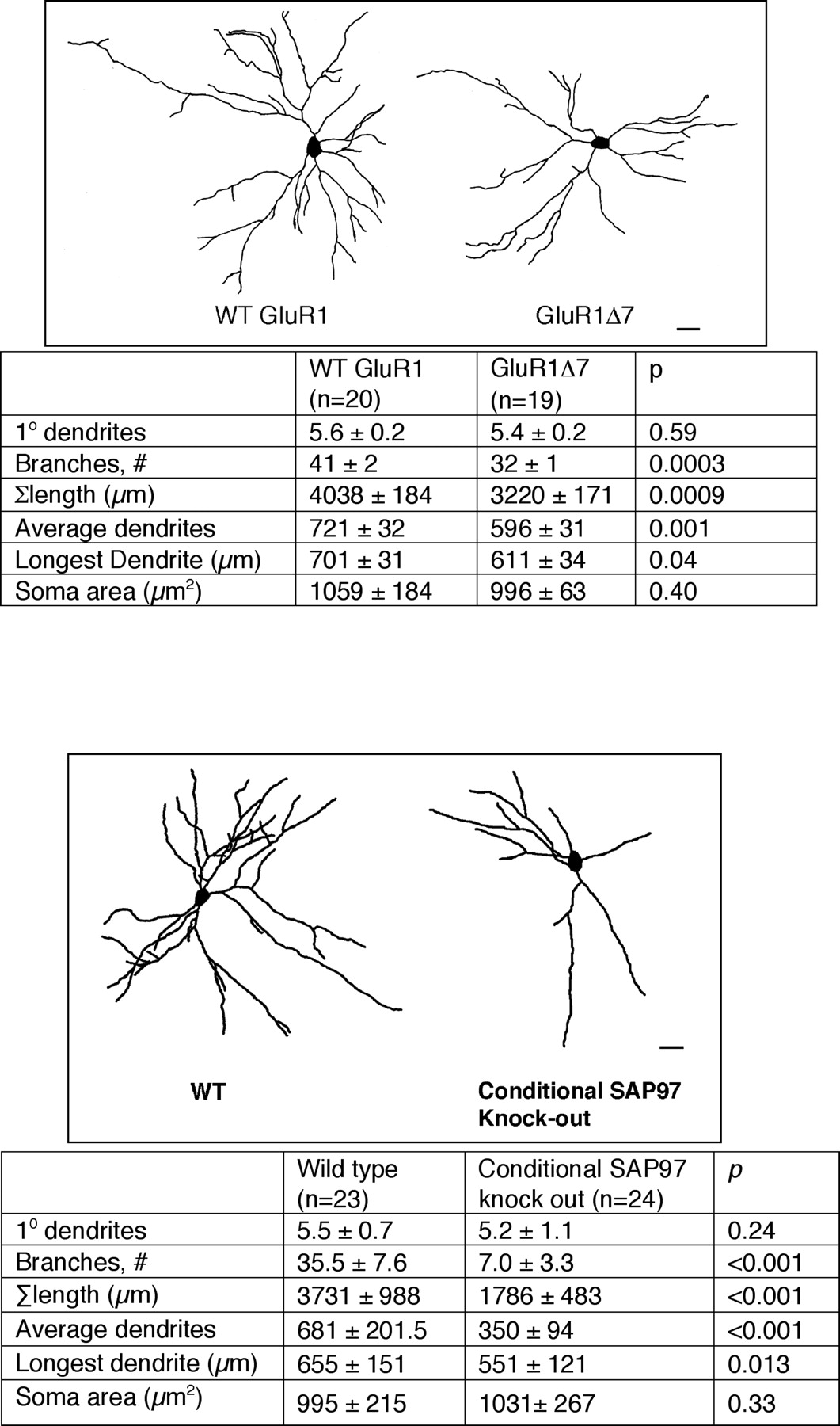
Comparison of motor neuron dendrites of WT mice versus homozygous GluR1Δ7 knock-in mice and versus conditional SAP97 knock-out mice. Top, Representative camera lucida images of motor neurons from WT mice and GluR1Δ7 knock-in mice. Scale bar, 22 μm. The chart below provides quantitative analysis of dendrites as well as statistical analysis using Student's t test. The number of neurons drawn is noted in parentheses next to the column title. In comparison with the WT animals, the motor neuron dendrites from GluR1Δ7 knock-in mice had fewer branches and a reduction in the overall arbor, the average dendrite length and the longest dendrite were also shortened. Bottom, Representative camera lucida images of motor neurons from WT mice and conditional SAP97 knock-out mice. Scale bar, 30 μm. The chart below provides quantitative analysis of dendrites as well as statistical analysis using Student's t test. The number of neurons drawn is noted in parentheses next to the column title. In comparison with the WT animals, the motor neuron dendrites from conditional SAP97 mice had fewer branches, smaller overall arbor size, reduction in average dendrite length, and shortening of the longest dendrite.
- Figure 9.
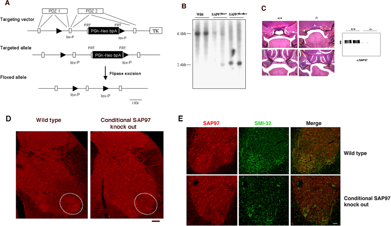
Generation and characterization of SAP97 conditional knock-out mice. A , Schematic representation of SAP97 conditional targeting strategy. B , Southern blot analysis of mouse genomic DNA from mutant mice. EcoRI digestion of wild-type genomic DNA generates a 6.8 kb fragment, and mutant genomic DNA with/without neor cassette produces a 2.4 kb fragment. C , Left, Coronal sections stained with hematoxylin and eosin of E18.5 wild-type (+/+) and CMV-Cre:SAP97LoxP/LoxP (−/−) embryos at two levels of the nasopharynx. The top row shows that the −/− mice have a cleft secondary palate as previously described in the null mice (Caruana and Bernstein, 2001). The bottom row shows that the palatal shelves are unfused in the −/− mice. Right, An immunoblot for SAP97 using brain from wild-type and conditional SAP knock-out mice. No SAP97 immunoreactivity is found in the −/− mice. D , Immunohistology of the spinal cord from wild-type and conditional SAP97 knock-out mice. SAP97 is expressed by motor neurons in wild-type but not the conditional knock-out mice (see regions denoted by ovals). There were no other discernible differences in SAP97-expressing cells between the two genotypes. Scale bar, 45 μm. E , The ventral horn of wild-type and conditional SAP97 knock-out mice stained for SAP97 (in red) and SMI32 (green). Colocalization of SAP97 in motor neurons is evident in the wild-type animals (see merged image). The conditional SAP97 motor neurons do not display SAP97 immunoreactivity. Scale bar, 55 μm.


Additional Files
Supplemental Data
Files in this Data Supplement:
- supplemental material - Supplemental Legend
- supplemental material - Supplemental Figure 1
- supplemental material - Supplemental Figure 2
- supplemental material - Supplemental Figure 3
- supplemental material - Supplemental Figure 4
- supplemental material - Supplemental Figure 5


