

Article Figures & Data
Figures
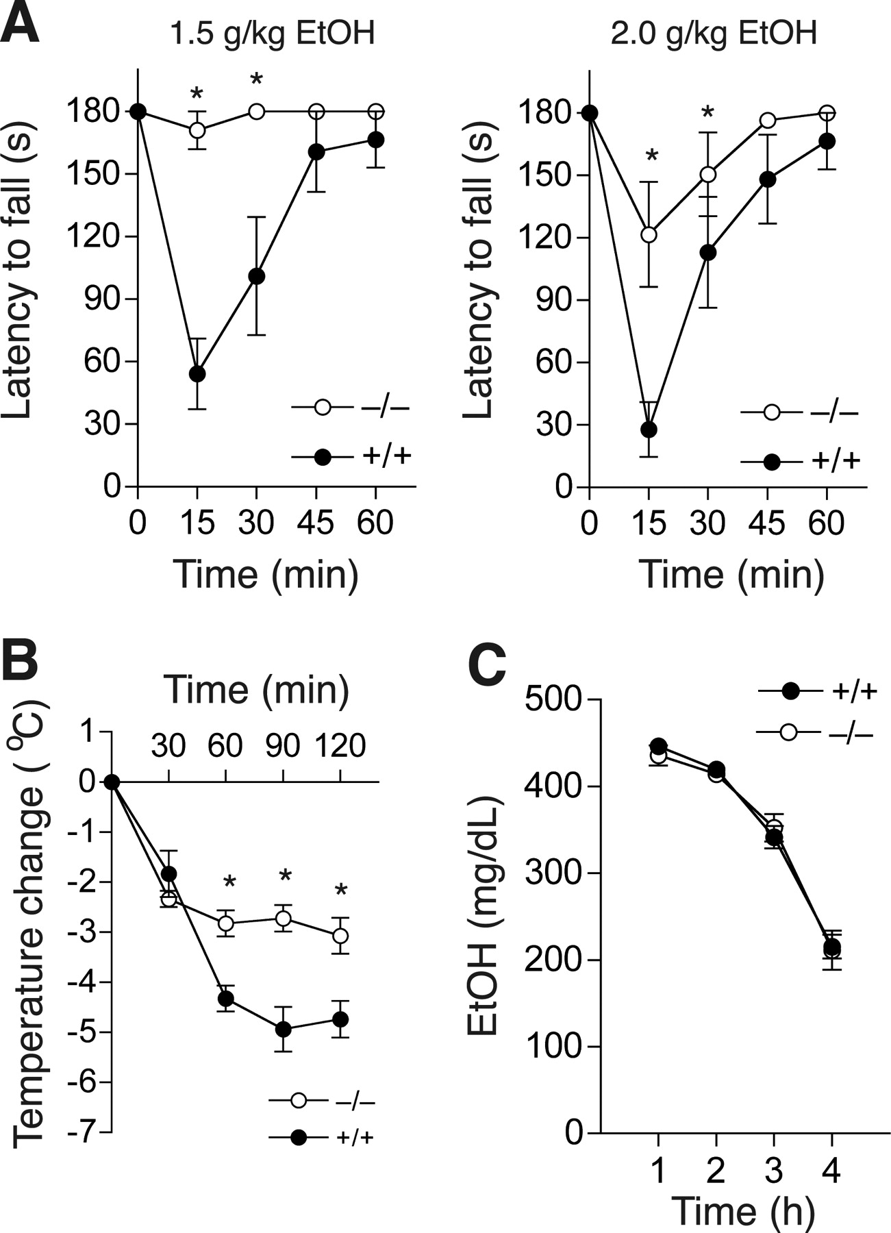
- Figure 1.
Acute responses to ethanol in PKCδ−/− mice. A , Ethanol induced much more ataxia in PKCδ+/+ mice compared with PKCδ−/− mice (n = 8 for each genotype; *p < 0.05 compared with PKCδ+/+ mice at the same time by Tukey's tests). B , Ethanol-induced hypothermia was greater in PKCδ+/+ mice compared with PKCδ−/− mice (n = 8 for each genotype; *p < 0.05 compared with PKCδ+/+ mice at the same time by Tukey's tests). C , Blood ethanol clearance after administration of 4.0 g/kg ethanol was similar in both genotypes (n = 12 for each genotype).
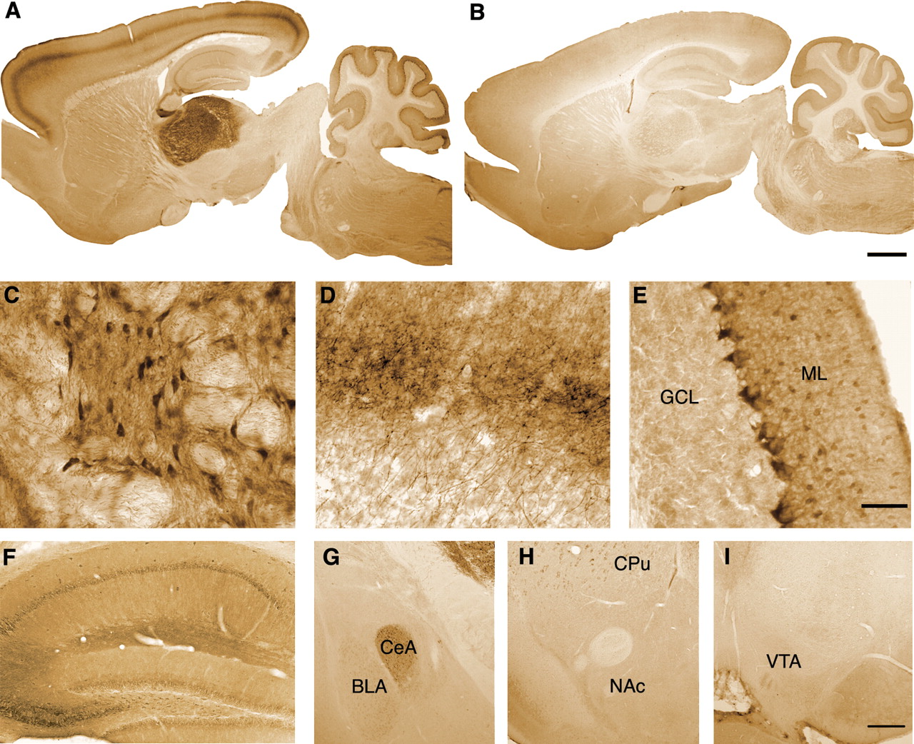
- Figure 2.
Expression of PKCδ in mouse brain. A , B , Immunoperoxidase staining for PKCδ immunoreactivity in sagittal brain sections from PKCδ+/+ ( A ) and PKCδ−/− ( B ) mice. C–E , High-power micrographs of ( C ) thalamus, ( D ) thalamocortical fibers in layer IV of cerebral cortex, and ( E ) cerebellar cortex from a PKCδ+/+ mouse. ML, Molecular layer; GCL, granule cell layer. F , G , PKCδ immunoreactivity was moderate in cell bodies of pyramidal neurons, dentate gyrus granule cells and molecular layer interneurons of the hippocampus ( F ), and in the central amygdala (CeA) ( G ). H , I , Scattered PKCδ immunoreactivity was observed in the caudate–putamen (CPu), but none was observed in the nucleus accumbens (NAc) or the ventral tegmental area (VTA). Scale bars: (in B ) A , B , 1 mm; (in E ) C–E , 50 μm; (in I ) F–I , 250 μm.
- Figure 3.
A–E , Ataxia induced by drugs that act at GABAA and NMDA receptors. Mice were tested for their ability to remain for 3 min on a rotarod treadmill rotating at a constant velocity of 20 rpm before and after intraperitoneal injection of pentobarbital ( A ), pregnanolone ( B ), flunitrazepam ( C ), ketamine ( D ), or MK-801 ( E ). n = 8 ( A , B , D ) and n = 16 ( C ) for each genotype. In E , n = 7 for PKCδ+/+ and n = 8 for PKCδ−/− mice.
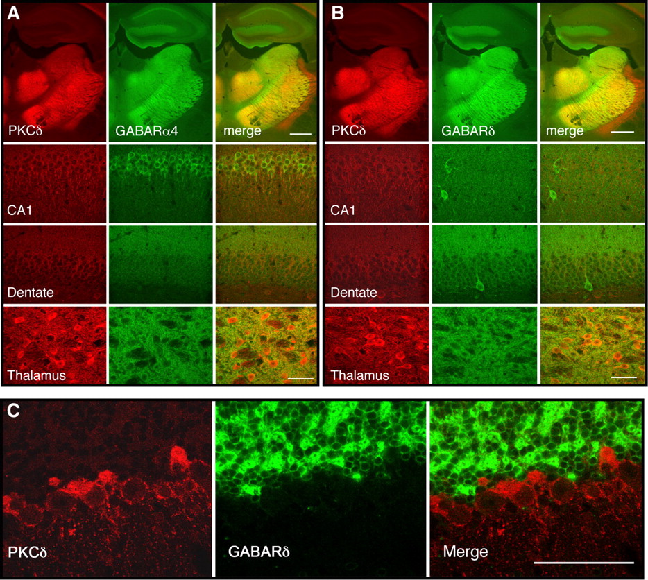
- Figure 4.
Colocalization of GABAA α4 and δ subunit immunoreactivity with PKCδ in mouse brain. A , Coronal sections through the hippocampus and thalamus showing immunoreactivity for PKCδ (red), α4 subunits (green), and their colocalization (yellow) in hippocampus and thalamus. B , Similar sections showing immunoreactivity for PKCδ (red), δ subunits (green), and their colocalization (yellow) in hippocampus and thalamus. C , Sections of cerebellar cortex showing PKCδ immunoreactivity in the Purkinje cell and molecular layers, which did not colocalize with δ subunit immunoreactivity present in the granule cell layer. Scale bars: A , B , 500 μm for low-power images, 50 μm for high-power images; C , 50 μm.
- Figure 5.
Ethanol enhances tonic inhibitory GABA currents in neurons from PKCδ+/+ but not PKCδ−/− mice. A , Sample tonic current traces recorded in a thalamic relay neuron from a PKCδ+/+ (left) and a PKCδ−/− (right) mouse. These currents were completely blocked by the nonselective GABAA receptor antagonist SR95531 (SR). B , Paired plots of tonic current amplitudes before and after the addition of 30 mm ethanol to the perfusate of thalamic neurons from PKCδ+/+ (left) and PKCδ−/− (right) mice. Filled circles (left) and squares (right) with error bars indicate mean ± SEM current values. C , Mean ± SEM values for tonic current amplitudes in PKCδ+/+ (n = 6) and PKCδ−/− (n = 12) thalamic relay (TR) neurons before (control) and after addition of 30 mm ethanol. D , Tonic current in PKCδ+/+ (n = 9) and PKCδ−/− (n = 7) dentate gyrus granule cell (DGGC) neurons and E , in PKCδ+/+ (n = 5) and PKCδ−/− (n = 9) ML interneurons (ML) before and after 30 mm ethanol. C–E , *p < 0.05 compared with the control condition in PKCδ+/+ neurons by Bonferroni tests.
- Figure 6.
PKCδ regulates ethanol sensitivity of α4β3δ GABAA receptors expressed in L(tk−) cells. A , Western blot analysis of PKCδ and actin immunoreactivity in HEK293, Neuro2A, L(tk−), and CHO cells showing less PKCδ abundance in L(tk−) cells relative to actin when compared with CHO and HEK293 cells. B , Western blot showing overexpression of PKCδ in L(tk−) cells after transfection with as-PKCδ. C , D , Ethanol enhancement of EC20 GABA-stimulated currents in L(tk−) cells that express α4β3δ receptors. *p < 0.05 compared with cells not transfected with as-PKCδ at the same concentration of ethanol by Bonferroni tests. E , Inhibition of as-PKCδ, a commercial mixture of PKC isozymes (PKC) (Calbiochem) and wild-type PKCδ by the PP1 analog 1NaPP1. Nonlinear regression analysis showed significant differences (< 0.0001) between log EC50 values (M) for 1NaPP1 inhibition of as-PKCδ (−6.81 ± 0.02) versus wild type PKCδ (−4.06 ± 0.04) or the mixture of PKC isozymes (−4.59 ± 0.04). F , L(tk−) cells that express α4β3δ receptors and as-PKCδ were treated with 0.1 μm GABA and 30 mm ethanol. Ethanol reversibly enhanced the GABA-stimulated current and addition of 10 μm 1NaPP1 reduced this effect of ethanol


Additional Files
Supplemental Data
Files in this Data Supplement:
- supplemental material - Supplemental Material
- supplemental material - Supplemental Legend
- supplemental material - Supplemental Table
- supplemental material - Supplemental Figure 1
- supplemental material - Supplemental Figure 2
- supplemental material - Supplemental Figure 3


